Why does chronic stress disrupt REM sleep? A subset of REM-active neurons in the lateral habenula
A mouse study separates two sleep-regulating outputs of the lateral habenula and shows that REM-active neurons become hyperexcitable after chronic restraint stress.
Increased rapid eye movement (REM) sleep duration, shortened REM latency, and heightened theta oscillations are among the most conserved polysomnographic endophenotypes of major depression and chronic stress models. Although the lateral habenula (LHb) is widely recognized as an anti-reward hub whose burst firing drives depressive-like states, how LHb neurons regulate physiological non-REM (NREM) versus REM sleep—and how they link chronic stress to pathological REM sleep excess and depressive behaviors—has remained unclear. On June 28, 2024, Prof. Hua Zhao's group at Jilin University together with Prof. Shuancheng Ren and Prof. Zhian Hu's group at Army Medical University (with first author Zehui Zhang) published a study in Current Biology titled "The lateral habenula regulates physiological sleep and mediates chronic stress-induced REM sleep disorder and depressive-like behaviors." Integrating polysomnographic EEG/EMG recordings, in vivo fiber photometry, tetrode single-unit recordings, activity-dependent genetic tagging (TRAP2), ex vivo slice patch-clamp electrophysiology, and pathway-specific chemo-/optogenetics, the team discovered that LHb glutamatergic neurons promote NREM sleep via projections to the rostromedial tegmental nucleus (RMTg), while a specialized subpopulation of REM-sleep-active LHb neurons projecting to the ventral tegmental area (VTA) selectively drives REM sleep. Under chronic restraint stress (CRS), membrane hyperpolarization and enhanced burst firing in these REM-active LHb neurons jointly mediate excess REM sleep and depressive-like behaviors.
This work demonstrates that the lateral habenula (LHb) regulates physiological NREM and REM sleep via distinct LHb–RMTg and LHb–VTA pathways, identifying a VTA-projecting REM-sleep-active LHb glutamatergic subpopulation whose stress-induced hyperpolarization and burst firing jointly drive REM sleep excess and depressive-like behaviors.
Contents · 7 sections
LHb Glutamatergic Neurons Bidirectionally Regulate Physiological NREM and REM Sleep
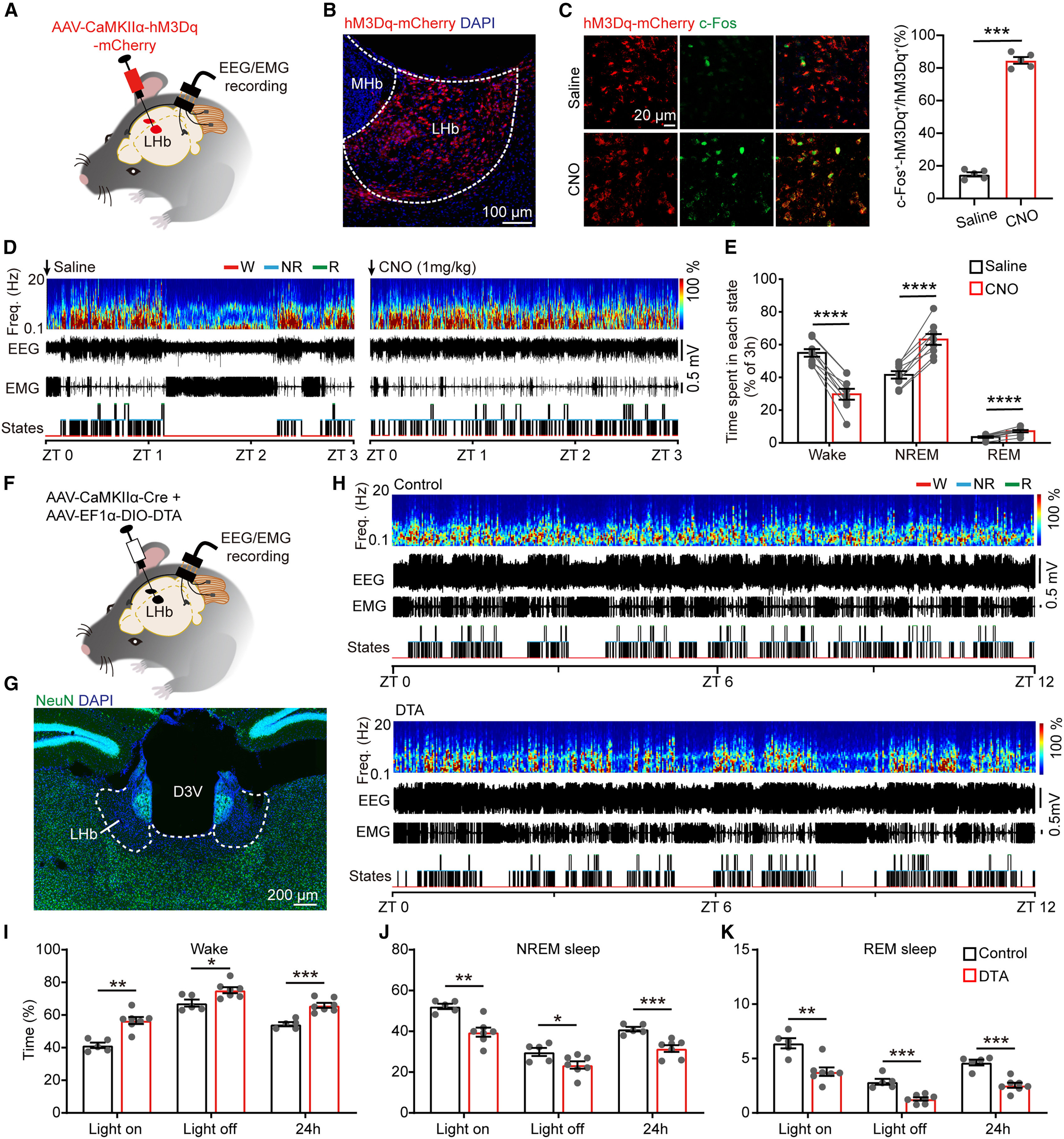
The researchers first injected AAV-CaMKIIα-hM3Dq-mCherry bilaterally into the LHb of adult male C57BL/6 mice and administered intraperitoneal CNO (1 mg/kg) at either light onset (ZT 0) or dark onset (ZT 12). Twenty-four-hour EEG/EMG monitoring demonstrated that chemogenetic activation of LHb glutamatergic neurons significantly increased both episode numbers and total duration of NREM and REM sleep across both circadian phases while reducing wakefulness.
Conversely, chemogenetic inhibition of LHb neurons via AAV-CaMKIIα-hM4Di-mCherry, chronic genetic ablation via co-injection of AAV-CaMKIIα-Cre and AAV-EF1α-DIO-DTA, or acute 590 nm optogenetic silencing during NREM sleep via AAV-CaMKIIα-ArchT-EGFP consistently reduced both NREM and REM sleep durations and promoted wakefulness, establishing that LHb glutamatergic neurons bidirectionally support physiological NREM and REM sleep.
Source: et al. · Current Biology · Original figure reproduced for academic literature review under fair scholarly commentary; copyright remains with the authors/publisher.
Original figure and English caption ↗
In Vivo Calcium Photometry and Single-Unit Recordings Reveal Peak LHb Activity During REM Sleep
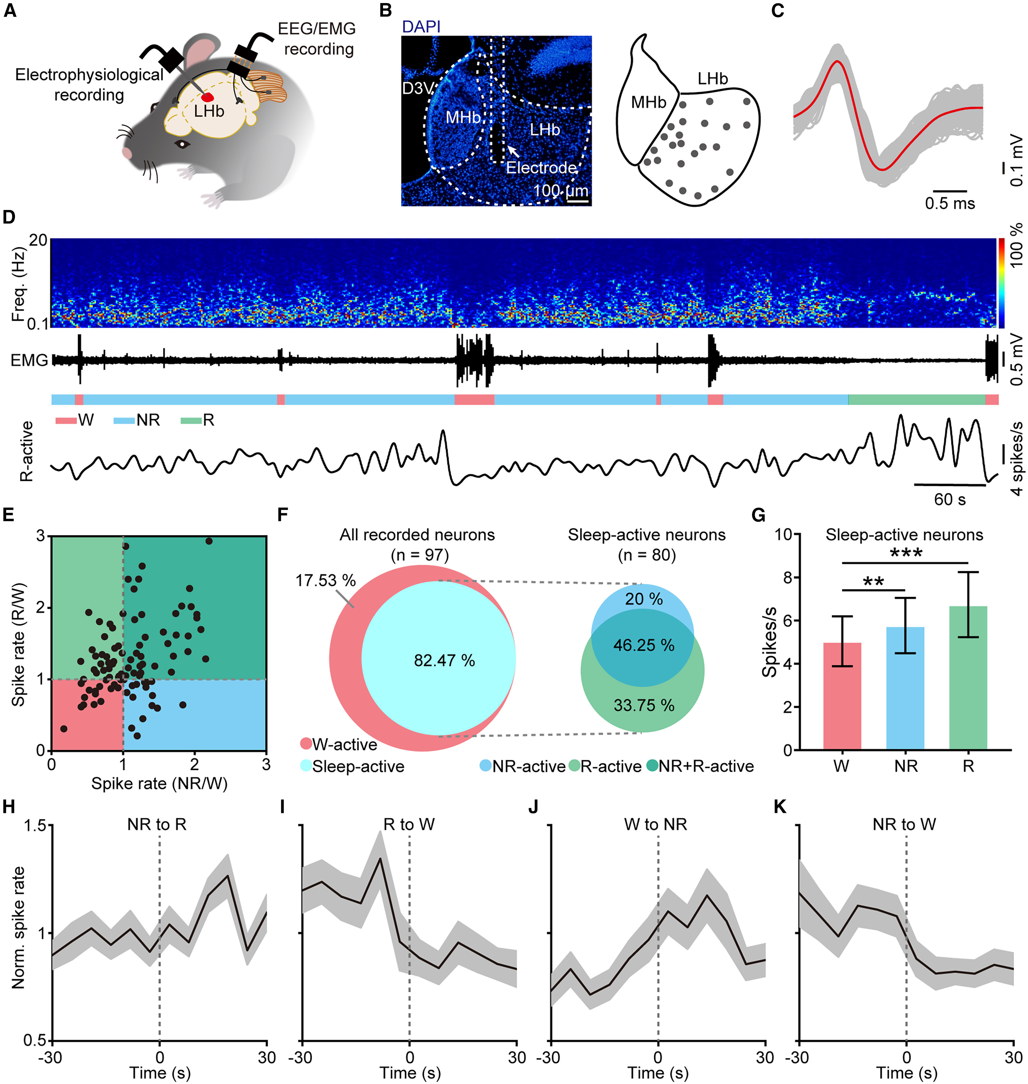
To characterize the natural dynamics of LHb neurons across sleep-wake states, the team injected AAV-CaMKIIα-GCaMP6s into the LHb, implanted ceramic optical fibers, and used an Inper dual-wavelength fiber photometry system (470 nm calcium excitation and 410 nm isosbestic control light, analyzed with Inper Data Process software) alongside simultaneous EEG/EMG recordings. Population calcium signals in the LHb peaked during REM sleep, surged robustly during NREM-to-REM transitions, and dropped sharply upon REM-to-wake transitions.
Because population photometry cannot resolve functionally heterogeneous subpopulations, the authors implanted microdrive tetrode arrays into the LHb of 14 mice and recorded 97 well-isolated single units. Clustering analysis revealed that 82.5% (80/97) of LHb units were sleep-active: among these, 33.75% (27/80) were selectively REM-sleep-active (R-active), 46.25% (37/80) were active during both NREM and REM sleep (NR+R-active), and 20% (16/80) were NREM-active (NR-active), identifying a dedicated REM-sleep-coding subpopulation within the LHb.
Source: et al. · Current Biology · Original figure reproduced for academic literature review under fair scholarly commentary; copyright remains with the authors/publisher.
Original figure and English caption ↗
Genetic Tagging of REM-Sleep-Active LHb Neurons Confirms Their Selective Control of REM Sleep
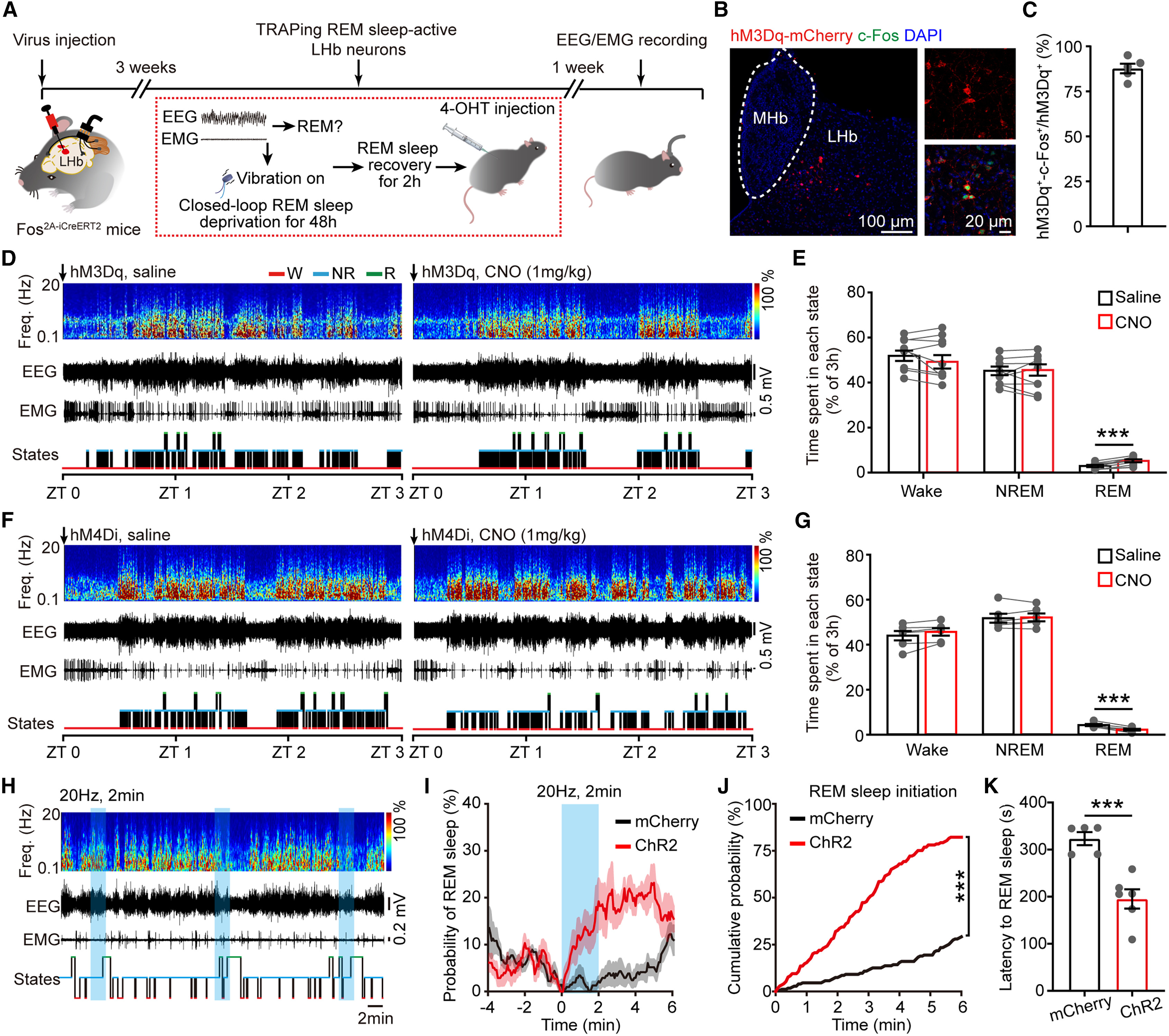
To selectively manipulate REM-sleep-active LHb neurons, the researchers used a closed-loop EEG/EMG-guided vibrational platform to subject mice to 48 hours of selective REM sleep deprivation (RSD), eliciting a pronounced REM sleep rebound during the subsequent 2-hour recovery window, and injected 4-OHT (50 mg/kg) into Fos2A-iCreERT2 (TRAP2) mice during this rebound to genetically capture REM-active neurons. Fluorescence in situ hybridization confirmed that approximately 88% of TRAPed mCherry-positive LHb neurons co-expressed c-Fos upon a second REM rebound and were almost exclusively Vglut2-positive glutamatergic neurons.
Expressing AAV-EF1α-DIO-hM3Dq-mCherry or AAV-EF1α-DIO-hM4Di-mCherry bilaterally in the LHb of TRAP2 mice revealed that CNO activation of TRAPed REM-active LHb neurons selectively increased REM sleep duration and episode numbers without altering NREM sleep or wakefulness, whereas CNO inhibition selectively reduced REM sleep. Furthermore, expressing AAV-EF1α-DIO-ChR2-mCherry and delivering 20 Hz blue-light stimulation during stable NREM sleep significantly shortened REM sleep initiation latency and increased the probability of transitioning into REM sleep.
Source: et al. · Current Biology · Original figure reproduced for academic literature review under fair scholarly commentary; copyright remains with the authors/publisher.
Original figure and English caption ↗
The LHb–RMTg Pathway Regulates NREM Sleep While the LHb–VTA Pathway Selectively Controls REM Sleep
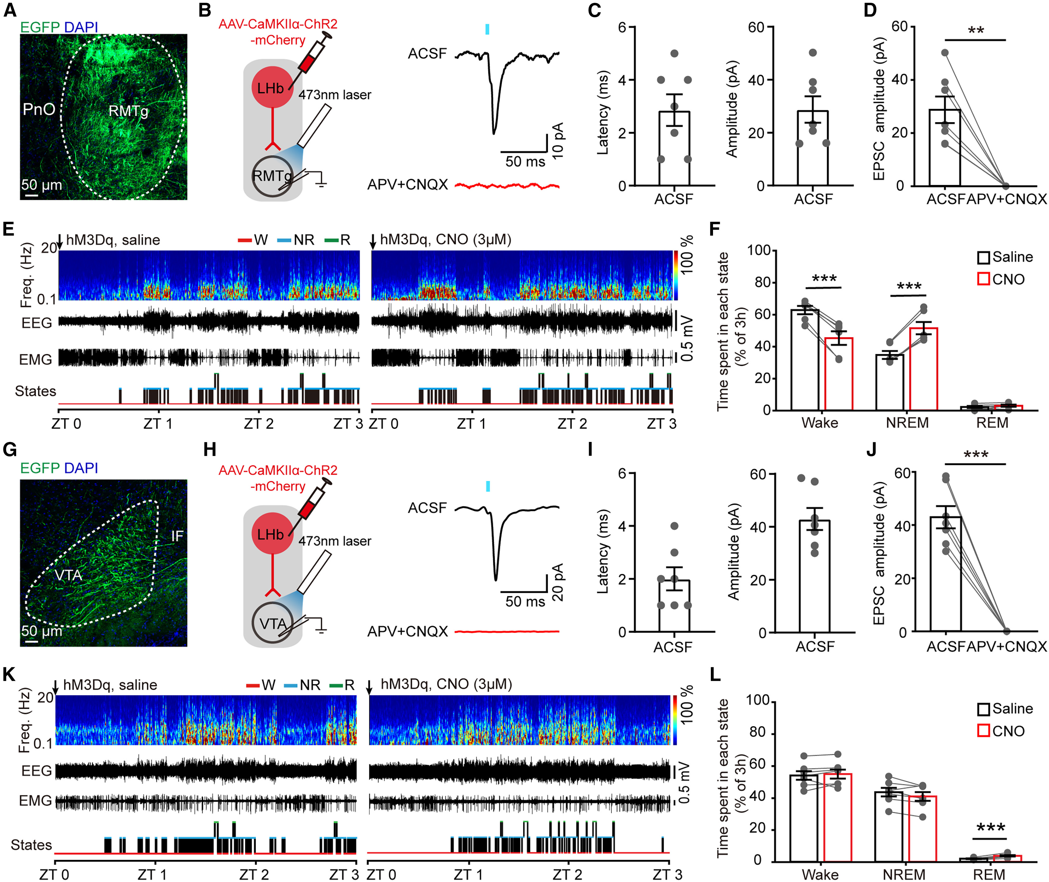
The RMTg and VTA are two major brainstem targets of the LHb. Ex vivo whole-cell slice recordings showed that photostimulating ChR2-expressing LHb axon terminals evoked APV- and CNQX-sensitive excitatory postsynaptic currents (EPSCs) in both RMTg and VTA neurons. When hM3Dq was expressed pan-neuronally in the LHb and CNO (3 μM) was locally microinfused via cannula into the RMTg to activate LHb–RMTg terminals, mice exhibited a marked increase in NREM sleep and reduced wakefulness, whereas REM sleep remained completely unchanged.
Tracing the axonal projections of TRAPed REM-active LHb neurons in TRAP2 mice revealed dense terminal arborizations in the VTA but virtually no fibers in the RMTg. Moreover, expressing hM3Dq in either pan-LHb neurons or TRAPed REM-active LHb neurons and locally infusing CNO into the VTA to activate LHb–VTA terminals selectively increased REM sleep duration and episode frequency without altering NREM sleep or wakefulness, dissecting parallel LHb output pathways for NREM versus REM sleep control.
Source: et al. · Current Biology · Original figure reproduced for academic literature review under fair scholarly commentary; copyright remains with the authors/publisher.
Original figure and English caption ↗
Chronic Restraint Stress Enhances Excitability and Burst Firing of REM-Active LHb Neurons and Induces REM Sleep Excess
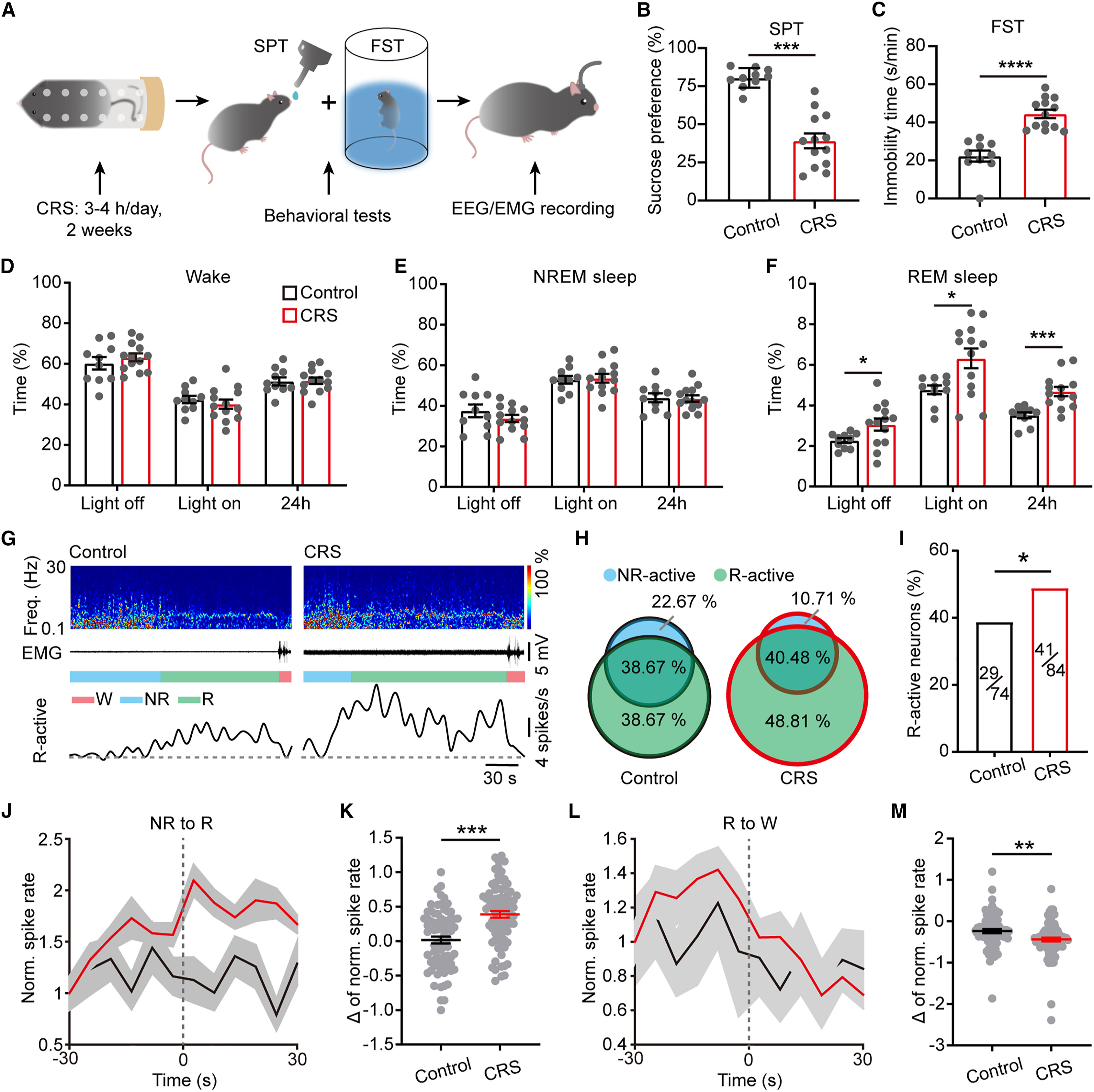
To investigate whether the LHb mediates chronic stress-induced REM sleep abnormalities, the authors subjected mice to 14 consecutive days of chronic restraint stress (CRS, 2–3 hours/day). CRS mice exhibited significantly reduced sucrose preference in the sucrose preference test (SPT) and prolonged immobility in the forced swim test (FST), accompanied by significant increases in 24-hour REM sleep duration, REM episode numbers, and REM theta-band (6–10 Hz) power.
Comparing in vivo tetrode recordings between 14 control mice (95 units) and 16 CRS mice (107 units) revealed that CRS markedly expanded the proportion of R-active LHb neurons and elevated the REM-state firing rates and NREM-to-REM transition surges of both R-active and NR+R-active units. Ex vivo patch-clamp recordings of TRAPed REM-active LHb neurons further demonstrated that CRS hyperpolarized their resting membrane potential, increased depolarizing current-evoked firing rates, and increased the fraction of spontaneously burst-firing neurons from ~10% in controls to ~46% in CRS mice.
Source: et al. · Current Biology · Original figure reproduced for academic literature review under fair scholarly commentary; copyright remains with the authors/publisher.
Original figure and English caption ↗
Inhibiting REM-Active LHb Neurons and Their VTA Projections Reverses Stress-Induced REM Sleep Excess
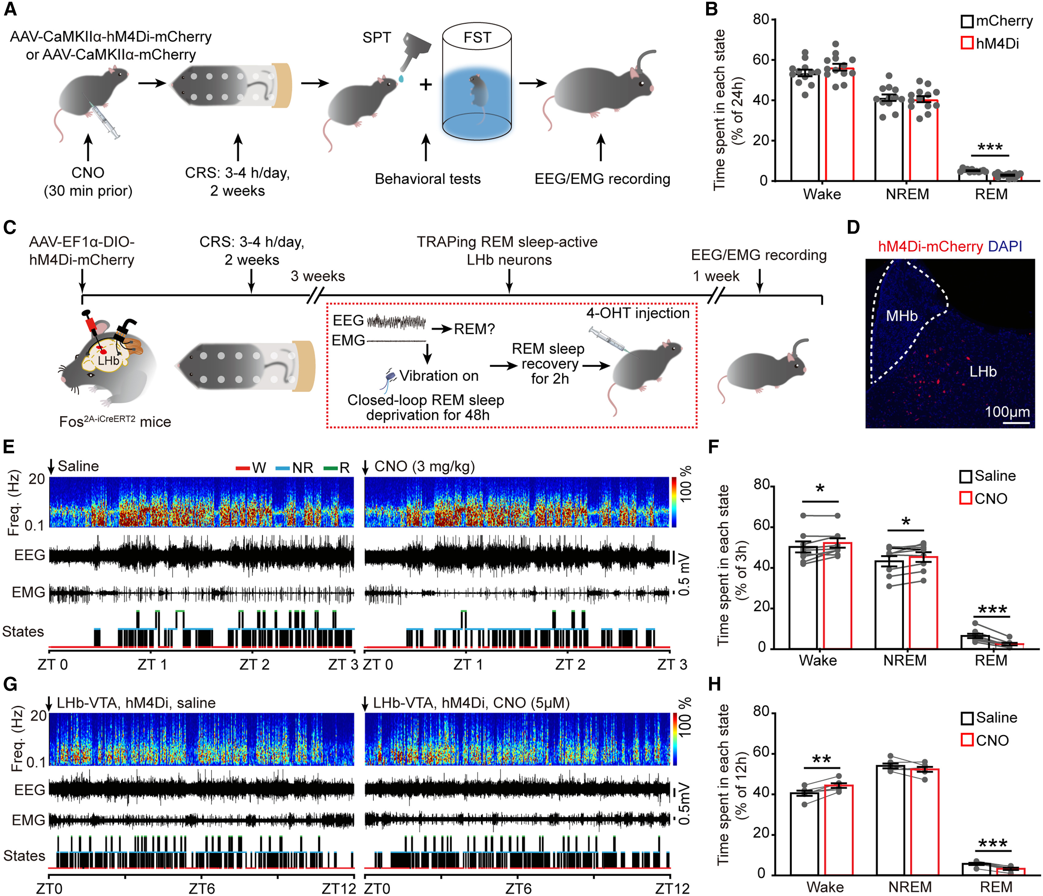
The team next tested whether inhibiting the LHb circuit could prevent or reverse CRS-induced REM sleep disturbances. First, chronically inhibiting hM4Di-expressing LHb neurons by administering CNO (3 mg/kg, i.p.) 30 minutes before each daily restraint session throughout the 14-day CRS paradigm effectively prevented the emergence of post-CRS REM sleep excess.
Second, in TRAP2 mice that had already completed 14 days of CRS, selectively inhibiting TRAPed REM-active LHb neurons via AAV-EF1α-DIO-hM4Di-mCherry—or locally microinfusing CNO (5 μM) into the bilateral VTA to inhibit LHb–VTA axon terminals—selectively normalized the pathologically elevated REM sleep duration without altering NREM sleep or wakefulness, demonstrating that hyperactivity of the REM-active LHb–VTA pathway is required to sustain post-stress REM sleep excess.
Source: et al. · Current Biology · Original figure reproduced for academic literature review under fair scholarly commentary; copyright remains with the authors/publisher.
Original figure and English caption ↗
REM-Active LHb Neurons Bidirectionally Regulate Chronic Stress-Induced Depressive-like Behaviors
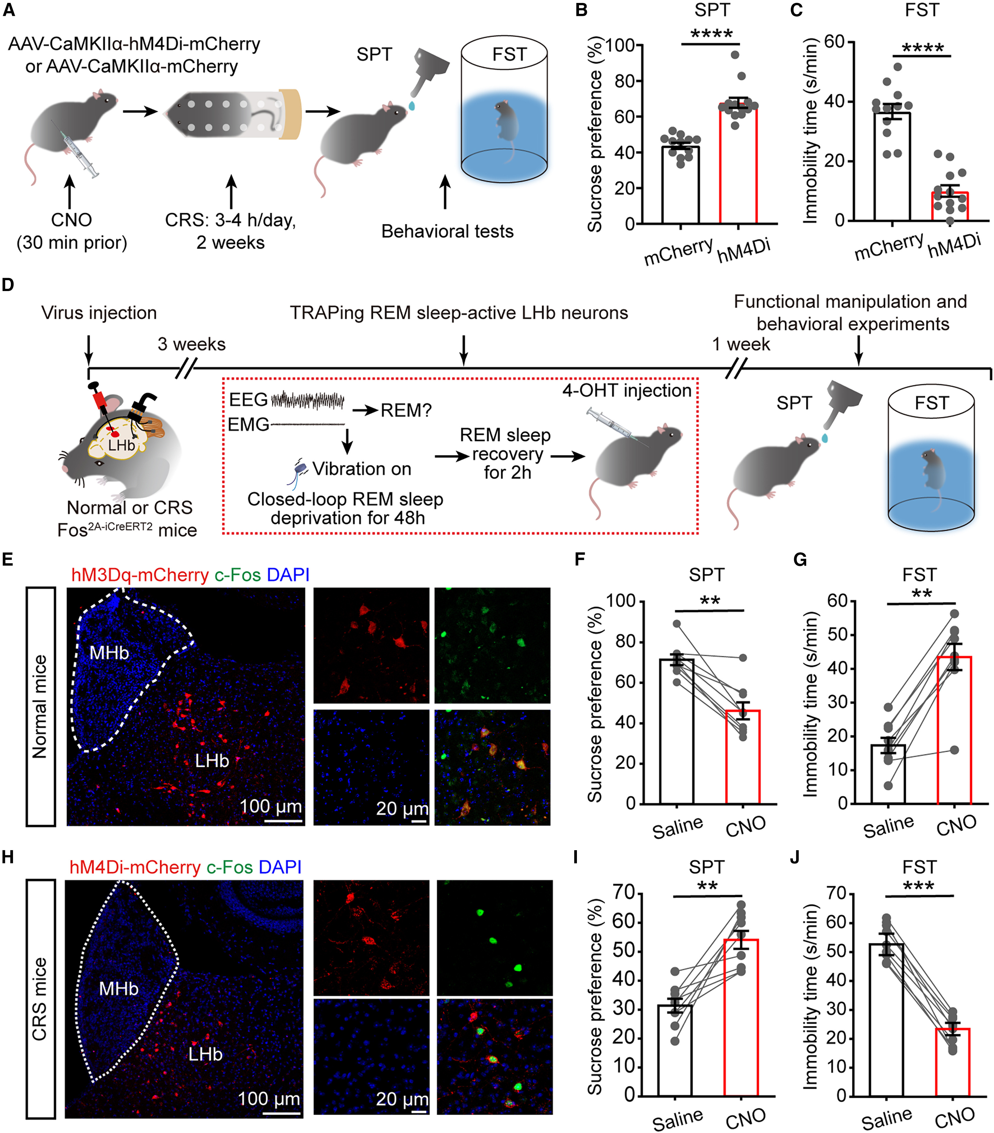
Finally, the researchers evaluated how manipulating REM-active LHb neurons impacts depressive-like behaviors. Chronic chemogenetic inhibition of LHb neurons during the 14-day CRS paradigm not only prevented REM sleep disturbances but also significantly ameliorated anhedonia in the sucrose preference test (SPT) and behavioral despair in the forced swim test (FST).
Crucially, in unstressed naive TRAP2 mice, chemogenetic activation of TRAPed REM-active LHb neurons (DIO-hM3Dq + CNO) was sufficient to induce a significant reduction in sucrose preference and prolonged immobility in the FST. Conversely, in CRS-exposed TRAP2 mice, acute chemogenetic inhibition of these REM-active LHb neurons (DIO-hM4Di + CNO) significantly restored sucrose preference and reduced FST immobility, establishing this REM-sleep-active LHb neuronal subpopulation as a shared circuit node linking REM sleep pathology and depressive-like behaviors.
Source: et al. · Current Biology · Original figure reproduced for academic literature review under fair scholarly commentary; copyright remains with the authors/publisher.
Original figure and English caption ↗