Three amygdala outputs divide arousal and exploration
In mice, distinct posteroventral medial-amygdala outputs differ in their effects on waking, exposed-space exploration and movement. Repeated sleep deprivation also changes their responses.
Is waking a mouse with an abrupt sound the same neural event as spontaneous waking? A 2024 JCI Insight study examines an excitatory-marked population in posteroventral medial amygdala, MePV, and finds clear responses to stimulus-evoked awakening. Increasing activity changes both waking and anxiety-like exploration, but following different outputs reveals distinct contributions. Repeated sleep deprivation introduces a further change of state.
MePV outputs to PMCo or BNST promote waking, while output to VMH mainly changes exploration and movement without significant sleep changes. After sleep deprivation, inhibiting the MePV population increases NREM sleep and improves some exploratory measures without significantly changing REM time.
Contents · 8 sections
External stimulation evokes a clear calcium response
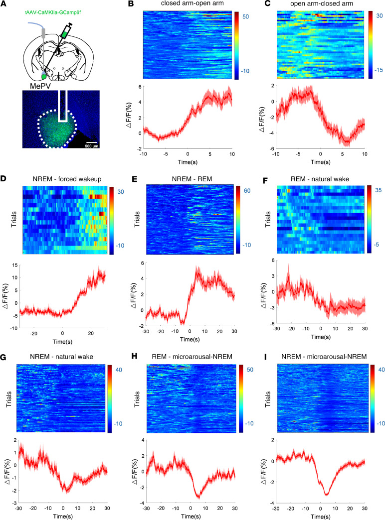
Population calcium recordings combined with EEG and EMG follow sleep-state transitions. External-stimulus awakening from NREM produces a clear MePV calcium increase, without the same prominent rise during spontaneous NREM awakening. The response depends on the waking context.
The population also increases calcium at NREM-to-REM transitions and decreases it during spontaneous REM-to-wake transitions. Approaching maze open arms raises signals, while returning to closed arms lowers them. Its event-dependent activity spans both sleep transitions and exploratory contexts.
Source: Li Y et al. · JCI insight · CC BY
Original figure and English caption ↗
Waking increases as exposed-area time declines
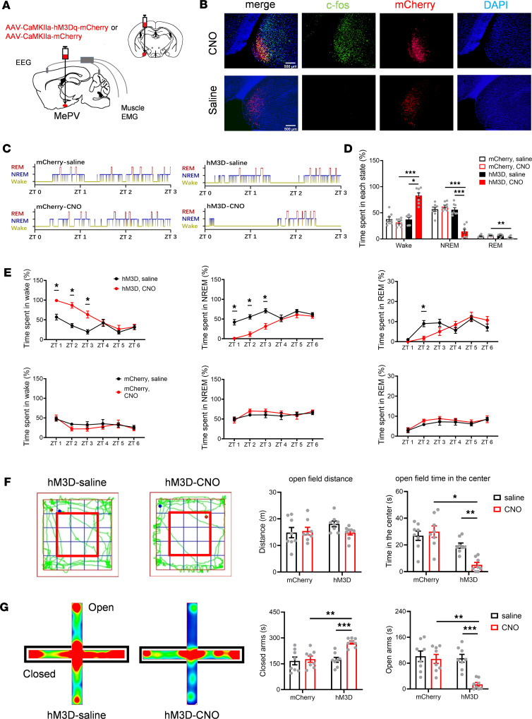
Increasing excitatory-marked MePV activity raises waking time and reduces NREM and REM sleep. Open-field center and maze open-arm time decrease without a significant total-distance change. Different activation approaches produce similar directions, connecting activity manipulation with behavior.
The main experiments use males, while supplementary female activation experiments also show reduced NREM and open-arm time. In this population, more waking can coexist with less exposed-area exploration, motivating examination of its outputs.
Source: Li Y et al. · JCI insight · CC BY
Original figure and English caption ↗
One output promotes waking and movement
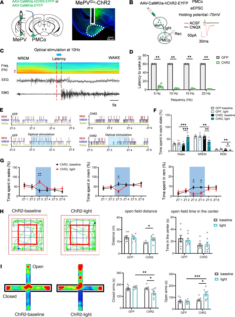
MePV projects to posteromedial cortical amygdala, PMCo. Terminal activation evokes excitatory currents locally and hastens waking from NREM. Over a longer observation window, waking increases while NREM and REM decline, showing that this branch influences sleep states.
Its behavioral effects differ from whole-population activation: total distance and open-arm time increase, while open-field center time does not significantly change. Waking, movement and exploratory choice form a particular combination.
Source: Li Y et al. · JCI insight · CC BY
Original figure and English caption ↗
Waking changes while exploratory measures remain similar
Another output reaches bed nucleus of the stria terminalis, BNST. Terminal activation likewise produces local excitatory responses and promotes NREM-to-wake transitions; waking increases and NREM and REM sleep decline over the observation window.
Open-field distance, center time and maze open-arm time do not significantly change. This branch separates waking promotion from the tested anxiety-like exploratory measures, indicating different output contributions to these outcomes.
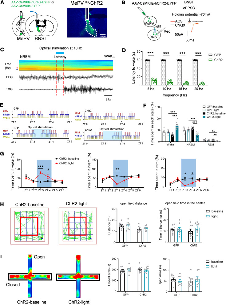
Source: Li Y et al. · JCI insight · CC BY
Original figure and English caption ↗
Exploration and movement decline without matching sleep changes
Output to ventromedial hypothalamus, VMH, produces a third pattern. Terminal activation evokes excitatory currents but does not significantly accelerate NREM awakening or change tested waking, NREM or REM time.
Distance traveled, open-field center time and open-arm time decrease. Sleep states and exploratory measures dissociate again, with lower movement contributing context for interpretation. The three recipient regions do not reproduce one uniform effect.
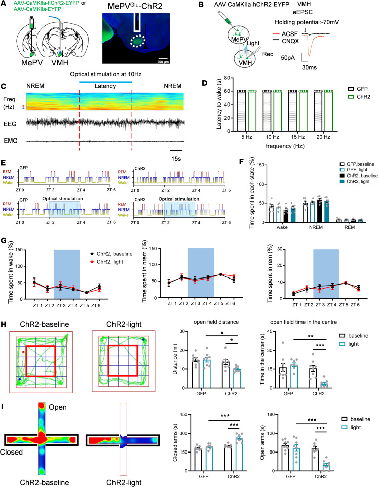
Source: Li Y et al. · JCI insight · CC BY
Original figure and English caption ↗
Exposed-area exploration increases while natural sleep is preserved
Reducing MePV-population activity increases center and open-arm time in normal-state mice without significantly changing movement distance. These exploratory changes coexist with no significant changes in tested waking, NREM or REM time.
Activation and inhibition produce different results: increasing activity promotes waking, while reducing activity in the normal state does not significantly alter sleep. Effects on waking and exposed-area exploration therefore depend on different aspects of the current state.
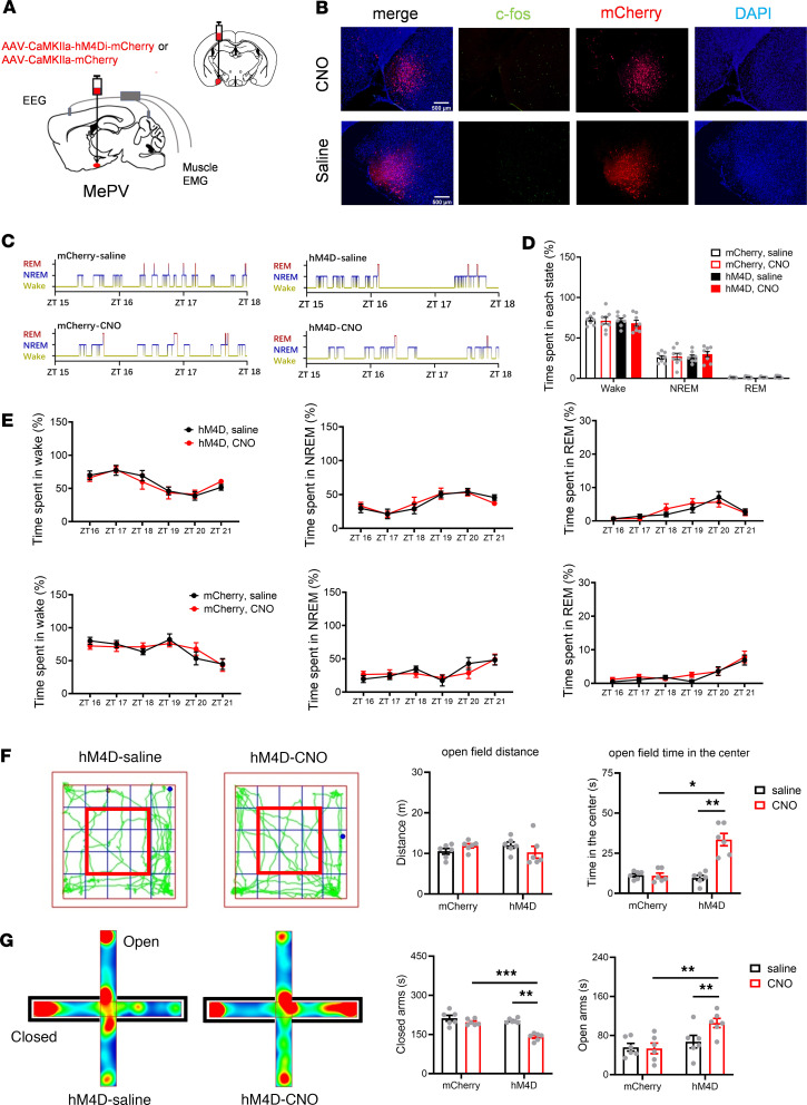
Source: Li Y et al. · JCI insight · CC BY
Original figure and English caption ↗
The same input meets different downstream responses
After repeated sleep deprivation, c-Fos labeling increases in MePV, PMCo and VMH, but not significantly in BNST. Under the same MePV activation condition, tested PMCo and VMH calcium responses increase while BNST responses decrease. Labeling and evoked responses describe different aspects of the change.
Sleep history does not strengthen every branch in the same direction. Recipient responses readjust, giving the node a different functional context after repeated sleep loss.
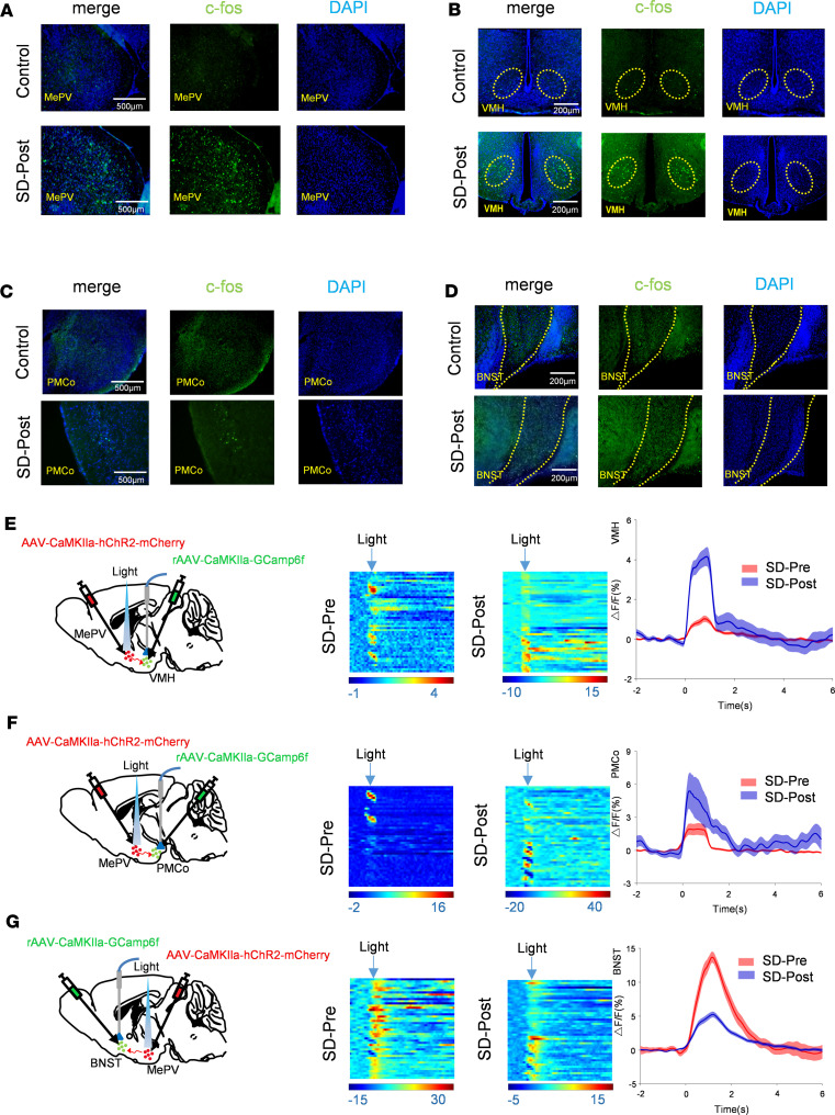
Source: Li Y et al. · JCI insight · CC BY
Original figure and English caption ↗
NREM rises while REM retains its separate pattern
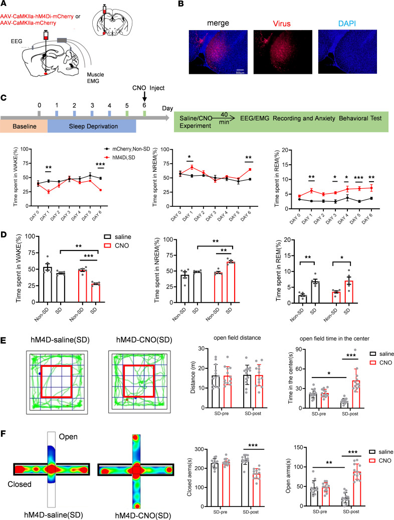
After the first deprivation day, mice show more rebound sleep; waking increases relative to this rebound phase during later observations. Inhibiting MePV after repeated deprivation reduces waking and increases NREM without significantly changing REM time.
Center and open-arm exploration also increase while overall movement remains similar. These predominantly male experiments show that amygdalar effects depend on output destination and prior sleep history, allowing one node to regulate different combinations of waking and exploration.
Source: Li Y et al. · JCI insight · CC BY
Original figure and English caption ↗