Stimulation patterns alter oxytocin-circuit responses in sleep-deprived mice
In male mice, chronic sleep-loss effects on exploration and fear extinction accompany altered PVN oxytocin-cell synapses. Stimulation patterns produce distinct aftereffects, with high-frequency intervention improving model outcomes.

After sustained sleep loss, mice may explore open areas less and show poorer extinction of learned threat responses. A CNS Neuroscience & Therapeutics study examines paraventricular, or PVN, oxytocin/OXT cells and their medial-prefrontal, or mPFC, connection. Excitatory synaptic responses weaken after sleep loss, while different stimulation patterns produce distinct subsequent cellular and behavioral outcomes.
Chronic sleep loss weakens PVN OXT AMPA-related currents. Prolonged low-frequency stimulation weakens related responses further; brief high-frequency stimulation one week after sleep loss improves exploration/extinction and restores selected prefrontal inhibitory measures.
Contents · 7 sections
Acute and chronic sleep loss produce different exploration patterns
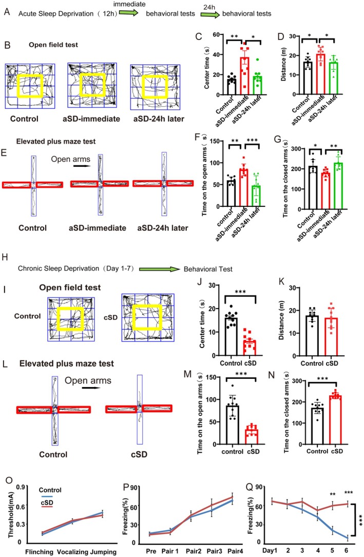
After twelve hours of acute deprivation, mice spend more time in centers and open arms and move more. Differences subside by twenty-four hours, without significant measured fear-extinction changes.
Seven days of partial daily deprivation reduce center/open-arm time and slow freezing decline during extinction. Total movement and shock-response thresholds do not significantly change, motivating investigation of neuropeptide cells.
Source: Wang Y et al. · CNS neuroscience & therapeutics · CC BY
Original figure and English caption ↗
Excitatory responses weaken in PVN oxytocin cells
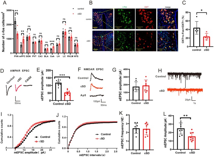
Chronic deprivation lowers PVN OXT c-Fos and increases mPFC/central-amygdala markers. PVN OXT AMPA-related currents decrease, while NMDA-related currents do not significantly change.
Miniature excitatory-event amplitudes decrease without frequency changes, while inhibitory-event amplitudes/frequencies remain unchanged. The measured alterations concentrate on excitatory synaptic responses.
Source: Wang Y et al. · CNS neuroscience & therapeutics · CC BY
Original figure and English caption ↗
Activating the population changes exploration in normal mice
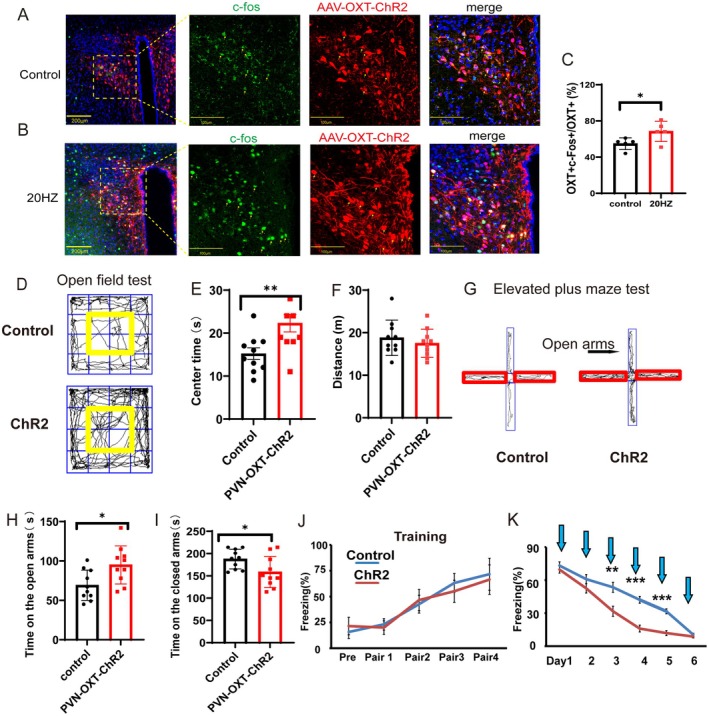
In normal males, immediate PVN OXT optical activation increases center/open-arm time without significantly changing movement distance, altering open-area exploration without a detected general activity increase.
Training freezing does not significantly differ, while activation yields faster decline during extinction. Effects on exploration and extinction motivate examination of downstream connections.
Source: Wang Y et al. · CNS neuroscience & therapeutics · CC BY
Original figure and English caption ↗
Different terminal targets yield different exploration outcomes
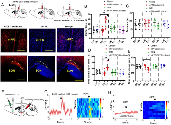
PVN OXT cells project to multiple regions. mPFC-terminal activation increases center/open-arm time; SON-terminal activation does not produce equivalent clear changes, distinguishing target-associated effects.
With an OXT-receptor antagonist, exploration changes during mPFC-terminal activation are nonsignificant. OXT sensors detect activation-linked prefrontal signals, connecting terminals, peptide signaling and exploration.
Source: Wang Y et al. · CNS neuroscience & therapeutics · CC BY
Original figure and English caption ↗
Prolonged low-frequency stimulation leaves weaker synaptic responses
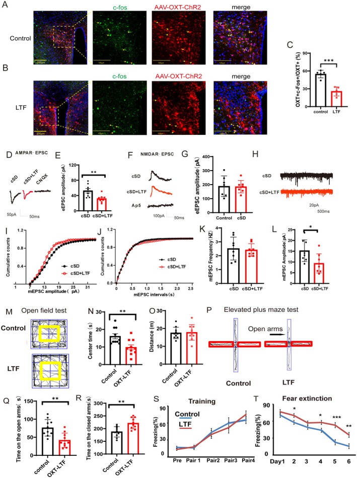
Comparing low-frequency aftereffects in sleep-deprived mice, next-day slices show weaker AMPA-related currents and miniature excitatory-event amplitudes in cells receiving prolonged stimulation. NMDA currents and event frequency do not change significantly. This differs from immediate activation effects and shows that stimulation can leave subsequent changes.
Low-frequency-treated normal mice explore centers/open arms less and freeze more during extinction, without significant movement or training-freezing changes. Effects involve both immediate activity and subsequent synaptic state.
Source: Wang Y et al. · CNS neuroscience & therapeutics · CC BY
Original figure and English caption ↗
Exploration remains modifiable after heart-rate differences subside
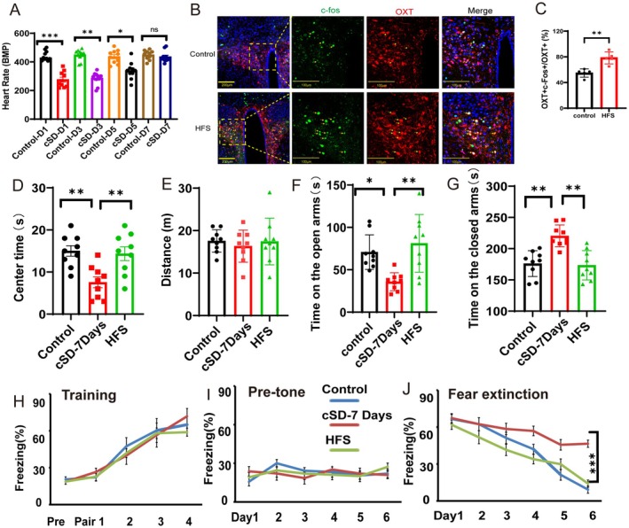
Seven days after deprivation ends, measured heart-rate differences are nonsignificant while exploration/extinction changes remain. Brief high-frequency PVN OXT stimulation increases center/open-arm time and reduces extinction freezing.
Movement, training freezing and pre-extinction baseline freezing remain nonsignificantly changed. Effects concern open-environment choice and extinction responses, motivating tests of synaptic aftereffects.
Source: Wang Y et al. · CNS neuroscience & therapeutics · CC BY
Original figure and English caption ↗
PVN excitation and prefrontal inhibition change together
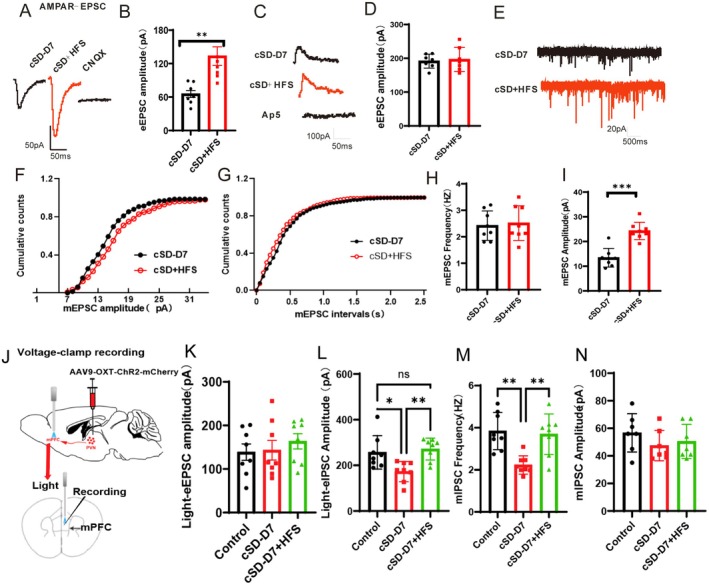
Next-day PVN OXT AMPA currents and miniature excitatory amplitudes increase, without frequency/NMDA changes. Prefrontal recordings show recovery of reduced light-evoked inhibition and inhibitory-event frequency, without significant excitatory-input changes.
After high-frequency treatment, PVN activation evokes larger prefrontal OXT-sensor responses than controls. With receptor antagonism, the exploratory improvement is absent, adding peptide-related signals alongside synaptic and behavioral outcomes.
This male-mouse study links post-sleep-loss behavior with peptide cells and synaptic states. Prolonged low-frequency and brief high-frequency interventions leave distinct aftereffects, showing how pattern and observation time shape circuit outcomes.
Source: Wang Y et al. · CNS neuroscience & therapeutics · CC BY
Original figure and English caption ↗