Grooming and waking: different roles for two zona-incerta cell populations
A mouse study links gene expression, cell location, and distant connections: rostral Ecel1 cells contribute to induced grooming, while medial Pde11a cells and their pontine projection promote waking.
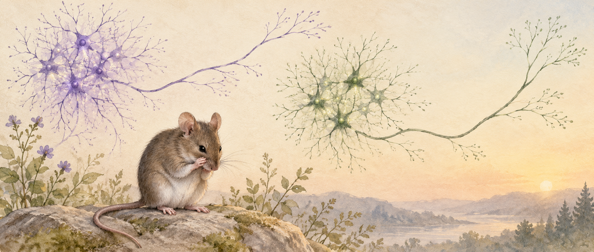
A mouse raises its forepaws to groom its face or wakes from sleep: ordinary actions involve different neural populations. The zona incerta is a widely connected region with varied functions, so its name alone says little about a specific role. A study in Nature Communications distinguishes its cells by gene expression and position, then follows their connections and behavioral effects. Even GABAergic inhibitory cells can do very different jobs.
Rostral Ecel1 cells promote grooming through a periaqueductal-gray projection, while medial Pde11a cells promote sleep-to-wake transitions through a pontine reticular projection. Their effects depend on behavioral context and circadian phase.
Contents · 7 sections
Different populations occupy the same region
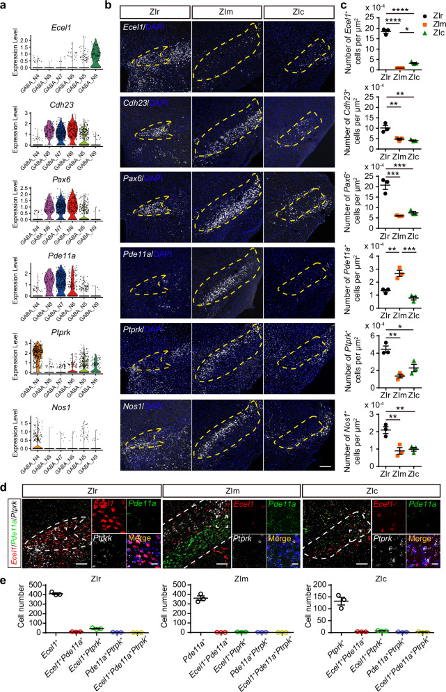
The team first distinguishes cell types using nuclear RNA expression and identifies six GABAergic clusters in the zona incerta. Mapping the markers reveals two especially clear populations: Ecel1-expressing cells are mainly rostral, Pde11a-expressing cells mainly medial, with little overlap.
This adds residents’ identities to a map previously showing only district names. Most cells in both populations carry a GABAergic marker, yet occupy different locations. Gene expression identifies the populations and gives the subsequent connection and behavioral analyses more specific targets.
Source: Zhu M et al. · Nature communications · CC BY-NC-ND
Original figure and English caption ↗
Inhibitory cells also send signals over long distances
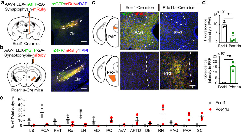
The axons of these populations extend beyond their neighborhood. Rostral Ecel1 cells project to regions including periaqueductal gray, while medial Pde11a cells project strongly to the oral pontine reticular formation. Both have multiple distant destinations, but their projection patterns differ substantially.
Periaqueductal gray contributes to sensory processing and diverse behaviors, whereas the pontine reticular formation relates to sleep and waking. These destinations suggest different functions, but a connection map is only part of the story. The team next studies adult male mice to link changes in cellular activity with behavior.
Source: Zhu M et al. · Nature communications · CC BY-NC-ND
Original figure and English caption ↗
Responses rise before the action begins
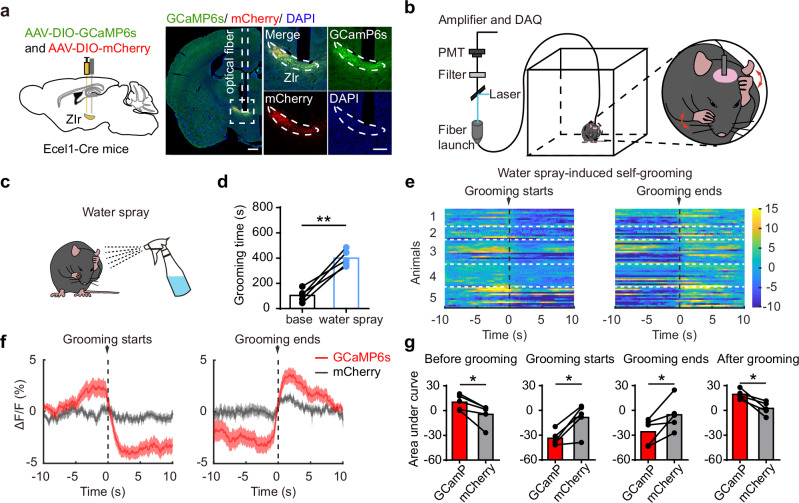
Increasing rostral Ecel1-cell activity increases grooming time and bouts. The signal does not remain elevated throughout grooming: during both spontaneous and water-spray-induced grooming, calcium rises before onset, falls during the action, and returns toward baseline afterward.
This timing is consistent with involvement in preparation and sensory responses preceding grooming rather than continuous drive of every movement. Activation also induces avoidance of the stimulation-associated place, while open-field center time and elevated-maze open-arm time do not change significantly.
Source: Zhu M et al. · Nature communications · CC BY-NC-ND
Original figure and English caption ↗
Induced grooming depends on these cells; spontaneous grooming changes little
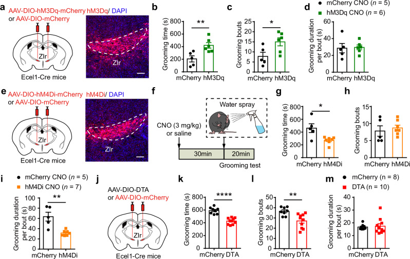
Suppressing or ablating Ecel1 cells reduces water-spray-induced grooming without a discernible change in spontaneous grooming. A population that can promote a behavior need not determine it in every setting; grooming following sensory stimulation is more sensitive to these cells.
Downstream, Ecel1 cells provide direct inhibitory input to periaqueductal gray, and activating this projection also increases grooming. Activating other projections to lateral septum, preoptic area, lateral hypothalamus, or superior colliculus does not produce the same change.
Source: Zhu M et al. · Nature communications · CC BY-NC-ND
Original figure and English caption ↗
Another population makes waking more likely
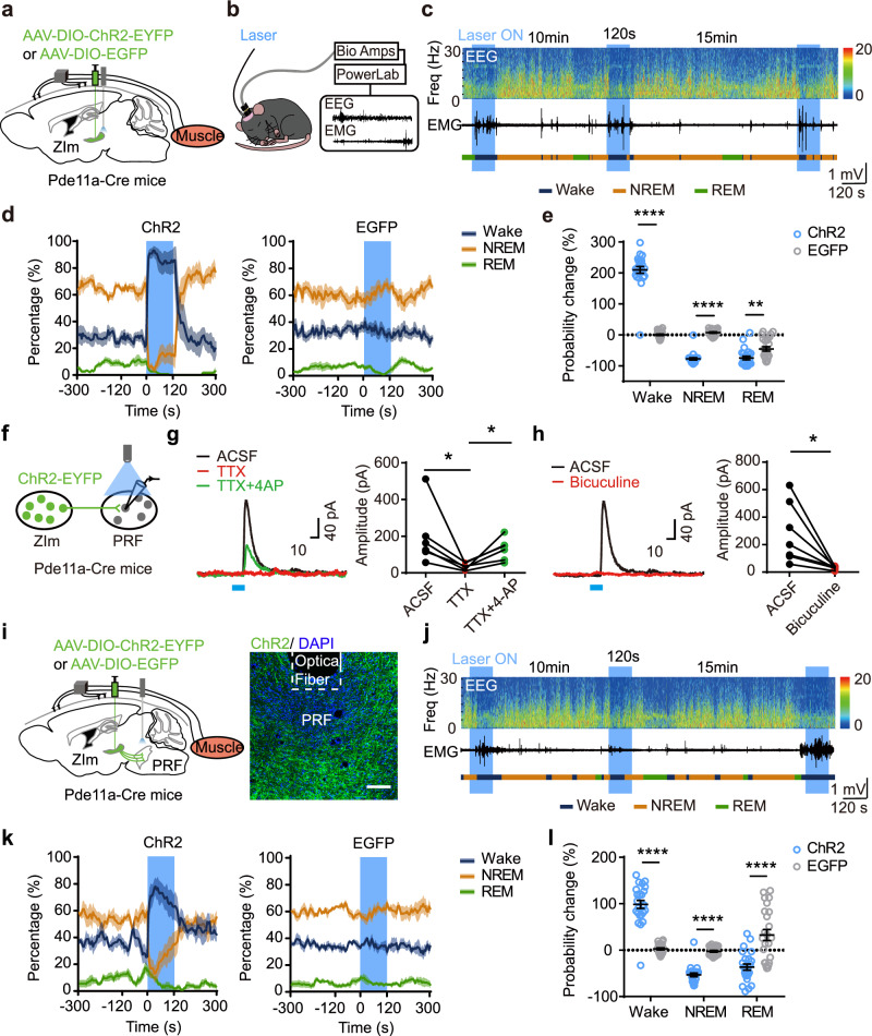
Medial Pde11a cells show higher activity during both REM sleep and wakefulness. Increasing their activity promotes transitions from REM or NREM sleep to waking. Population activity in natural states and activation outcomes together show their involvement in state transitions.
These cells provide direct inhibitory input to the oral pontine reticular formation. Enhancing the projection increases transitions from NREM sleep to waking and reduces transitions into NREM and from NREM to REM. The final behavioral effect of an inhibitory signal depends on the receiving network.
Source: Zhu M et al. · Nature communications · CC BY-NC-ND
Original figure and English caption ↗
Promoting waking also changes sleep episodes
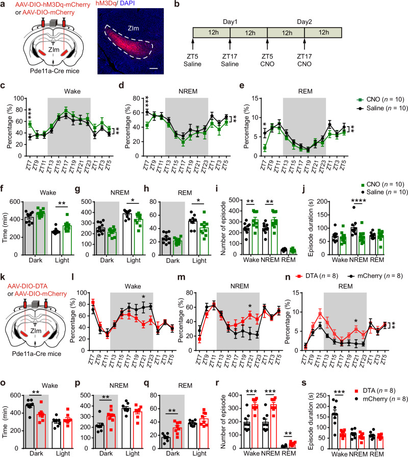
During the normally resting light phase, increasing Pde11a-cell activity increases waking, reduces both sleep types, and shortens NREM episodes. Acute inhibition produces no significant sleep-state change. After ablation, changes occur mainly during the dark phase: waking decreases, sleep increases, and waking episodes shorten.
The division of roles also undergoes cross-comparison: Pde11a-cell ablation does not discernibly alter spontaneous or induced grooming, and Ecel1-cell ablation does not significantly alter sleep–wake measures. Differences in position and gene expression thus extend to observable behavioral differences.
Source: Zhu M et al. · Nature communications · CC BY-NC-ND
Original figure and English caption ↗
A shared transmitter does not imply a shared job
The region’s name stays the same, but its functional picture becomes finer. Two populations releasing the same inhibitory transmitter differ in location, distant connections, and behavioral involvement. The study moves from asking what a region does to asking which cells and connections contribute.
The sensory context of grooming and the day–night phase of waking add timing and context to that division. An apparently simple mouse action involves different populations acting under different conditions, rather than all cells switching together.