How does melatonin act in the paraventricular thalamus to promote non-REM sleep?
Studying melatonin-proficient C3H/HeJ mice, researchers show that melatonin suppresses paraventricular thalamic excitability via MT1 and MT2 receptors to promote non-REM sleep.
Melatonin (MLT), a neuroendocrine hormone secreted by the pineal gland, plays an important role in coordinating circadian rhythms and sleep homeostasis, and exogenous melatonin and its receptor agonists are widely used clinically to treat insomnia and circadian sleep disorders. Yet the precise brain nuclei through which endogenous melatonin directly promotes sleep have remained elusive—in part because commonly used laboratory mouse strains such as C57BL/6J carry a natural truncation in arylalkylamine N-acetyltransferase (AANT) and lack endogenous melatonin synthesis. Published in Current Biology in August 2024, Shuancheng Ren, Zhian Hu, Chao He, and colleagues at the College of Basic Medical Sciences of Army Medical University investigated melatonin-proficient C3H/HeJ mice and identified the paraventricular thalamus (PVT) as a direct central target mediating the somnogenic actions of melatonin. The authors show that PVT neurons abundantly express MT1 and MT2 receptors (MT1R and MT2R) with diurnal oscillation, and that melatonin directly hyperpolarizes and suppresses wake-promoting PVT neurons via postsynaptic MT1R/MT2R signaling to promote non-rapid eye movement (NREM) sleep.
In melatonin-proficient C3H/HeJ mice, melatonin directly inhibits paraventricular thalamus (PVT) neurons via postsynaptic MT1R and MT2R activation, reducing in vivo calcium activity and single-unit firing to promote and consolidate non-rapid eye movement (NREM) sleep.
Contents · 6 sections
PVT Neurons Abundantly Express MT1R and MT2R with Diurnal Oscillation
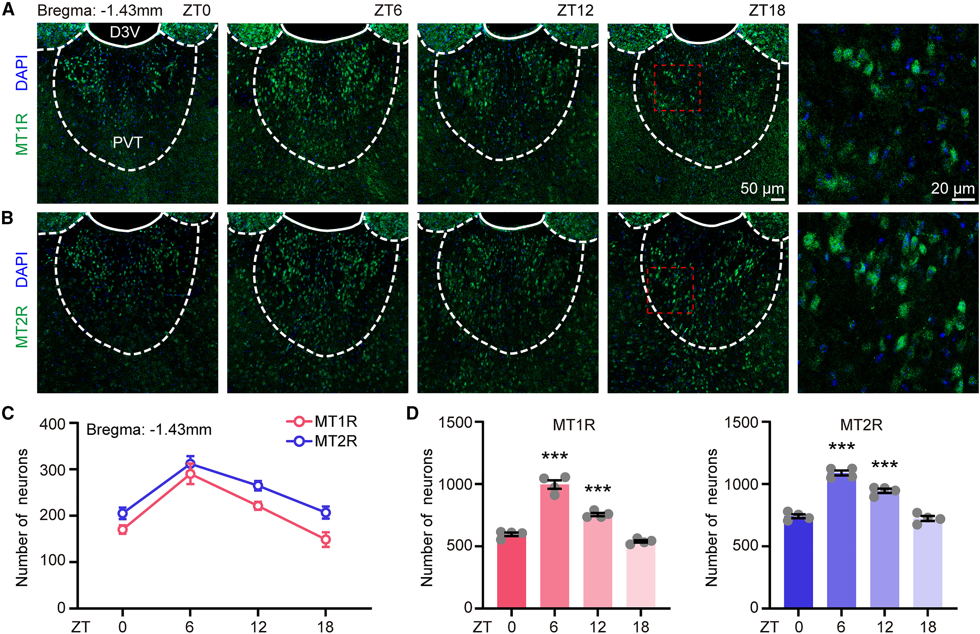
The paraventricular thalamus (PVT) is a critical thalamic wake-promoting hub that receives direct inputs from the suprachiasmatic nucleus (SCN) and borders the third ventricle, placing it in close contact with pineal melatonin in the cerebrospinal fluid. To determine whether the PVT possesses the molecular machinery to sense melatonin directly, the authors examined adult male C3H/HeJ mice housed under a 12-hour light/12-hour dark cycle across four zeitgeber time points—light onset (ZT0), mid-light (ZT6), dark onset (ZT12), and mid-dark (ZT18)—spanning four rostrocaudal coronal planes of the PVT (Bregma -1.23, -1.43, -1.79, and -2.03 mm).
Immunostaining revealed dense expression of both G-protein-coupled melatonin receptors, MT1R and MT2R, throughout the rostrocaudal extent of the PVT, with pronounced diurnal variation: receptor protein fluorescence peaked at mid-light (ZT6) and was significantly higher than at ZT0 and ZT18. Complementary RNAscope fluorescent in situ hybridization (targeting MTNR1A and MTNR1B) and qRT-PCR confirmed that receptor transcript levels likewise peaked at ZT6, with MTNR1A exhibiting substantially higher abundance than MTNR1B in the PVT. This diurnal regulation indicates that the sensitivity of PVT neurons to melatonin is gated across the sleep-wake cycle.
Source: et al. · Current Biology · Original figure reproduced for academic literature review under fair scholarly commentary; copyright remains with the authors/publisher.
Original figure and English caption ↗
Melatonin Directly Hyperpolarizes and Inhibits PVT Neurons via Postsynaptic MT1R and MT2R
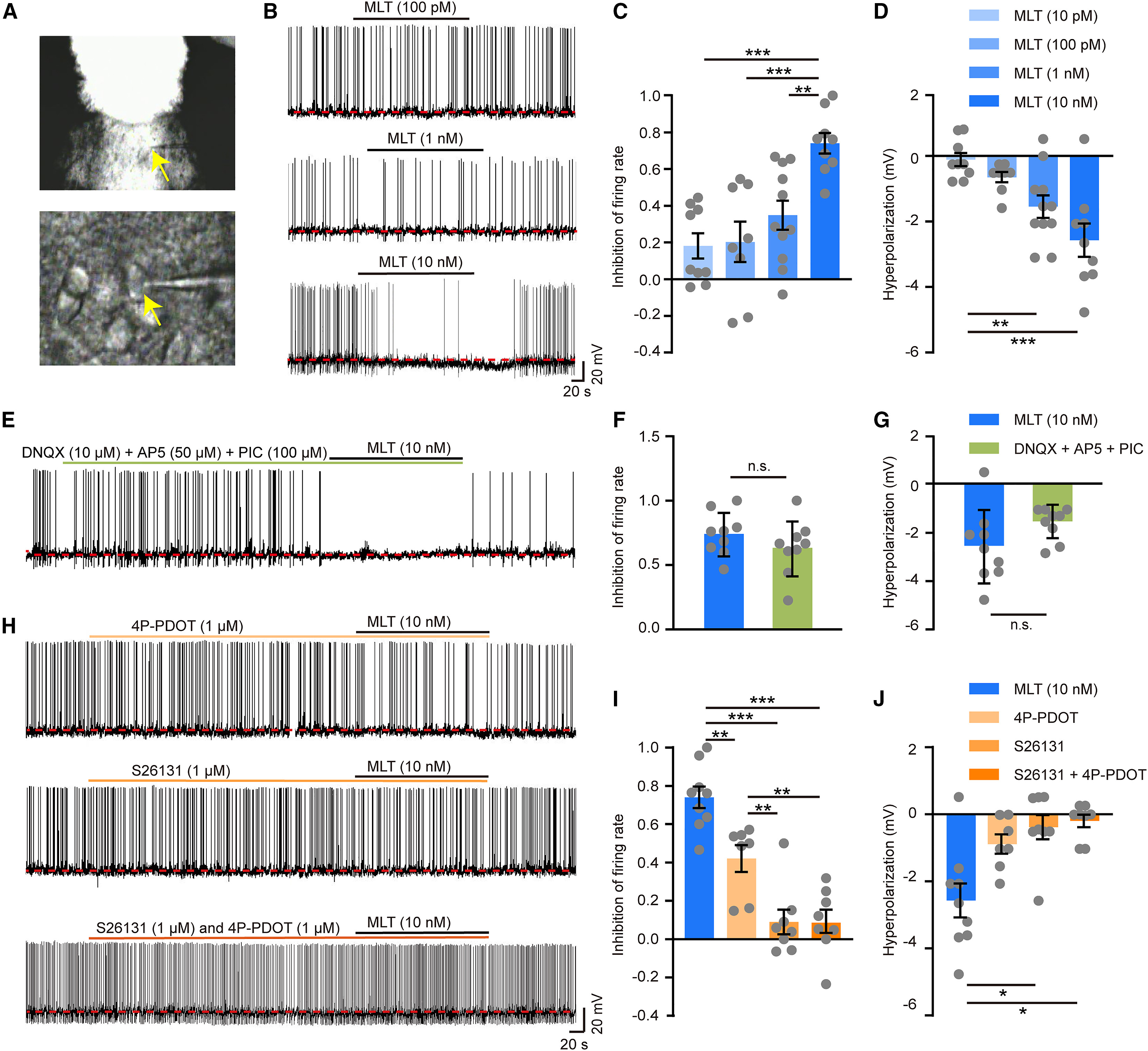
To directly examine how melatonin modulates PVT neuronal excitability, the researchers performed whole-cell current-clamp recordings in acute coronal PVT brain slices from C3H/HeJ mice. Bath application of melatonin across graded concentrations (10 pM, 100 pM, 1 nM, and 10 nM) concentration-dependently suppressed spontaneous action potential firing rates and hyperpolarized the resting membrane potential of PVT neurons, with 10 nM eliciting the most robust inhibition before recovery upon washout.
To distinguish direct postsynaptic action from presynaptic network effects, the authors applied melatonin (10 nM) in the presence of synaptic transmission blockers—DNQX (10 μM), AP5 (50 μM), and picrotoxin (100 μM). Melatonin-induced firing suppression and membrane hyperpolarization persisted unabated, confirming a direct postsynaptic mechanism. Pharmacological dissection further revealed that pre-application of the selective MT2R antagonist 4P-PDOT (1 μM) partially attenuated melatonin's inhibitory effect, whereas the selective MT1R antagonist S26131 (1 μM) alone or co-applied with 4P-PDOT completely abolished both firing suppression and hyperpolarization, indicating a predominant role for MT1R alongside cooperative MT2R signaling.
Source: et al. · Current Biology · Original figure reproduced for academic literature review under fair scholarly commentary; copyright remains with the authors/publisher.
Original figure and English caption ↗
Local Melatonin Infusion Suppresses Spontaneous PVT Calcium Activity and Single-Unit Firing in Freely Behaving Mice
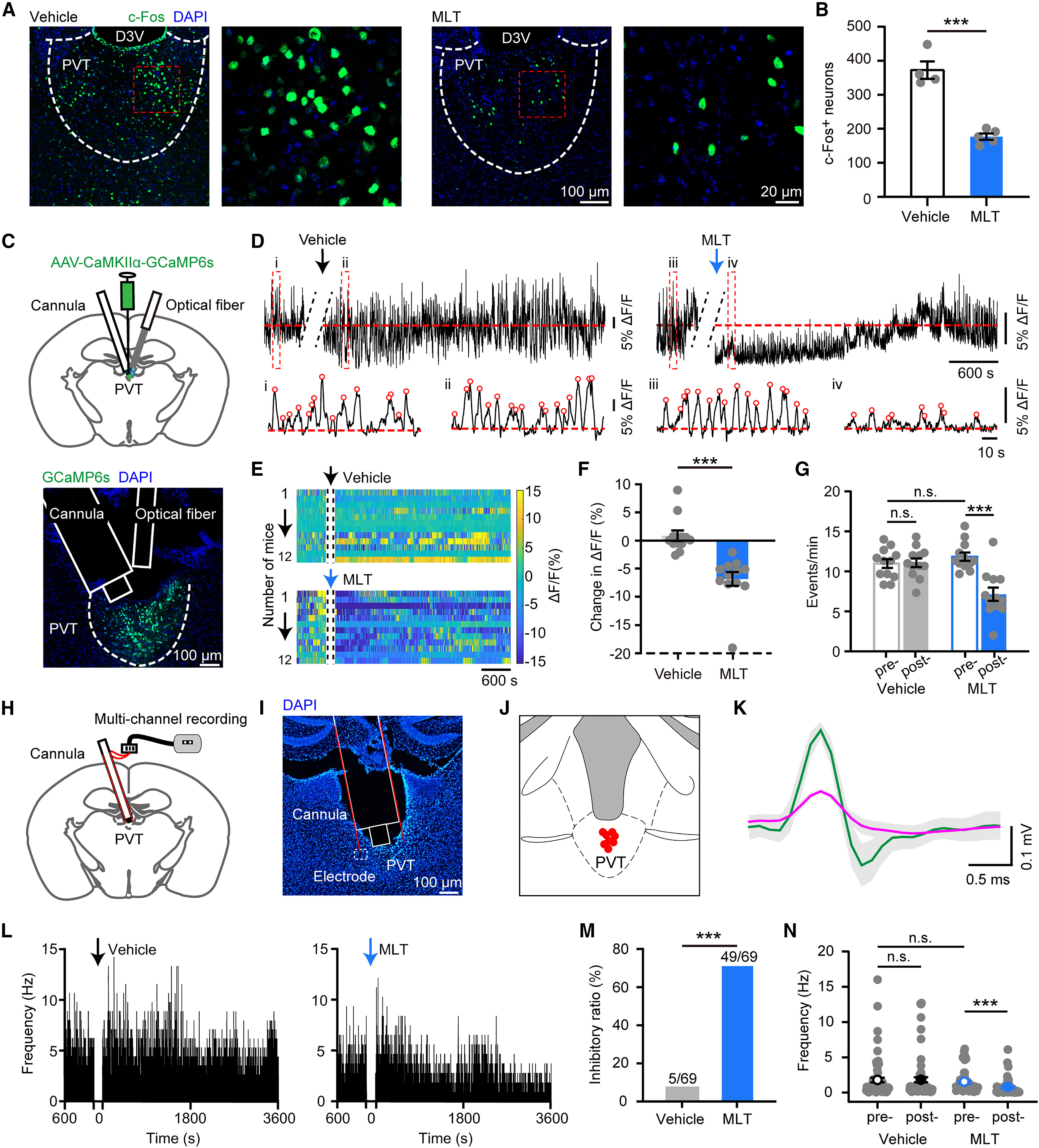
To verify whether melatonin suppresses PVT activity in freely behaving animals, the authors first microinfused melatonin (2 pmol in 200 nL) locally into the PVT at dark onset (ZT12), which reduced the number of c-Fos-positive PVT neurons 3 hours later from 373 ± 26 in vehicle controls to 177 ± 9 (p < 0.001). To monitor real-time population dynamics, the team expressed AAV-CaMKIIα-GCaMP6s in the PVT and implanted an integrated cannula-optic fiber assembly to perform simultaneous intra-PVT drug delivery and in vivo fiber photometry (recording 470 nm GCaMP6s and 410 nm isosbestic control signals). Local melatonin infusion significantly decreased mean ΔF/F calcium signal amplitude (-6.85% ± 1.23% vs. +0.85% ± 0.93% in vehicle controls, p < 0.001) and reduced spontaneous Ca2+ transient event frequency from 11.86 ± 0.51 events/min to 7.14 ± 0.83 events/min (p < 0.001).
Complementary in vivo multi-channel tetrode recordings coupled to a local infusion cannula captured 69 single units in the PVT of freely moving mice. Following intra-PVT melatonin infusion, 49 of 69 neurons (71.0%) exhibited marked firing suppression, whereas only 7 (10.1%) were excited and 13 (18.9%) remained unchanged; overall mean firing frequency dropped from 1.7 ± 0.3 Hz to 0.7 ± 0.2 Hz (p < 0.001). Together, c-Fos mapping, in vivo fiber photometry, and single-unit electrophysiology consistently demonstrate potent melatonin-mediated suppression of PVT neuronal activity in vivo.
Source: et al. · Current Biology · Original figure reproduced for academic literature review under fair scholarly commentary; copyright remains with the authors/publisher.
Original figure and English caption ↗
Intra-PVT Melatonin Agonism and Antagonism Bidirectionally Regulate NREM Sleep and Wakefulness
Having established that melatonin suppresses wake-promoting PVT neurons, the team used polysomnographic EEG/EMG recordings to evaluate how local PVT melatonin signaling regulates sleep-wake architecture. Microinfusion of exogenous melatonin (2 pmol) into the PVT at dark onset (ZT12) increased cumulative NREM sleep time by 51.01% (p = 0.004) and decreased wakefulness by 22.24% (p = 0.002) over the subsequent 3 hours without altering REM sleep duration or EEG spectral power. Architecture analysis revealed that melatonin promoted NREM sleep primarily by elongating mean NREM episode duration (p = 0.049) rather than increasing episode numbers. By contrast, control infusion of the same melatonin dose into the adjacent thalamic ventrobasal (VB) complex—despite local MT1R/MT2R expression—had no effect on sleep-wake states, underscoring the anatomical specificity of the PVT.
Conversely, to test whether endogenous melatonin signaling in the PVT maintains physiological sleep, the authors microinfused an MT1R/MT2R antagonist mixture (S26131 + 4P-PDOT, 0.1 mM) into the PVT at light onset (ZT0). Blocking endogenous melatonin receptors in the PVT increased cumulative wakefulness by 51.42% (p < 0.001) and reduced NREM sleep by 29.03% (p < 0.001) over the ensuing 3 hours, driven by lengthened wake episode duration (p = 0.013), with no significant change in REM sleep. These complementary gain- and loss-of-function pharmacological experiments establish bidirectional control of NREM sleep by PVT melatonin signaling.
Knockdown of PVT Melatonin Receptors Impairs 24-Hour NREM Sleep Especially at Light-Dark Transitions
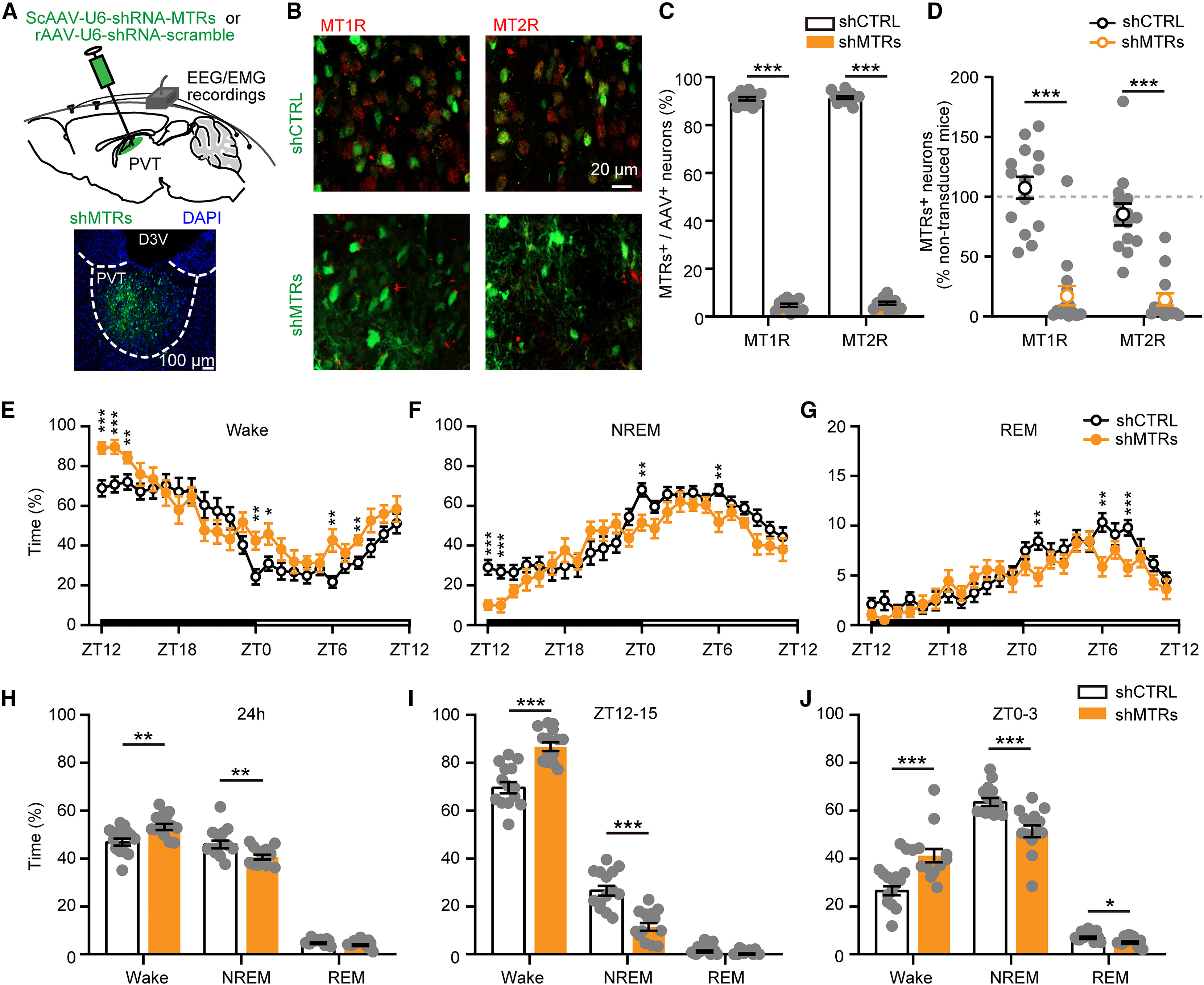
To determine the chronic physiological contribution of endogenous PVT melatonin receptors across the full 24-hour cycle, the authors co-injected self-complementary AAVs carrying shRNAs against MTNR1A and MTNR1B (ScAAV-U6-shRNA-MTR1A-CMV-EGFP and ScAAV-U6-shRNA-MTR1B-CMV-EGFP) into the PVT of C3H/HeJ mice (shMTR group) to knock down both MT1R and MT2R. Across 24-hour EEG/EMG recordings, shMTR mice exhibited a significant increase in total wakefulness (54.07% ± 1.25% vs. 47.69% ± 1.48% in scrambled shCTRL mice, p = 0.003) and a corresponding reduction in total NREM sleep (48.53% ± 4.17% vs. 56.73% ± 2.50%, p = 0.009).
Time-resolved analysis revealed that sleep deficits in shMTR mice were particularly pronounced during the 3-hour windows following light-dark transitions and around peak receptor expression. During the first 3 hours of the dark phase (ZT12–15, when endogenous pineal melatonin secretion begins to rise), shMTR mice displayed markedly increased wakefulness and decreased NREM sleep (p < 0.001), characterized by prolonged wake bout duration and fewer NREM bouts. Similarly, during the first 3 hours after light onset (ZT0–3, when mice transition into their rest phase) and around ZT6 (when PVT MT1R/MT2R expression peaks), shMTR mice showed robust increases in wakefulness and reductions in NREM sleep (along with a modest reduction in REM sleep during ZT0–3, p = 0.011). Thus, endogenous melatonin acts on PVT receptors to brake the wakefulness system during critical circadian transitions.
Source: et al. · Current Biology · Original figure reproduced for academic literature review under fair scholarly commentary; copyright remains with the authors/publisher.
Original figure and English caption ↗
Bridging Neuroendocrine Melatonin Signaling and Thalamic Wakefulness Gating
Although melatonin is not strictly required for sleep generation in mammals, how endogenous melatonin finely shapes sleep-wake transitions has remained poorly understood. By deliberately utilizing melatonin-proficient C3H/HeJ mice rather than melatonin-deficient C57BL/6J mice, this study demonstrates that the periventricular PVT integrates daily pineal melatonin rhythms with intrinsic diurnal oscillations in MT1R and MT2R expression. At mid-light (ZT6), peak receptor abundance acts in concert with basal melatonin to consolidate daytime NREM sleep, whereas during light-dark transitions (ZT12–15 and ZT0–3), endogenous melatonin signaling tempers excessive PVT arousal drive to prevent pathologically prolonged wake bouts.
From a translational perspective, the PVT not only governs baseline arousal maintenance but also drives hyperarousal and sleep disturbances associated with chronic stress, anxiety, pain, and withdrawal. Defining the postsynaptic MT1R/MT2R mechanism through which melatonin directly silences PVT glutamatergic neurons provides a precise anatomical substrate for the clinical efficacy of exogenous melatonin and MT1R/MT2R agonists, while offering new avenues for treating hyperarousal-related insomnia.