Parabrachial input alters transitions under sevoflurane anesthesia
In mice, activating parabrachial input to a tested extended-amygdala region delays loss of righting and accelerates recovery after anesthetic cessation. Suppression only shortens induction, revealing differences across phases.
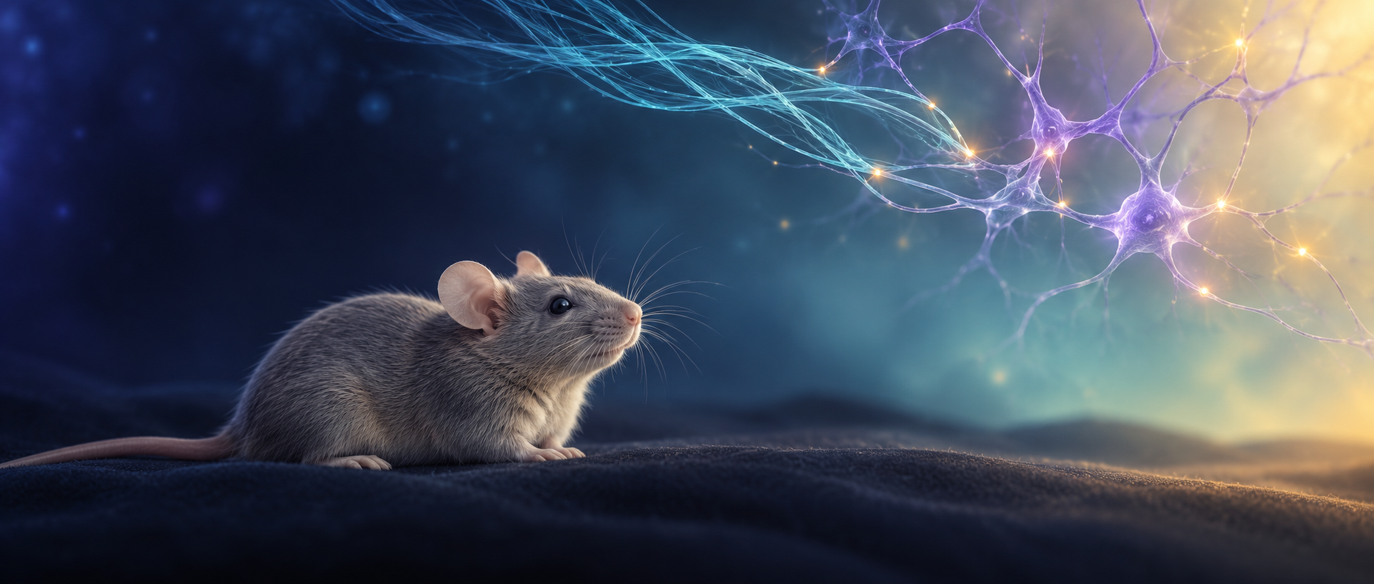
Entering anesthesia and recovering after it stops appear opposite, yet need not be regulated symmetrically by the same circuits. A 2024 CNS Neuroscience & Therapeutics study follows glutamatergic parabrachial, PBN, input to a tested extended-amygdala, EA, region in mice. Terminal calcium, righting responses and EEG changes are observed alongside female–male comparisons. The input alters behavioral transitions under sevoflurane, with distinct results for activation versus suppression and induction versus recovery. Righting measures an animal’s ability to restore its posture; its loss and return serve as behavioral indices of anesthetic transitions.
Terminal activation delays loss of righting and accelerates recovery after anesthetic cessation, alongside EEG-band changes. Suppression shortens time to loss of righting but changes recovery time and measured EEG bands nonsignificantly. Female–male differences also vary by phase and intervention condition.
Contents · 6 sections
An input reaches the tested extended-amygdala region
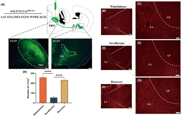
PBN participates in sensory and state-related regulation, with glutamatergic cells sending signals to forebrain regions. Vglut2 genetic tracing reveals terminals in the tested EA region. EA comprises several related structures, while recording and intervention focus on a local target.
Histological comparisons across wakefulness, sevoflurane anesthesia and recovery show less local c-Fos labeling during anesthesia and an increase afterward. This slower readout describes state-associated changes; terminal calcium recording then follows brief fluctuations during transitions.
Source: Yan Y et al. · CNS neuroscience & therapeutics · CC BY
Original figure and English caption ↗
An initial rise precedes a fall, followed by recovery increases
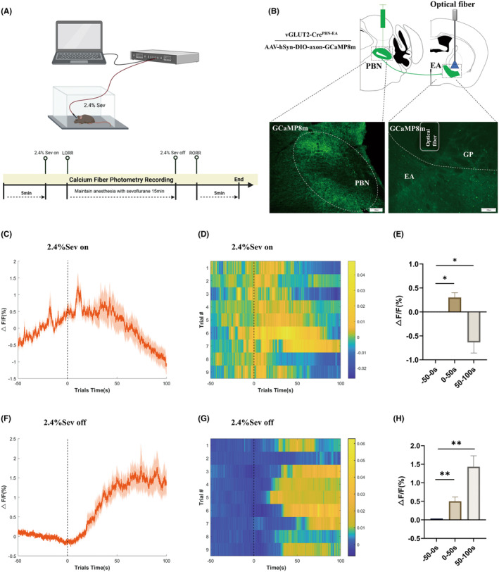
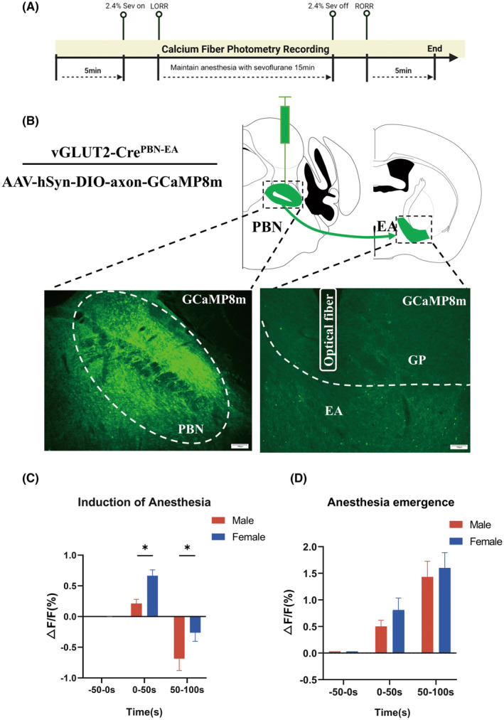
A calcium indicator expressed in PBN axons is recorded from the EA terminal region. Sevoflurane inhalation first produces a transient calcium rise, followed by a decline during early anesthesia. Entry into anesthesia therefore includes an initial response rather than a continuously descending signal.
After anesthetic inhalation stops, terminal calcium rises again, with both earlier and later recovery windows above the preceding baseline. The readout reflects population calcium in input terminals, rather than all PBN or EA cells, and does not directly quantify transmitter release.
Population recordings use anesthetic onset and cessation as temporal references. The same type of input fibers shows distinct profiles: a transient initial induction peak and increasing signals across recovery windows. Following transition dynamics adds information beyond an average before–after comparison.
Source: Yan Y et al. · CNS neuroscience & therapeutics · CC BY
Original figure and English caption ↗
Later loss of righting and faster recovery
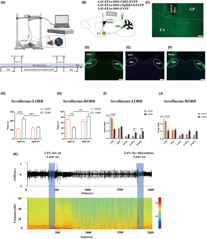
Activating PBN terminals at the EA target prolongs time to loss of righting. Activating the input after sevoflurane stops shortens time to recovery of righting. The first measure describes induction, and the second describes behavioral recovery after anesthetic cessation.
The changes accompany redistribution of cortical EEG power. Induction comparisons show lower relative delta and higher beta and gamma power; recovery comparisons also show lower relative delta power. Together, behavior and electrical activity indicate that the input can influence the measured transitions.
Source: Yan Y et al. · CNS neuroscience & therapeutics · CC BY
Original figure and English caption ↗
Faster induction without an opposite recovery effect
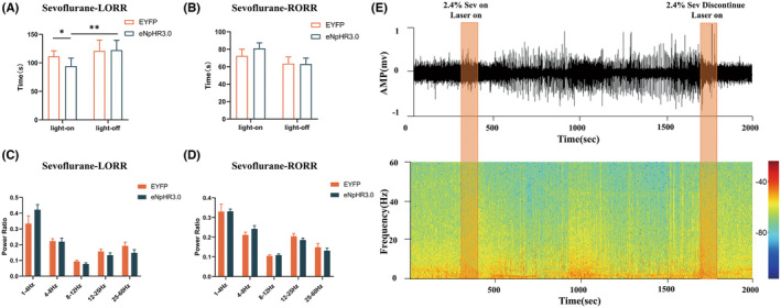
Suppressing PBN terminals in EA instead brings earlier loss of righting. During recovery after cessation, the same suppressive approach does not significantly prolong recovery. An ability to accelerate recovery does not make its dependence on the input identical to that of induction.
Under suppression, measured EEG bands change nonsignificantly in both induction and recovery comparisons. Behavioral timing changes without a fully opposite EEG-power response to activation. Contributions within the anesthesia network thus depend on phase and intervention direction.
Source: Yan Y et al. · CNS neuroscience & therapeutics · CC BY
Original figure and English caption ↗
Calcium differs during induction but not significantly during recovery
Terminal calcium in female and male mice is compared across matched time windows. Females have higher mean signals during initial induction. Both groups subsequently shift downward in early anesthesia, with a larger male decline. Sex-associated differences appear during entry into anesthesia.
Female–male calcium differences are nonsignificant in the two post-cessation recovery windows. Unmanipulated recordings therefore do not show the same distinction in every phase. Hormonal regulation is discussed as a possibility, without direct estrogen or receptor intervention testing that explanation.
Source: Yan Y et al. · CNS neuroscience & therapeutics · CC BY
Original figure and English caption ↗
Activation reveals a different behavioral and EEG comparison
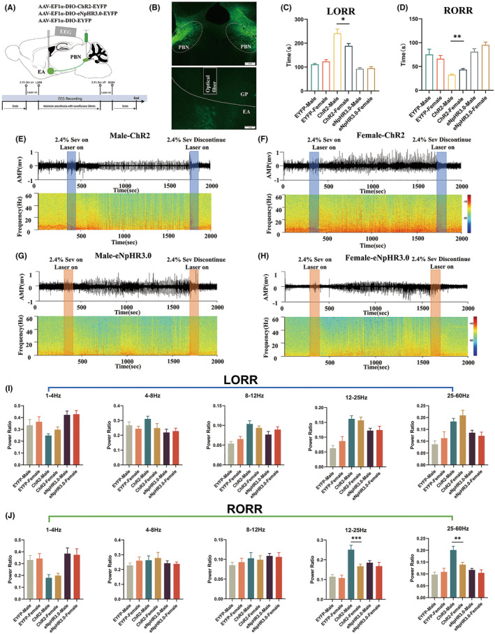
Under terminal activation, males lose righting later and recover earlier after cessation than females. Corresponding female–male timing differences under suppression are nonsignificant. Higher female terminal calcium during induction does not correspond to a longer induction time in the activation condition. The readouts describe different components.
EEG-band differences between sexes are nonsignificant during induction after activation, while male beta and gamma relative power is higher during recovery. In adult female and male mice, the study examines induction and post-cessation recovery as distinct processes. Righting responses, terminal signals and cortical EEG can be associated while retaining different dynamics.
Source: Yan Y et al. · CNS neuroscience & therapeutics · CC BY
Original figure and English caption ↗