How can one septal input push anesthetized brain activity in different directions?
A sevoflurane mouse study finds that VTA recipient identity and anesthetic depth shape cortical activation and suppression.
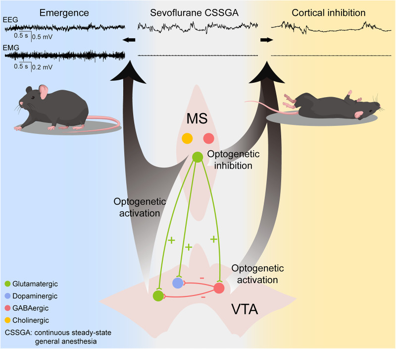
Under anesthesia, does increasing an excitatory population simply promote emergence? The answer also depends on its targets and the brain’s current depth of anesthesia. A 2026 Journal of Neuroscience study follows glutamatergic medial septal, or MS, cells into three VTA recipient classes. Under sevoflurane, the same upstream connection reaches effects in different directions: some cells promote cortical activation, while others enhance suppression during deeper anesthesia.
MS-associated VTA glutamatergic cells activate cortex at both tested depths, dopamine cells mainly in lighter anesthesia, and GABA cells enhance suppression mainly during burst suppression.
Contents · 6 sections
EEG states differ after loss of righting
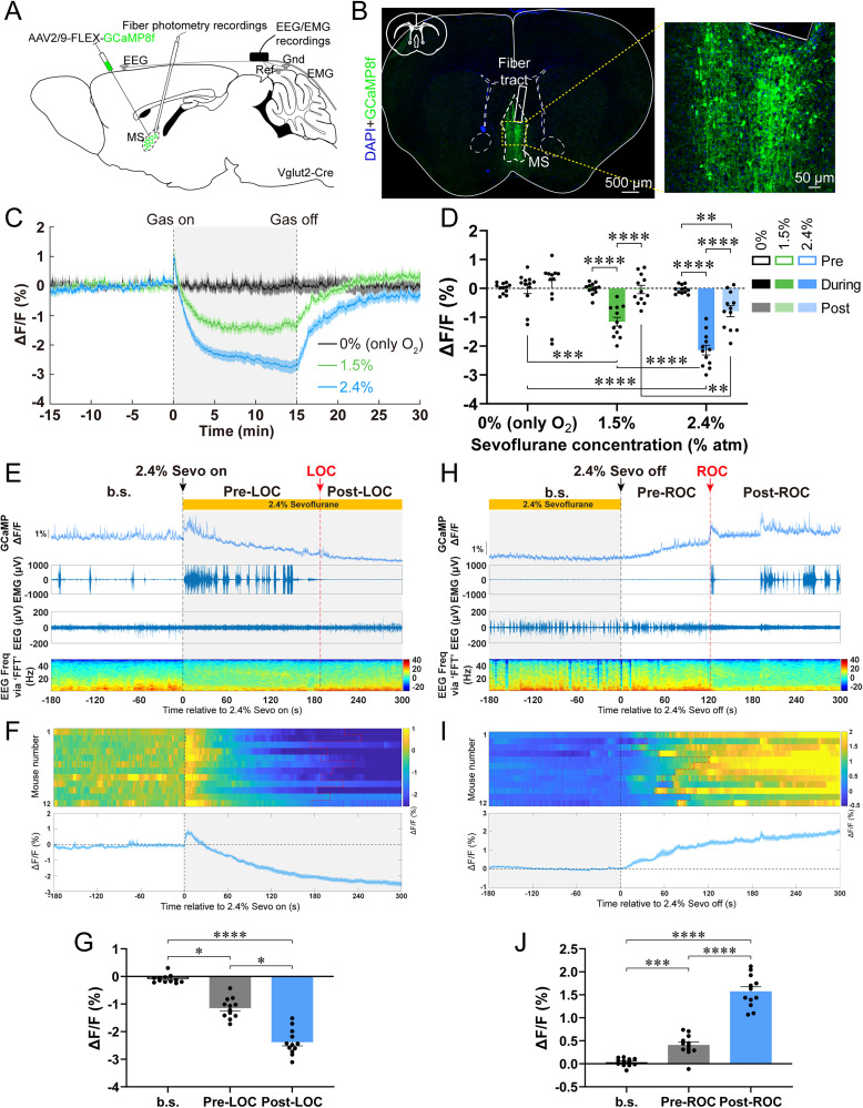
In the lighter steady state, mice maintained loss of righting; deeper conditions produced stable EEG burst suppression, alternating active periods with low-activity intervals. MS VGLUT2 calcium decreased with sevoflurane concentration and induction and rose during emergence, linking the population with state transitions.
The study followed righting, other movements, EMG and cortical spectra. These describe state from different angles: movement or cortical activation can appear without restored righting. Following both behavior and EEG allowed cellular-modulation effects to be examined at two levels.
Source: Wu JY et al. · The Journal of neuroscience : the official journal of the Society for Neuroscience · CC BY
Original figure and English caption ↗
Increasing MS activity shifts transitions toward emergence
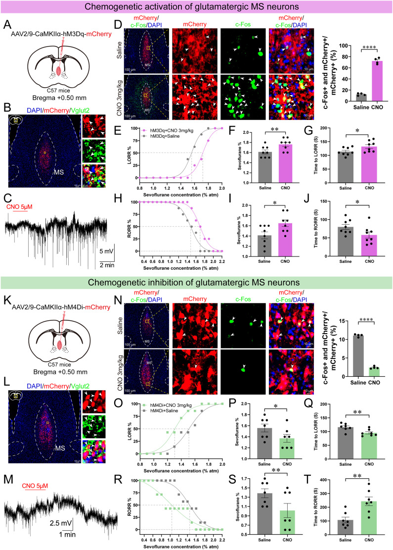
Increasing MS population activity raised the concentration needed for loss of righting, delayed induction and shortened recovery; suppression reversed these directions. Optical stimulation during maintenance changed spectra and EMG, with some lighter-state righting and deeper-state EEG activation.
Different measures showed different effect sizes, including a nonsignificant lighter-state decrease in burst-suppression ratio. Broad MS modulation influenced anesthetic sensitivity and transitions. The study then followed its VTA output to examine how much of the effect one connection could reproduce.
Source: Wu JY et al. · The Journal of neuroscience : the official journal of the Society for Neuroscience · CC BY
Original figure and English caption ↗
The VTA connection changes cortex with more limited righting
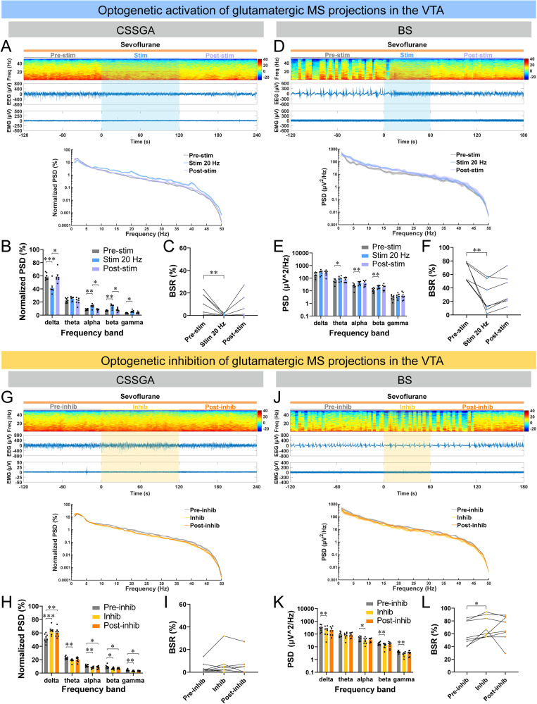
Increasing MS→VTA terminal activity promoted cortical activation at both depths; suppression deepened cortical inhibition. Compared with broad MS effects, righting changes after projection activation were less certain, and emergence-related terminal calcium increases were not significant. The distinction between cortical activation and fuller behavioral recovery became clearer at the output level.
The projection placed VTA within septal modulation of anesthesia, while MS also reaches other regions. Broad upstream effects can involve several outputs, and different cells within a target may act in opposing directions. The study therefore resolved VTA recipient populations.
Source: Wu JY et al. · The Journal of neuroscience : the official journal of the Society for Neuroscience · CC BY
Original figure and English caption ↗
One input reaches glutamatergic, dopamine and GABA cells
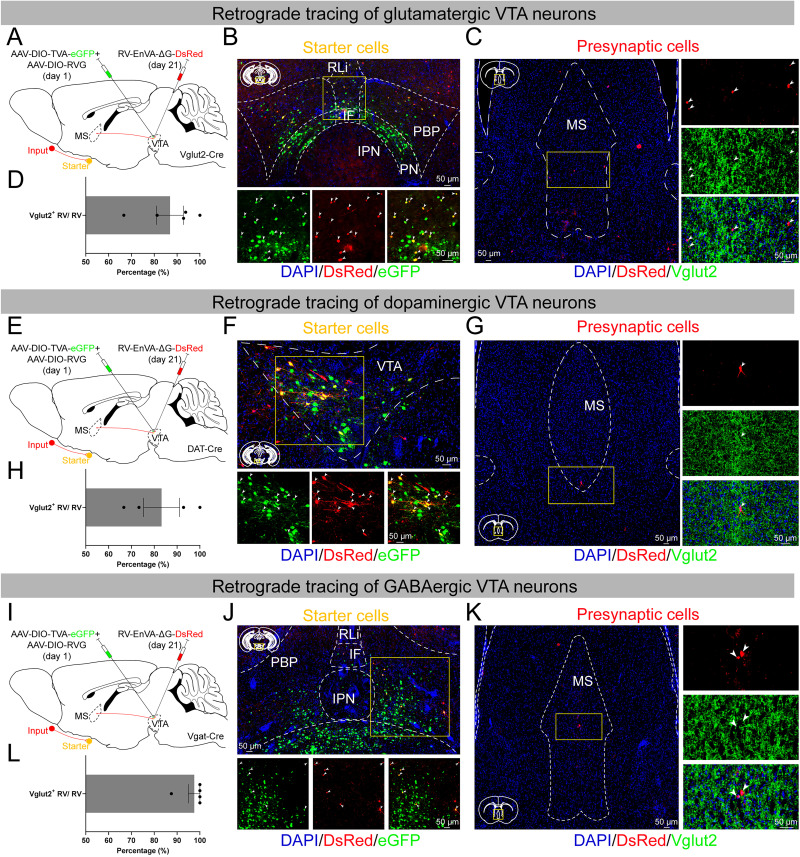
Monosynaptic tracing identified MS VGLUT2-associated inputs to VTA glutamatergic, dopamine and GABA cells. Anesthesia suppressed overall calcium, but relative depth effects differed, with higher relative GABA activity at greater depth. A shared upstream input did not produce identical state responses across classes.
Separating populations by input relationship and identity unpacked the regional effect. A septal connection reaching different recipients could have different directions of influence on cortex and behavior. The functional results illustrated this internal diversity.
Source: Wu JY et al. · The Journal of neuroscience : the official journal of the Society for Neuroscience · CC BY
Original figure and English caption ↗
Different functional patterns at lighter and deeper states
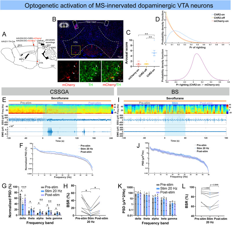
Increasing MS-recipient VTA glutamatergic activity activated cortex at both depths, with four of seven lighter-state animals righting. Dopamine cells mainly activated cortex in lighter anesthesia, with few animals righting and no significant deep-state EEG or EMG change. GABA effects were limited in lighter anesthesia but enhanced burst suppression at depth.
The lighter-state dopamine result particularly illustrates separation among readouts: EEG became more active, yet few animals regained righting. Righting was more common in the glutamatergic population. Cortical changes in the same direction can therefore accompany different behavioral outcomes; observing movement, EMG and righting together captures this distinction.
The three input-associated classes therefore formed different patterns across depths. Glutamatergic activation was broader, dopamine effects were stronger in the lighter state, and GABA suppression effects stood out at depth. Shorter deep-state stimulation also remains relevant to interpreting the dopamine result.
Source: Wu JY et al. · The Journal of neuroscience : the official journal of the Society for Neuroscience · CC BY
Original figure and English caption ↗
Knowing the source also requires knowing its recipients
The study gives excitatory upstream input a more specific network meaning. One source reaches activating and suppressing populations, and current anesthetic state changes their influence. Recipient composition and network state are key to understanding the outcome alongside regional activation.
The experiments included both sexes, mainly during sevoflurane maintenance. Cortical activity could rise before righting returned, while glutamatergic, dopamine and GABA populations had different windows of influence. Brain activity under anesthesia thus behaved less like a single volume control and more like the combined work of cell populations whose contributions changed with state.
Source: Wu JY et al. · The Journal of neuroscience : the official journal of the Society for Neuroscience · CC BY
Original figure and English caption ↗