Cooling the body or changing brain networks? Two hypothalamic routes in anesthesia
A mouse study finds that two populations within thermoregulatory networks influence brain activity and emergence from isoflurane on different timescales.
Anesthetic delivery has stopped, yet an animal has not regained righting. The reason may involve brain networks or falling body temperature. How can these contributions be separated? A 2026 iScience study compares two hypothalamic populations: LPO BDNF cells and DMH VGLUT2 cells. Both belong to thermoregulatory networks but show different routes. LPO effects mainly track cooling, whereas DMH can change cortical activity before a concurrent shift in core temperature.
LPO BDNF anesthetic effects mainly relate to temperature. DMH VGLUT2 cells and their RPa output also alter EEG during maintained isoflurane without concurrent core-temperature changes.
Contents · 6 sections
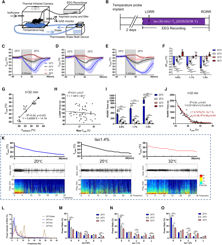
Colder bodies recover more slowly under similar anesthesia
Ambient temperature changed core temperature in isoflurane-anesthetized mice. After delivery stopped, lower temperature related to slower righting recovery, especially in the colder range. Initial temperatures were stable, and time to lose righting lacked the same association. Temperature therefore had different relationships with entry into and emergence from anesthesia.
Temperature may influence metabolism, circulation and drug clearance, so recovery after withdrawal can reflect several bodily processes. Slower emergence in colder states adds physiological context to circuit effects: a population changing righting time may influence both brain activity and body temperature. The temperature comparisons reveal that context.
The finding introduced a question about neuronal effects: when a population slows recovery, has it first cooled the body? Righting time alone leaves the two routes difficult to separate. Following temperature alongside EEG reveals their ordering, motivating study of two thermoregulatory populations.
Source: Cai S et al. · iScience · CC BY
Original figure and English caption ↗
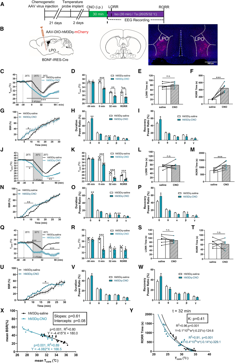
LPO effects emerge alongside cooling
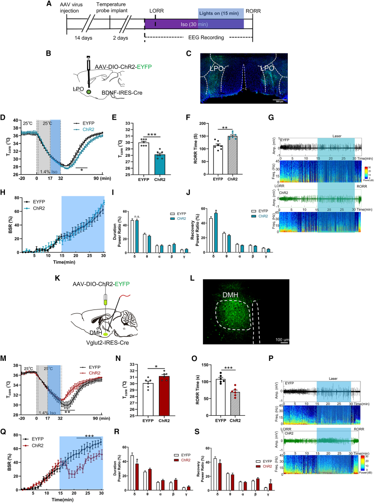
Increasing LPO BDNF activity before anesthesia lowered temperature through the anesthetic period. EEG burst suppression and delta power increased, and recovery slowed, with little change in induction time. A body already in a colder state showed different cortical and recovery outcomes; under warmer conditions, the recovery delay was not significant.
When the population was enhanced only during maintenance, immediate temperature and EEG lacked the same changes, but cooling and delayed emergence appeared after withdrawal. Recruiting the same cells at different times shifted when effects appeared, tying LPO outcomes more closely to the thermal process.
Source: Cai S et al. · iScience · CC BY
Original figure and English caption ↗
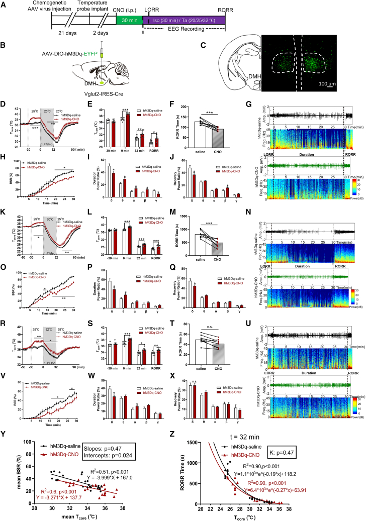
DMH warms the body and reduces cortical suppression
Increasing DMH VGLUT2 activity before anesthesia raised temperature, reduced burst suppression and delta, and shortened recovery, with generally opposite effects after suppression. Magnitudes differed across thermal environments, with some warmer-condition recovery comparisons nonsignificant. DMH outcomes, like LPO outcomes, carried a thermal context.
The stories diverged here. LPO had limited immediate effects during anesthesia, whereas DMH enhancement rapidly changed cortical activity. Maintenance-phase tests allowed observation of whether EEG responded before temperature changed.
Source: Cai S et al. · iScience · CC BY
Original figure and English caption ↗
Burst suppression falls while core temperature remains similar
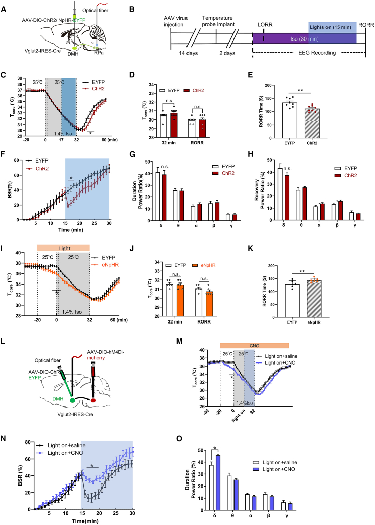
DMH enhancement during maintained isoflurane immediately lowered burst suppression without a concurrent core-temperature change. The EEG effect also appeared in thermoneutral conditions maintaining normal temperature. Similar whole-body temperature with an earlier cortical change provided a temporal clue to another DMH contribution.
Temperature differences could still appear during recovery, so thermal and network effects could coexist. The temporal separation revealed a contribution beyond warming followed by emergence: DMH influenced cortex before the measured core-temperature shift.
Source: Cai S et al. · iScience · CC BY
Original figure and English caption ↗
The RPa route carries part of the effect
Enhancing DMH→raphe pallidus, or RPa, terminals during anesthesia shortened recovery and lowered burst suppression, with similar maintenance and righting temperatures and no significant delta change. Suppression delayed recovery, with early cooling no longer evident at the endpoint. Directional findings placed part of the effect in a brainstem connection.
Suppressing RPa cells only partly weakened DMH effects, indicating contribution by this output with possible participation of others. EEG, core temperature and emergence changed differently, showing several processes through which a thermoregulatory network can influence anesthesia.
Source: Cai S et al. · iScience · CC BY
Original figure and English caption ↗
Temperature and cortex follow different time courses
LPO cooling accompanied deeper suppression, while DMH could warm the body and change EEG before core temperature shifted. The populations clarified different processes behind emergence speed. Timing of cellular recruitment and EEG responses matters alongside body temperature for understanding behavior.
The findings come from adult male mice under isoflurane. Core temperature reflects whole-body state, while brain temperature, circulation and drug clearance may also contribute. The temporal differences reveal distinguishable routes of thermoregulatory-cell influence and clarify relationships between bodily physiology and brain networks.