From sleep to anesthesia: how does the bed nucleus contribute to arousal?
A bed-nucleus inhibitory population promotes sleep-to-wake transitions in mice. Comparing activation, chronic cell reduction and anesthetic states reveals different conditions for behavioral and EEG arousal.
Wakefulness, sleep, dreaming and drug-induced anesthesia are distinct brain states. A study published in Communications Biology on March 19, 2024 examines how GABAergic cells in the bed nucleus of the stria terminalis contribute to their transitions. Activation promoted waking from sleep, while chronic cell reduction altered nighttime sleep allocation without significantly changing anesthetic induction or emergence timing, revealing state-dependent contributions.
BNST GABAergic cells promote natural waking and selected anesthesia-related arousal effects. Chronic reduction changes dark-phase sleep architecture, while VTA-directed output contributes to only part of the activation effects.
Contents · 6 sections
Active during both waking and REM sleep
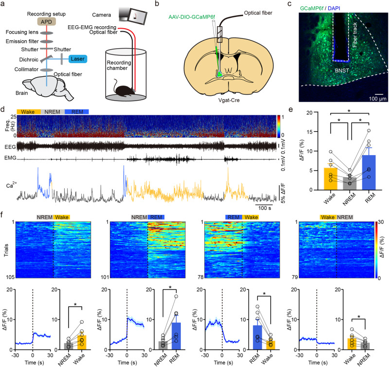
Population calcium, EEG, EMG and video recordings compared wakefulness, NREM and REM sleep. BNST GABAergic signals were higher during waking than NREM and higher still during REM, so activity did not simply mark being awake.
Signals rose with NREM-to-wake and NREM-to-REM transitions but declined from REM to waking. These relative changes reveal a richer pattern than a single arousal label. Population averaging leaves open how distinct cellular subsets contribute.
Source: Li M et al. · Communications biology · CC BY
Original figure and English caption ↗
Population signals decline under isoflurane
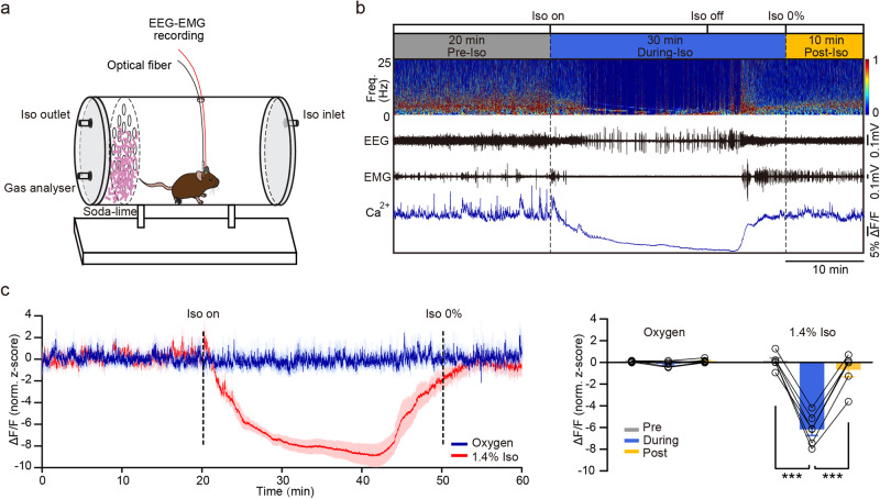
Under isoflurane, population calcium signals declined and returned toward their earlier level after anesthesia ended. Oxygen controls did not show the same change, and slice recordings likewise showed reduced firing during exposure and recovery after washout.
High REM activity and lower activity under isoflurane illustrate the distinct network states involved. These observations motivated a functional question: could changing this population influence transitions?
Source: Li M et al. · Communications biology · CC BY
Original figure and English caption ↗
Faster waking from both sleep states
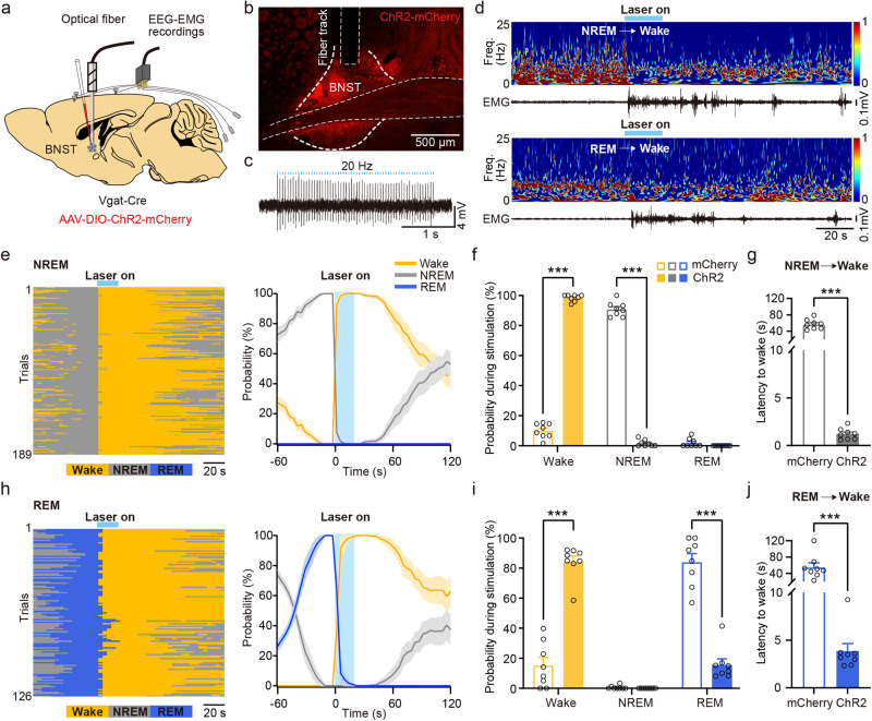
Activating the population promoted rapid waking from stable NREM, increasing waking probability and shortening latency. Activation during REM likewise accelerated waking, with EEG and EMG providing complementary state measures.
Prolonged activation during a sleep-prone period increased waking and reduced NREM without significantly changing REM amount. Rapid transitions and sustained state allocation thus revealed related but distinct effects under cellular activation.
Source: Li M et al. · Communications biology · CC BY
Original figure and English caption ↗
EEG activation and behavioral arousal are distinct outcomes
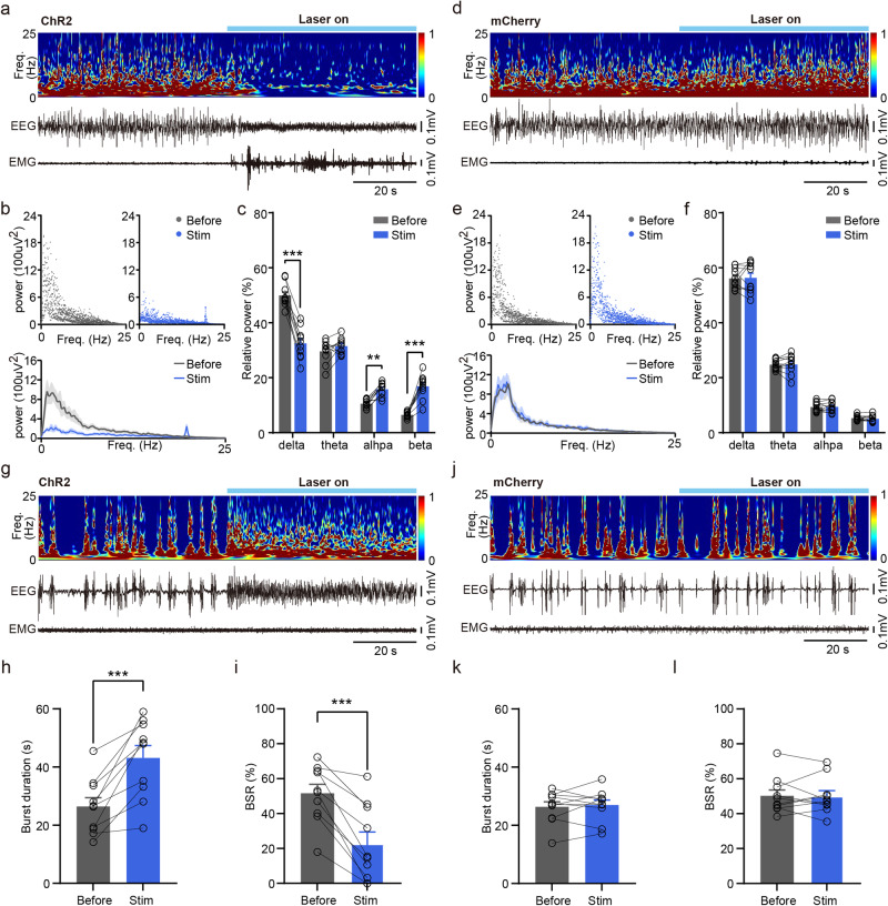
During steady-state isoflurane anesthesia, activation increased EMG and body movement while reducing relative delta and increasing alpha and beta power, linking the population to behavioral arousal-related changes.
Under deeper anesthesia, activation prolonged EEG bursts and lowered the burst-suppression ratio without a corresponding EMG increase. This was an EEG activation effect without matching behavioral evidence, distinguishing its scope from the steady-state result.
Source: Li M et al. · Communications biology · CC BY
Original figure and English caption ↗
Nighttime sleep changes without altered anesthetic timing
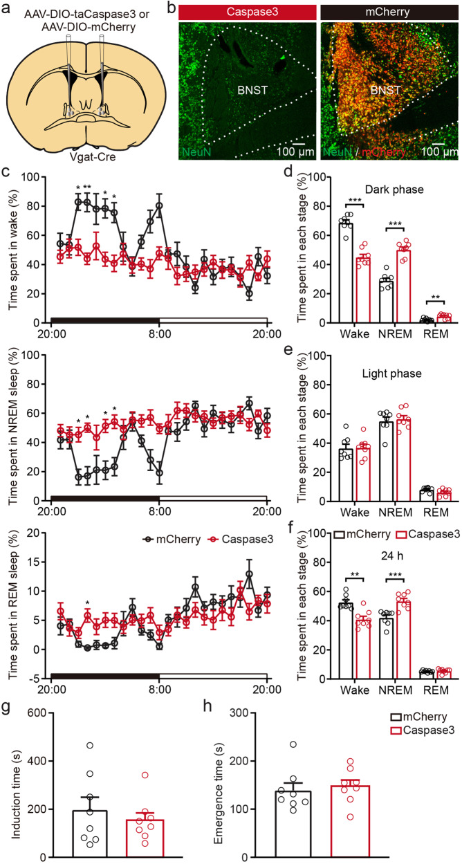
Chronic cell reduction decreased dark-phase waking and increased NREM and REM, without significant corresponding light-phase differences. Across the full day, waking decreased and NREM increased while REM amount did not significantly change.
Isoflurane induction and emergence timing did not differ significantly after chronic reduction. Promoting arousal through activation and changing anesthetic timing after chronic loss are distinct questions. How networks redistribute function after prolonged change remains unresolved.
Source: Li M et al. · Communications biology · CC BY
Original figure and English caption ↗
VTA-directed output accounts for part of the effect
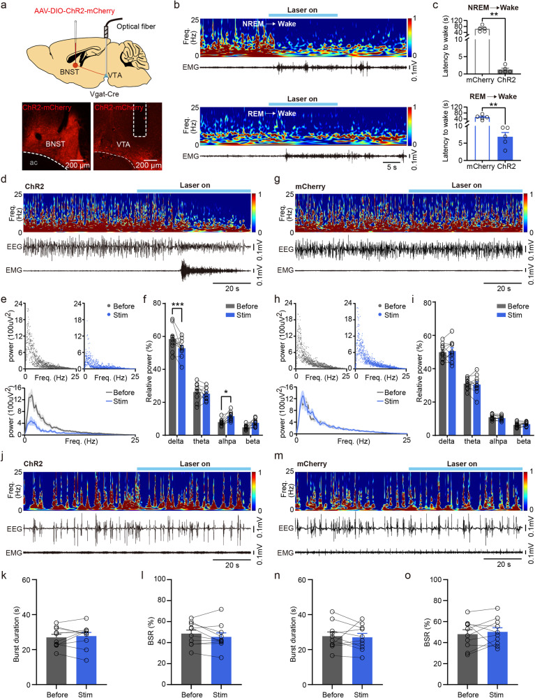
Activating BNST terminals in VTA shortened waking latency from NREM and REM and produced EEG, EMG and behavioral arousal-related changes during steady-state anesthesia. Relative delta decreased and alpha increased, echoing part of the population-activation findings.
Under deep anesthesia, terminal activation did not significantly alter burst duration or suppression ratio and did not reproduce the population-level EEG effect. This output therefore contributes without explaining every outcome. Both sexes were used without establishing equivalent effects in each; finer cellular and downstream resolution will help clarify state-dependent arousal.
Source: Li M et al. · Communications biology · CC BY
Original figure and English caption ↗