Cortical adenosine connects flicker with sleep
A mouse study connects flicker-related adenosine changes, ENT2 transport and increased sleep. A two-night self-controlled observation in 49 children adds preliminary clinical clues awaiting further testing.
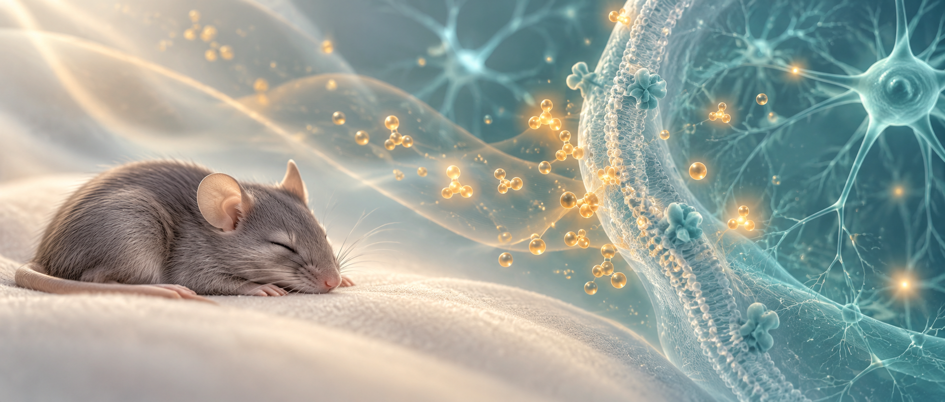
Light is commonly associated with wakefulness, yet rhythmic flicker can produce a different outcome. A 2024 Cell Research study follows mouse brains after flickering-light stimulation and finds elevated adenosine signals in primary visual cortex, V1, persisting after stimulation ends. Adenosine’s links with energy metabolism and sleep need connect visual stimulation, cellular metabolism and sleep. The paper also reports preliminary observations in children, providing a different level of evidence from the animal mechanisms.
Tested 40-Hz flicker increases V1 adenosine signals and subsequent sleep in mice. Increased sleep is absent after ENT2 deletion or V1-neuron removal. Children’s before-and-after night comparisons show faster sleep onset and longer sleep, remaining preliminary self-controlled observations.
Contents · 8 sections
Adenosine signals persist after flicker stops
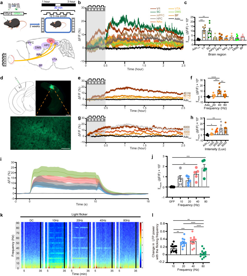
Extracellular adenosine-related fluorescence rises most prominently in V1 and superior colliculus, with smaller changes elsewhere and little increase in dorsomedial striatum. V1 signals continue rising after flicker ends and take hours to return to baseline.
Among tested frequencies, 40 Hz produces larger, longer adenosine responses, while faster flicker evokes stronger neuronal calcium signals. Activity amplitude and adenosine therefore do not correspond simply; rhythm, network activity and metabolism may contribute together.
Source: Zhou X et al. · Cell research · CC BY
Original figure and English caption ↗
Neuronal involvement and a glial intervention with no detected effect
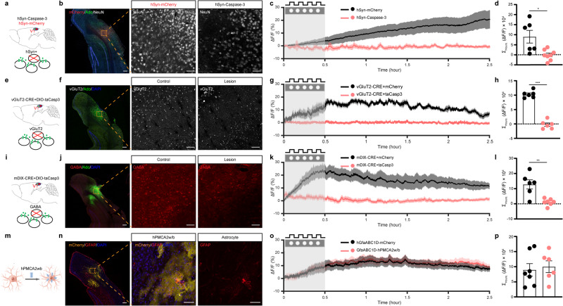
Removing V1 neurons markedly weakens flicker-related adenosine increases. Reducing glutamatergic or GABAergic cells also attenuates the response, supporting neuronal-network involvement. Interactions between excitatory and inhibitory cells may affect signal generation.
A separate intervention partially reduces astrocytic calcium responses without significantly changing the adenosine increase. This tested intervention, alongside neuronal removal, helps locate cellular components of the flicker response.
Source: Zhou X et al. · Cell research · CC BY
Original figure and English caption ↗
Adenosine moves from inside cells to the extracellular space
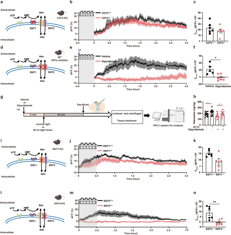
Adenosine can arise from extracellular ATP breakdown or be produced inside cells and exported. Mice lacking the extracellular conversion enzyme CD73 retain a clear response. Blocking nucleoside transport markedly reduces extracellular signals while tissue-total adenosine still increases.
Comparing transporters shows persistent responses after ENT1 deletion, with somewhat lower later amplitudes, whereas ENT2 deletion greatly attenuates them. Intracellular generation and transmembrane export provide linked explanations for the sustained change.
Source: Zhou X et al. · Cell research · CC BY
Original figure and English caption ↗
Energy-related activity leaves metabolic traces
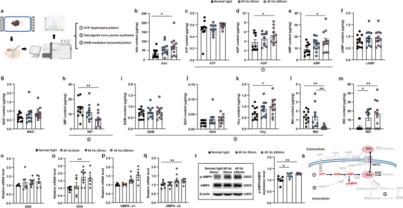
Across V1 tissue sampling times, adenosine, ADP and AMP change without a significant decrease in total ATP. Breakdown and replenishment can occur together, so tissue totals need not directly reflect moment-to-moment consumption.
AMPK-related expression and phosphorylation also change, providing clues to energy-metabolic involvement. The study links adenosine increases with stepwise ATP metabolism. These tissue observations describe metabolic state without separately testing whether AMPK determines the sleep effect.
Source: Zhou X et al. · Cell research · CC BY
Original figure and English caption ↗
Sleep increases depend on the tested rhythm and timing
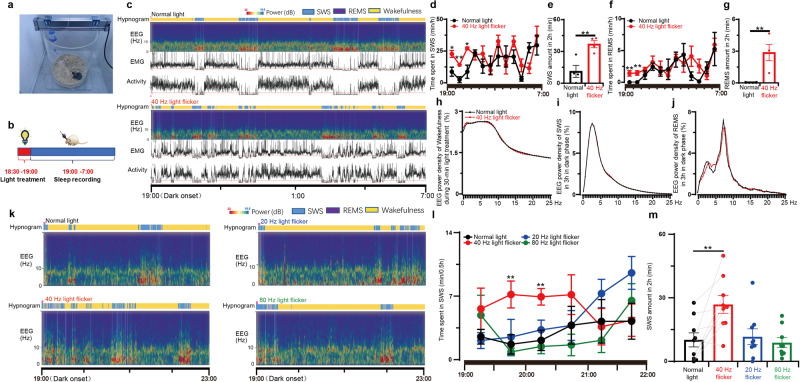
Mice receiving flicker near the light-phase end show increased non-REM and REM during the first hours of darkness. The same frequency early in the light phase lacks the same effect, as do the other tested frequencies, highlighting context and timing.
Increased sleep amount is not accompanied by significant changes in tested sleep-EEG power density. Recordings distinguish duration from spectral properties, rather than treating both as a single measure of deeper sleep.
Source: Zhou X et al. · Cell research · CC BY
Original figure and English caption ↗
Increased sleep is absent when ENT2 is deleted
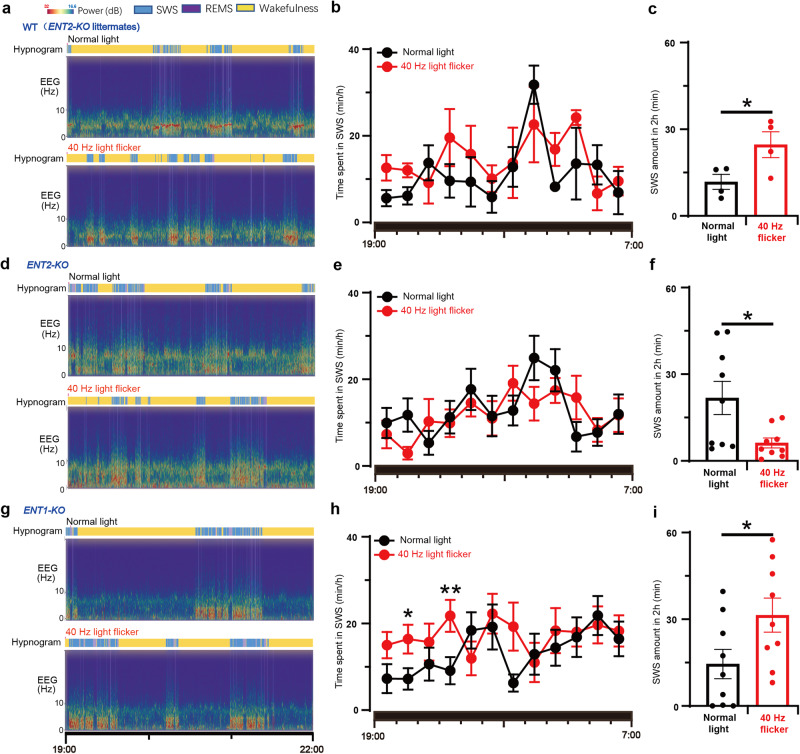
ENT2-deficient mice no longer show post-flicker non-REM increases; sleep is instead lower than under ordinary light within the tested window. ENT1-deficient mice retain the increase. This transporter contrast links extracellular adenosine changes more closely with sleep outcomes.
The comparison uses transporter-deficient animals, describing changes at the whole-animal level. Alongside local V1 recordings, it supports ENT2-related adenosine export and motivates examining visual cortex itself.
Source: Zhou X et al. · Cell research · CC BY
Original figure and English caption ↗
Visual cortex does more than convey light
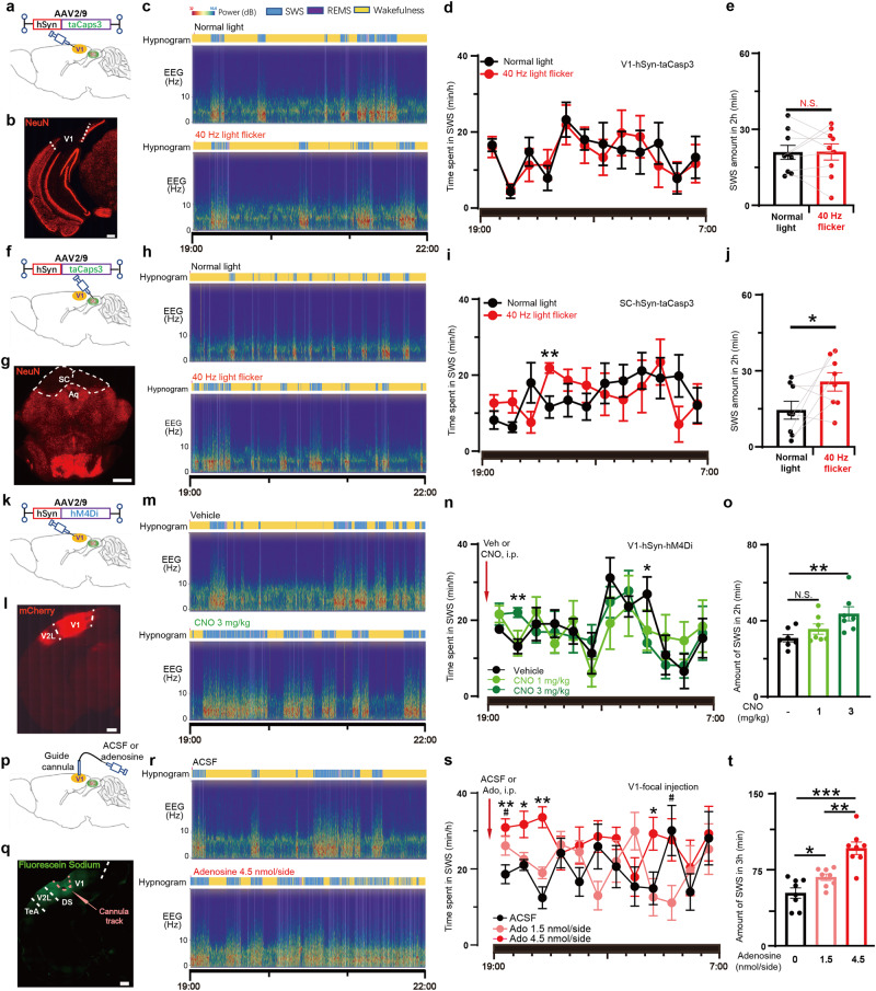
Removing V1 neurons eliminates the flicker-related sleep increase, while tested non-REM increases remain after superior-colliculus neuronal reduction. Although both regions show adenosine responses, their outcomes differ in these sleep experiments.
Directly suppressing V1 neurons or locally elevating V1 adenosine also increases tested sleep. Visual-cortical state therefore enters the sleep-regulation story, with the receptor subtype carrying this effect still to be established.
Source: Zhou X et al. · Cell research · CC BY
Original figure and English caption ↗
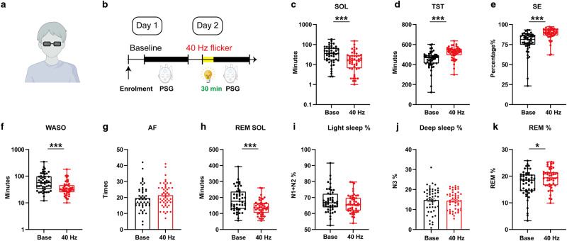
Observations in children provide preliminary clinical clues
Forty-nine children with insomnia symptoms undergo recordings on two consecutive nights, with the first as baseline and flicker included on the second. Sleep onset is sooner, total sleep and efficiency increase, and waking after sleep onset decreases. Awakening count and light/deep-sleep proportions do not significantly change; REM proportion increases.
This sequential two-night self-controlled study lacks a randomized sham condition and does not assess long-term effects. It offers clinical clues, while the animal work provides a more specific insight: energy metabolism and adenosine export after visual stimulation can contribute to subsequent sleep changes.
Source: Zhou X et al. · Cell research · CC BY
Original figure and English caption ↗