An anesthesia-recruited sensory region has selective effects
Anterior piriform glutamatergic-related cells bidirectionally alter induction with two inhaled anesthetics in male mice. Emergence and propofol outcomes remain largely unchanged; odor conditions reveal additional sensory effects.

Anesthesia diminishes behavioral responses, yet not every brain cell becomes quiet. Piriform cortex, closely associated with smell, shows increased activity markers during anesthesia. As a major olfactory cortex, it receives direct olfactory-bulb input and participates in learning and emotion-related processes. Does it merely follow the state or alter entry into it? A study published online in Fundamental Research on January 3, 2023 compares piriform subdivisions, cellular manipulations and three anesthetics in male mice. Using loss and return of righting reflex alongside EEG, the team finds that anterior piriform glutamatergic-related cells can delay or accelerate induction with two inhaled anesthetics without similarly changing propofol outcomes. This selectivity complicates the relationship between sensory regions and anesthesia.
Anterior piriform manipulation affects induction with isoflurane and sevoflurane without changing tested emergence times. Odor-related explanations coexist with other possible output mechanisms.
Contents · 6 sections
Increased markers identify a candidate region
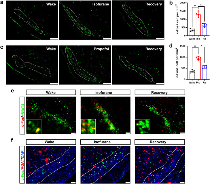
During isoflurane anesthesia, piriform c-Fos-positive cells increase and then decline relative to anesthesia during recovery. Propofol also produces marker accumulation. Isoflurane-associated increases chiefly colocalize with glutamatergic-related labeling, without a corresponding increase in the labeled GABAergic population.
c-Fos reflects activity-related changes over a time interval. Both agents increase the marker, indicating responses to different anesthetic states. Altering cellular activity then reveals different functions beneath the similar marker changes.
Source: Zhou L et al. · Fundamental research · CC BY-NC-ND
Original figure and English caption ↗
Anterior and posterior regions contribute differently
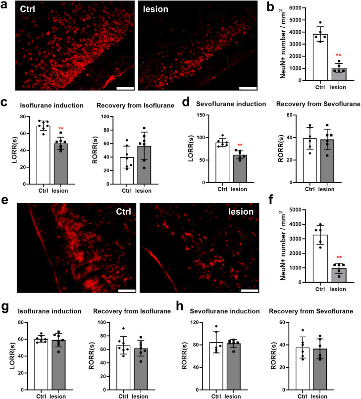
After neuronal lesions in anterior piriform cortex (APC), loss of righting occurs sooner with isoflurane and sevoflurane, without a significant change in return time. Posterior piriform lesions do not significantly change either timing measure for the two agents.
Subdivisions of this sensory region are not interchangeable. Righting reflects a mouse’s ability to restore its body position and operationally defines induction and recovery here. The anterior result narrows attention from an entire cortex to selected populations within it.
Source: Zhou L et al. · Fundamental research · CC BY-NC-ND
Original figure and English caption ↗
Increasing activity delays induction without changing emergence time
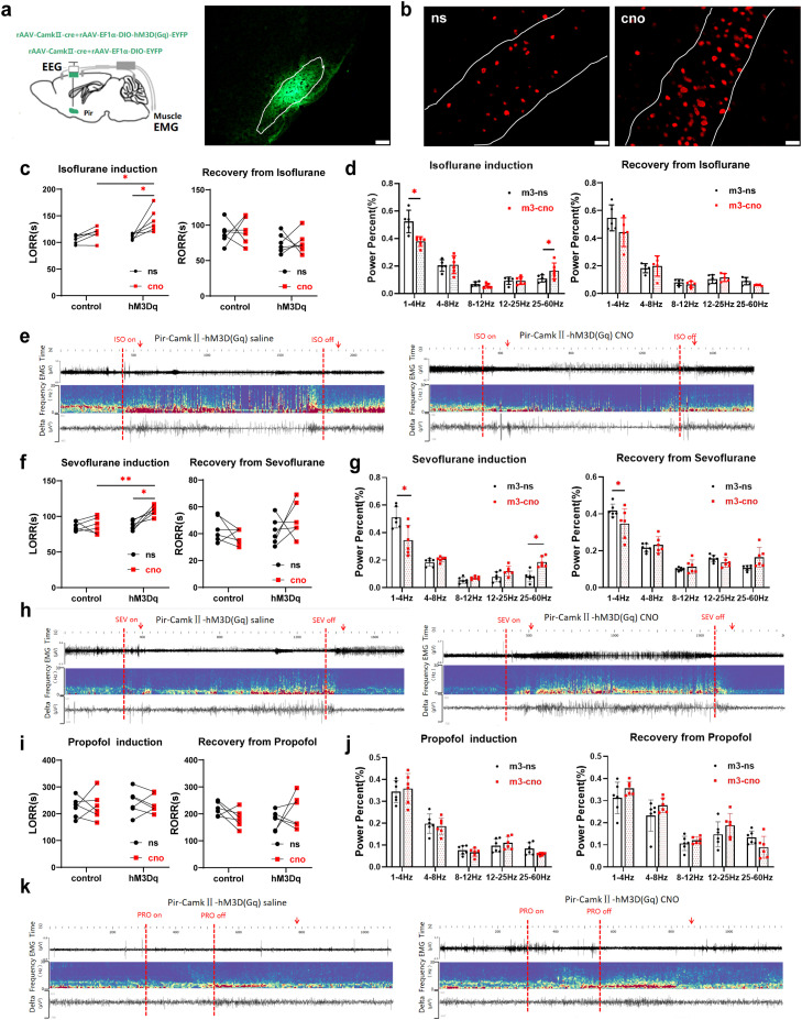
Increasing APC glutamatergic-related activity prolongs induction with isoflurane and sevoflurane. During induction, EEG delta-power proportion falls and gamma proportion rises, linking behavioral timing with spectral changes. Propofol induction, emergence and tested EEG comparisons show no significant effects.
Righting-return times with the inhaled agents remain largely unchanged, although sevoflurane emergence still shows a lower delta proportion. Stable behavioral timing can coexist with a spectral difference; induction and emergence are distinct phases.
Source: Zhou L et al. · Fundamental research · CC BY-NC-ND
Original figure and English caption ↗
Reducing activity shifts induction oppositely
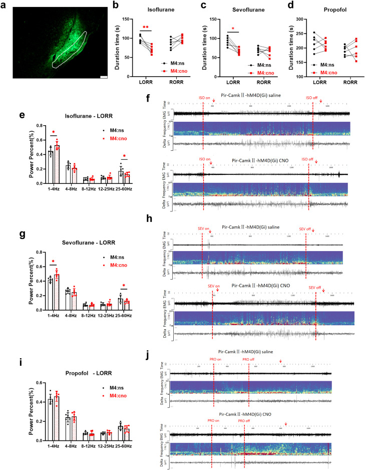
Inhibiting the related APC population shortens isoflurane and sevoflurane induction, raises induction delta proportion and lowers gamma proportion, opposite to activation. Righting-return times for both agents remain nonsignificant, as do propofol timing and tested EEG outcomes.
Opposing manipulations clarify participation in the tested inhaled-agent induction process rather than a uniform change in sensitivity across every anesthetic phase. The population’s influence depends on the agent and transition stage.
Source: Zhou L et al. · Fundamental research · CC BY-NC-ND
Original figure and English caption ↗
A different manipulation supports the same main pattern
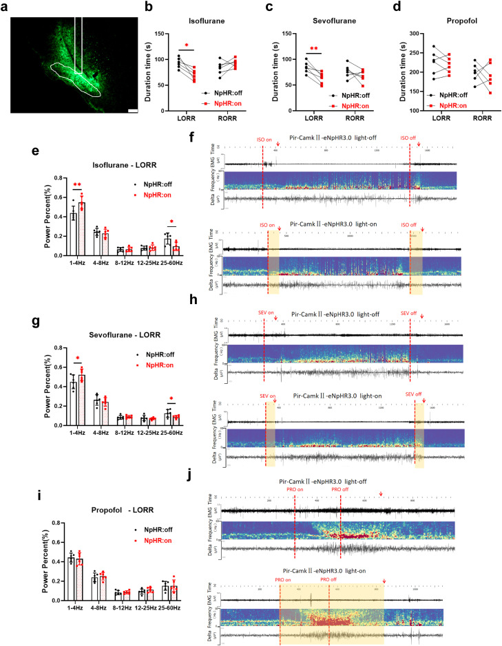
Optogenetic activation likewise delays induction with both inhaled agents and lowers delta while raising gamma proportion; inhibition accelerates induction with opposite spectral changes. Righting-return and tested propofol timing comparisons remain nonsignificant.
Convergent main results support an influence of APC activity on this transition. Reflex timing and spectra provide complementary behavioral and population-level views of the transition. The specific distant projection responsible remains to be identified.
Source: Zhou L et al. · Fundamental research · CC BY-NC-ND
Original figure and English caption ↗
Odor context also changes induction
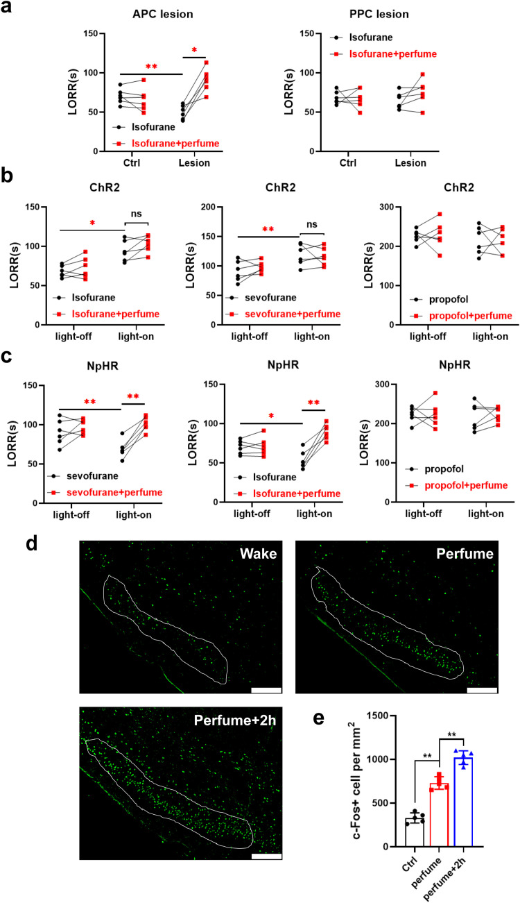
Under an odor condition, inhaled-anesthetic induction becomes longer in APC-lesioned or inhibited groups, changing the earlier shortening pattern. Adding odor does not significantly prolong timing further in activated populations, while unstimulated controls also show prolongation. Sensory context therefore influences outcomes across conditions.
c-Fos increases after odor exposure and increases further at the observation point after odor removal, unlike its decline during isoflurane recovery. The authors discuss odor processing alongside possible other outputs. These male-mouse findings concern the tested agents, populations and reflex measures, informing relationships between sensory circuitry and induction.
Source: Zhou L et al. · Fundamental research · CC BY-NC-ND
Original figure and English caption ↗