Why do neighboring inhibitory populations differ during anesthetic emergence?
Two neighboring hypothalamic regions contain GABAergic cells but show different effects under sevoflurane. Male-mouse research links EEG, righting recovery and cellular activity to dorsomedial hypothalamic arousal.
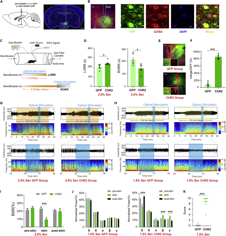
Do inhibitory-transmitter-marked cells in neighboring regions play the same role under anesthesia? A study published in iScience on December 30, 2022 compared GABAergic populations in dorsomedial, DMH, and ventromedial, VMH, hypothalamus. Enhancing DMH-related activity delayed loss of righting, accelerated recovery and altered maintenance-phase EEG and movement. VMH activation mainly changed EEG without significant changes in the tested behavioral transitions, revealing regional and state-dependent cellular contributions.
DMH GABAergic activity has complementary functional evidence: activation delayed induction and accelerated emergence, while suppression did the reverse. VMH activation did not significantly alter transitions or deep-state burst suppression, although spectral changes occurred under lighter anesthesia.
Contents · 6 sections
Medial hypothalamic activity influences anesthetic transitions
Both DMH and VMH participate in bodily-state regulation. Prior work links DMH with circadian and sleep–wake control and VMH activity with sleep states. These observations motivated their examination in anesthesia, where their actual contributions required separate functional evidence.
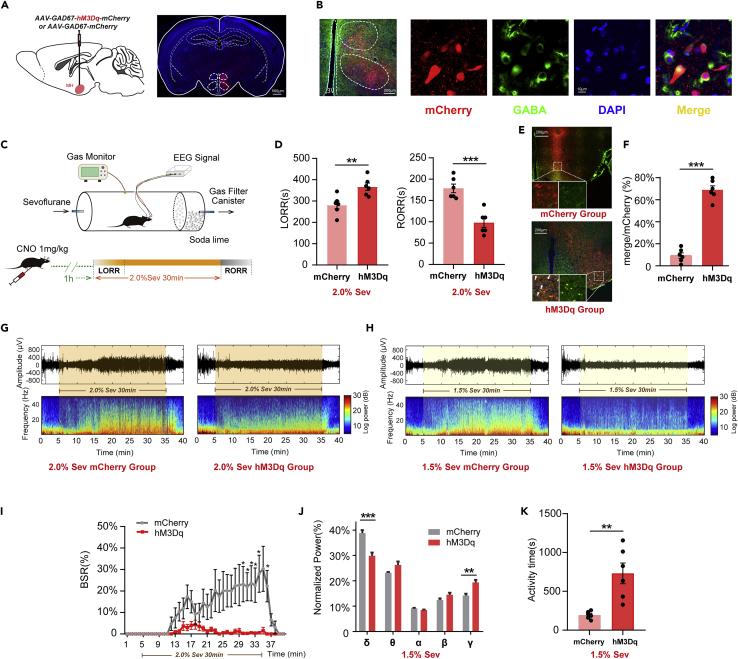
Enhancing a GABA-associated population across medial hypothalamus, including DMH and VMH, delayed loss of righting and shortened recovery. Maintenance-phase burst suppression decreased; lighter conditions showed a lower delta and higher gamma power fraction with more movement time.
The broader regional result linked inhibitory cells with overall arousal, while leaving open which neighboring nucleus accounted for the behavioral effects. Anatomical location therefore became central to the next comparison.
Source: Wang Y et al. · iScience · CC BY-NC-ND
Original figure and English caption ↗
DMH activation changes both behavior and EEG
Focusing activation on DMH again delayed induction and accelerated emergence. In a deeper burst-suppression state, activation lowered the suppression ratio with subsequent return. Under lighter anesthesia, movement increased, delta power fraction fell and beta and gamma fractions rose.
Righting, bodily movement and EEG provide different aspects of state. Their convergence supports DMH participation in anesthetic transitions, without directly measuring every subjective aspect of consciousness.
Source: Wang Y et al. · iScience · CC BY-NC-ND
Original figure and English caption ↗
VMH EEG changes lack the same behavioral effects
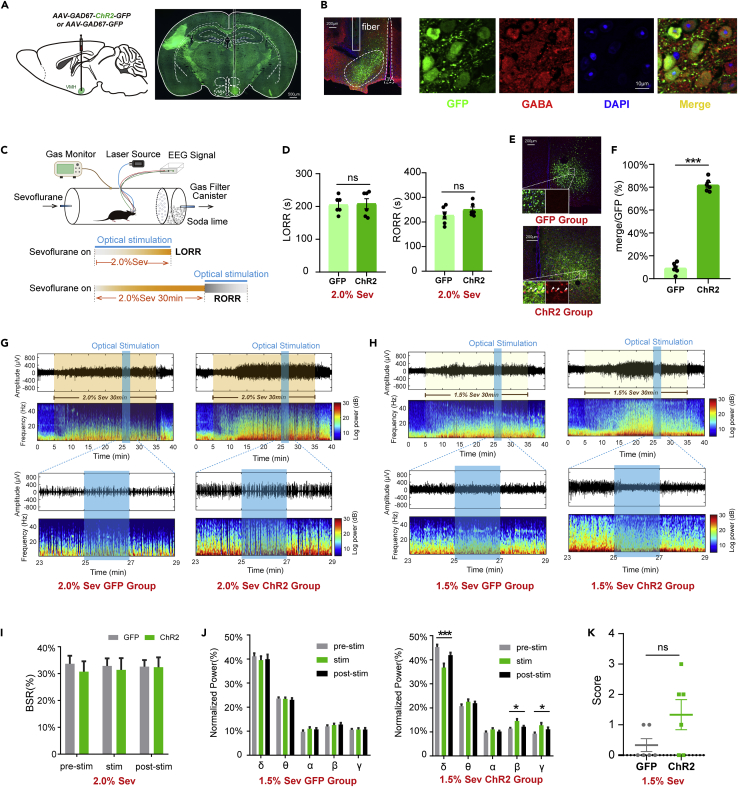
Activating the neighboring VMH GABA-associated population did not significantly change induction, emergence or deep-state burst suppression. Under lighter anesthesia, delta fraction decreased and beta and gamma increased, but behavioral arousal scores did not significantly rise.
Spectral changes could therefore occur separately from obvious movement. VMH influenced brain activity without producing the DMH-like behavioral outcomes under the tested conditions, revealing heterogeneity within the broader medial region.
Source: Wang Y et al. · iScience · CC BY-NC-ND
Original figure and English caption ↗
Activity falls during induction and rises during recovery
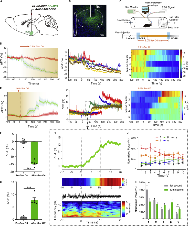
DMH GABA-associated calcium changed during anesthetic transitions: it declined after sevoflurane began and rose after withdrawal. A brief stronger rise accompanied rapid recovery, disappearance of burst suppression and shifts in low- and high-frequency power fractions.
The timing links cellular activity with recovery but does not by itself establish causal order. Recovery-related movement or restlessness could also contribute to calcium changes, making the functional manipulations a complementary source of evidence.
Source: Wang Y et al. · iScience · CC BY-NC-ND
Original figure and English caption ↗
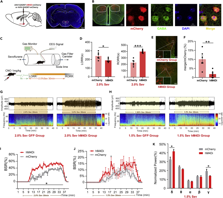
DMH suppression speeds induction and delays recovery
Suppressing the DMH GABA-associated population accelerated loss of righting and delayed recovery. Burst suppression increased under two anesthetic conditions; an earlier window without suppression showed a higher delta and lower gamma power fraction.
Activation and suppression provided complementary evidence that this population supports arousal under the tested conditions. Burst-suppression ratios and spectral fractions describe different quantities measured in distinct state windows.
Source: Wang Y et al. · iScience · CC BY-NC-ND
Original figure and English caption ↗
A shared marker can have different network roles
A further DMH activation comparison likewise delayed induction and accelerated emergence. These male-mouse findings combine regional, cellular and phase-specific evidence to distinguish neighboring nuclei. The responsible outputs and GABAergic subtypes remain untested, and proposed downstream disinhibition requires more specific connectivity evidence.
Brief activation did not significantly alter measured temperature, while cardiorespiratory contributions, longer durations and other anesthetics require further comparisons. The findings inform anesthetic networks without establishing clinical benefit from manipulating this population in humans.