A brainstem astrocyte population helps sustain waking
Rodent experiments link parafacial-zone astrocyte calcium to arousal. Activation increases adenosine signals, with A1 receptors and a sleep-promoting GABA pathway implicated in local waking promotion.
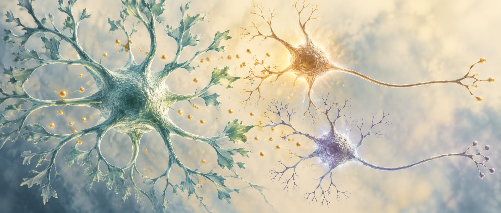
Within a sleep-promoting brainstem region, some cells can help animals wake and remain awake longer. A 2024 Advanced Science study examines astrocytes in the parafacial zone, PZ. Although not neurons, these cells influence surrounding networks through calcium activity and extracellular signals. Adenosine, often associated with sleep pressure, has a wake-promoting role here. Following this seemingly unexpected result, the team examines receptors, signal sources and a GABAergic connection to parabrachial nucleus, PB.
PZ astrocyte activation promotes and prolongs waking, while lowering calcium activity reduces waking and increases NREM during the active period. Activation raises adenosine and reduces PZ-to-PB GABA-terminal calcium and local PB GABA signals; A1 blockade weakens arousal.
Contents · 7 sections
Astrocyte calcium changes with vigilance state
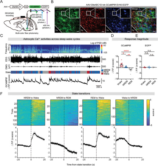
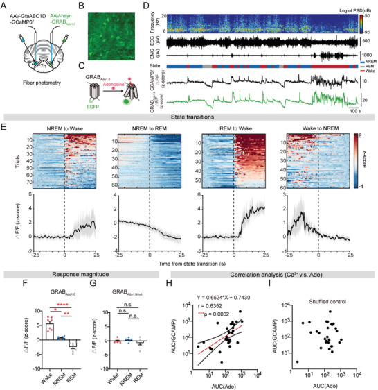
Combining PZ astrocyte calcium recordings with EEG and EMG shows the highest signals during waking and the lowest during REM sleep. Calcium rises at transitions from NREM or REM to waking and falls at wake-to-NREM and NREM-to-REM transitions.
A calcium-insensitive fluorescent control lacks the same state differences. This places support-cell activity at sleep–wake transitions, while intervention is needed to test whether the activity contributes to maintaining waking.
Source: Zhu Y et al. · Advanced science (Weinheim, Baden-Wurttemberg, Germany) · CC BY
Original figure and English caption ↗
Activation produces longer waking episodes
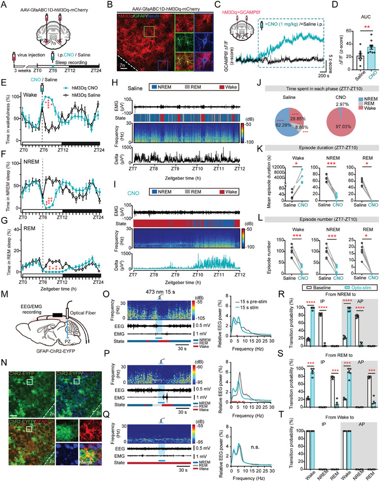
Increasing PZ astrocyte activity in mice markedly increases waking over the subsequent hours while reducing NREM and REM. Waking rises in total and in episode duration, with fewer sleep episodes; sleep rebounds as the intervention effect subsides.
Optical activation in rats further shows rapid promotion of waking from NREM or REM during both active and inactive periods. The same type of activation does not act uniformly across regions: lateral hypothalamus also shows waking promotion, whereas the tested basal-forebrain effect is nonsignificant.
Source: Zhu Y et al. · Advanced science (Weinheim, Baden-Wurttemberg, Germany) · CC BY
Original figure and English caption ↗
Lower calcium makes waking less sustained
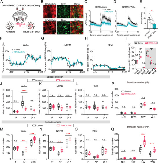
The team uses a calcium pump to lower intracellular calcium in PZ astrocytes. During the mouse active period, waking decreases and NREM increases, without a significant total-REM change. Waking episodes shorten and transitions between waking and NREM become more frequent.
The inactive period lacks the same significant state-duration changes. Alongside activation findings, this suggests particular involvement in sustaining higher waking levels. Lower activity makes waking episodes harder to maintain rather than increasing every sleep state.
Source: Zhu Y et al. · Advanced science (Weinheim, Baden-Wurttemberg, Germany) · CC BY
Original figure and English caption ↗
An extracellular signal rises at waking
Recording adenosine-sensor and astrocyte-calcium signals on opposite sides of PZ reveals a positive correlation. Adenosine rises at sleep-to-wake transitions, is highest during waking and lowest during REM. A nonbinding mutant sensor shows no significant state differences.
Their coordinated timing makes adenosine a candidate intercellular signal. Its action needs to be understood within the local network: whether a molecule promotes sleep or waking depends on its receptors and how recipient cells regulate that network.
Source: Zhu Y et al. · Advanced science (Weinheim, Baden-Wurttemberg, Germany) · CC BY
Original figure and English caption ↗
A1 receptors and ATP hydrolysis provide different clues
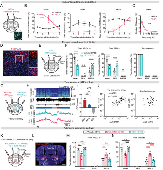
A local adenosine analog increases waking, as does A1-receptor activation; tested A2A-related activation has no significant effect. Blocking A1 before activating PZ astrocytes reduces waking probability from NREM or REM, supporting an A1-related contribution.
ATP and adenosine sensor signals also covary with state. Inhibiting extracellular ATP-hydrolysis-related processes reduces astrocyte-evoked NREM-to-wake transitions, while equilibrative nucleoside-transporter inhibition lacks the same significant blockade. Transport intervention can affect baseline sleep–wake behavior without producing the same effect on induced arousal.
Source: Zhu Y et al. · Advanced science (Weinheim, Baden-Wurttemberg, Germany) · CC BY
Original figure and English caption ↗
Changing astrocyte calcium alters adenosine
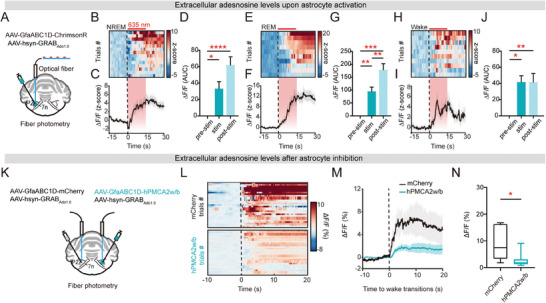
Direct PZ astrocyte activation increases adenosine-sensor signals from NREM, REM and waking alike, without the same change in light-only controls. Lowering astrocyte calcium also reduces waking-period adenosine signals.
These interventions strengthen the link between the signals, supporting astrocyte activity in local adenosine regulation. Local sensing can integrate multiple sources; the study does not trace every ATP molecule to its releasing cell. It next examines a sleep-promoting connection potentially influenced by the signal.
Source: Zhu Y et al. · Advanced science (Weinheim, Baden-Wurttemberg, Germany) · CC BY
Original figure and English caption ↗
Weaker inhibitory input accompanies waking promotion
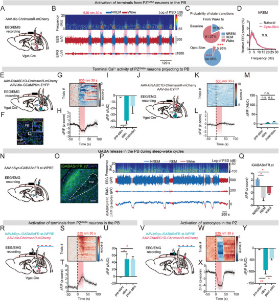
PZ GABA cells project to PB, where terminal activation promotes NREM. Astrocyte activation lowers calcium signals in these terminals and reduces local PB GABA-sensor signals. Astrocytes are thus linked with weakening of a sleep-promoting inhibitory input. This is consistent with reduced inhibition of a wake-promoting network, illustrating how support cells can influence state by regulating signals between neurons.
A1 receptors are expressed in both GABAergic and glutamatergic PZ cells, so local adenosine may influence multiple cell populations. The main mouse experiments use males. Together with rat optical-activation results, they support a local arousal model in which astrocyte activity accompanies increased adenosine and weakened sleep-promoting inhibitory input, helping waking persist.
Source: Zhu Y et al. · Advanced science (Weinheim, Baden-Wurttemberg, Germany) · CC BY
Original figure and English caption ↗