How does one arousal-promoting input reshape two sleep states?
Raphe serotonergic input to VTA can awaken mice from NREM and REM sleep. Downstream mean firing responses differ between states, revealing a diverse rather than uniform recruitment pattern.
How does the brain switch from sleeping to waking? Serotonin participates, but cellular source, projection destination and sleep state make the story more specific. A mouse study published online in CNS Neuroscience & Therapeutics on November 26, 2024 examines dorsal raphe nucleus (DRN) serotonergic input to ventral tegmental area (VTA). These terminals were more active during wakefulness, and recruiting the related cells or terminals promoted waking. VTA units did not respond uniformly: some increased and others decreased their firing, with different mean patterns during NREM and REM sleep.
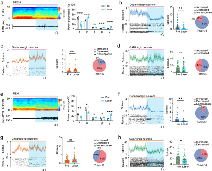
DRN serotonergic input to VTA promotes arousal. During NREM recruitment, units classified as glutamatergic increased mean firing and dopaminergic units decreased it. During REM recruitment, dopaminergic and putative GABAergic units decreased mean firing.
Contents · 5 sections
An input more active during waking
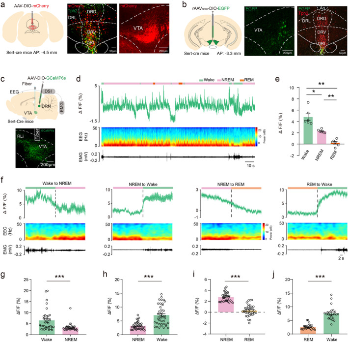
Tracing identified serotonergic DRN fibers reaching VTA, with related VTA-projecting cells mainly in the interfascicular raphe region. Terminal calcium signals were higher during wakefulness than during NREM or REM sleep, linking the input to behavioral state.
VTA is widely studied in reward and motivation, and its cell populations also contribute to sleep–wake regulation. Examining raphe cells by their destination makes the question more specific: when is the VTA-directed input active, and how can it change behavioral state?
Signals decreased at wake-to-NREM and NREM-to-REM transitions and increased at transitions from either sleep state to waking. This state-linked terminal activity provides a clue for subsequent manipulation.
Source: Wang Y et al. · CNS neuroscience & therapeutics · CC BY
Original figure and English caption ↗
Recruiting projection-defined cells increases waking
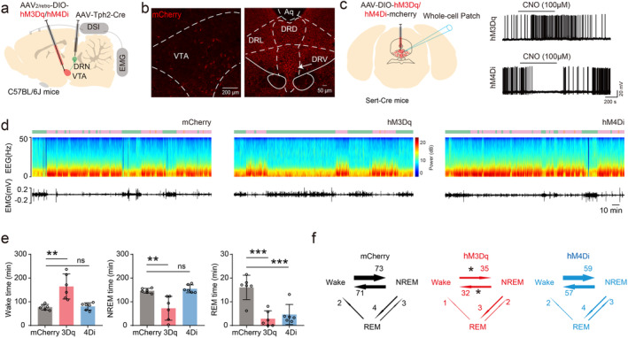
Recruiting VTA-projecting serotonergic DRN cells increased waking and reduced NREM and REM time during observation. Transitions in both directions between wake and NREM decreased, yielding more waking with fewer switches.
Suppressing the projection-defined population did not significantly alter waking or NREM time but reduced REM. Opposite manipulations therefore were not mirror images. The identified cells can also have other outputs; local terminal recruitment subsequently focuses on the VTA input.
Source: Wang Y et al. · CNS neuroscience & therapeutics · CC BY
Original figure and English caption ↗
Recruiting VTA terminals shortens awakening latency
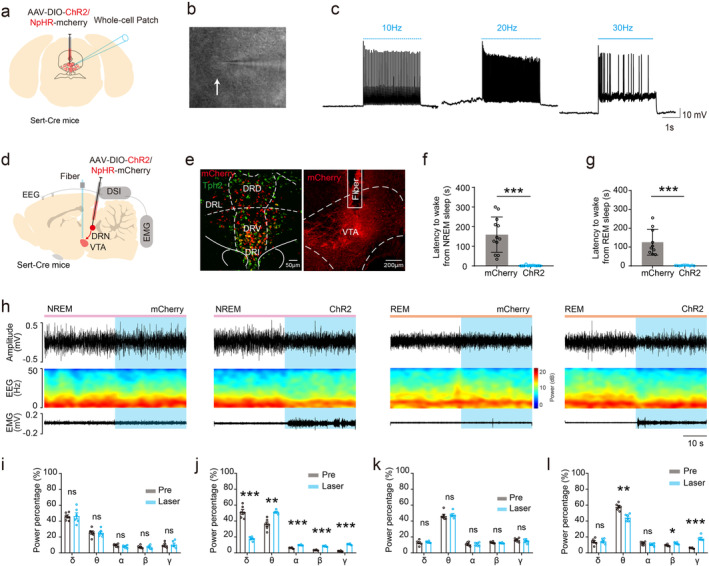
Recruiting serotonergic DRN terminals within VTA shortened awakening latency from NREM and REM sleep. EEG and EMG showed corresponding state transitions, linking local input recruitment to waking.
The result demonstrates that input recruitment can promote waking. NREM and REM retain different baseline activity patterns, so downstream cells may respond differently to the same incoming signal. Examining VTA responses helps explore that distinction.
Source: Wang Y et al. · CNS neuroscience & therapeutics · CC BY
Original figure and English caption ↗
Not all VTA units change together across states
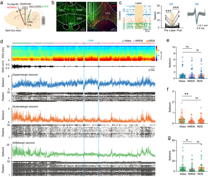
The study recorded VTA units. Dopaminergic units were identified using TH-related labeling and responses to optical inhibition; the other groups were classified mainly from waveform and electrophysiological properties as glutamatergic or GABAergic. Mean firing patterns differed among groups.
Dopaminergic units did not show significant mean-firing differences among wake, NREM and REM. Putative glutamatergic and GABAergic groups had higher mean firing in wake than NREM, while REM-to-NREM comparisons were not significant.
Mean firing rate is one aspect of activity; timing and participation of individual units also shape network output. An animal can awaken without every population increasing its rate, providing context for responses to the incoming pathway.
Source: Wang Y et al. · CNS neuroscience & therapeutics · CC BY
Original figure and English caption ↗
Similar arousal, different response combinations
During NREM terminal recruitment, putative glutamatergic units increased mean firing and dopaminergic units decreased it. Mean firing in the putative GABAergic group did not significantly change, although individual units included both increases and decreases.
During REM recruitment, dopaminergic and putative GABAergic units decreased mean firing, while the glutamatergic-group mean change was not significant. Recruitment promoted waking from both states but accompanied different downstream mean responses.
The study links an arousal-promoting input with heterogeneous downstream responses. Relevant receptors, interactions among cells and which responses are necessary for waking remain to be examined. Both sexes were included without establishing equivalent effects; functions in anesthetic emergence and human sleep conditions also require separate investigation.
Source: Wang Y et al. · CNS neuroscience & therapeutics · CC BY
Original figure and English caption ↗