A nanoscale pair in a magnetic field: millisecond responses in cultured neurons
MagTIES couples magnetic nanodiscs with piezoelectric particles. Voltage imaging shows fast responses in cultured cells, while mouse amygdala calcium-signal spectra vary with field frequency.
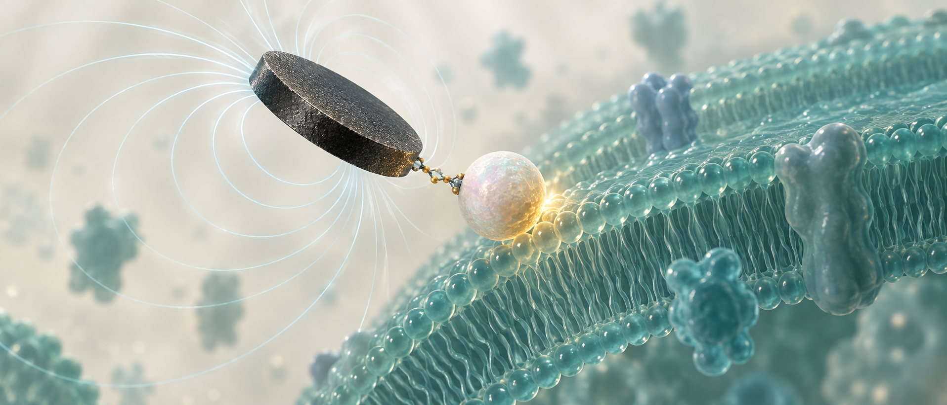
How can an external magnetic field recruit rapid local neural responses? An Advanced Healthcare Materials study proposes magnetic-driven torque-induced electrical stimulation, or MagTIES. Magnetic nanodiscs couple to barium titanate particles, with torque proposed to engage piezoelectric effects near neuronal membranes. Tests move from material arrangement and cultured-cell responses to activity in mouse amygdala, connecting nanoscale design with response timing and frequency.
Cultured-cell voltage peaks occur about 12.3 milliseconds after control signals. Amygdala markers and calcium signals increase in vivo; fields at 10, 11 and 12 Hz associate with fluorescence-spectrum increases at 20, 22 and 24 Hz.
Contents · 7 sections
Magnetic discs and piezoelectric particles require a suitable arrangement
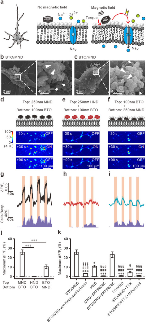
MND magnetic discs connect to BTO particles positioned near the membrane, with MNDs outside. Alternating fields evoke cultured-neuron calcium responses; reversing the arrangement produces much smaller responses, linking geometry to efficacy.
Microscopy places the particles near neuronal surfaces, and fluorescence energy transfer supports proximity. Linking the magnetic component with the membrane-adjacent component is central to the proposed design.
Unlinked materials, nonpiezoelectric replacements and alternative magnetic particles do not reproduce the same effect. Responses persist under the tested TRPC-blocking condition but disappear with voltage-gated sodium/calcium channel blockers, supporting the proposed transduction design.
Source: Cheng CC et al. · Advanced healthcare materials · CC BY
Original figure and English caption ↗
The two components show different size relationships
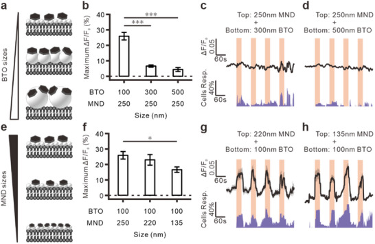
With the same magnetic discs, 100-nanometer BTO particles evoke stronger calcium responses than 300- or 500-nanometer particles. The authors relate this to nanoscale piezoelectric properties, beyond size reduction for tissue access.
The magnetic component shows the opposite trend: 250-nanometer discs yield stronger responses than 135-nanometer discs. Torque-generating and piezoelectric components have different size-dependent contributions.
Source: Cheng CC et al. · Advanced healthcare materials · CC BY
Original figure and English caption ↗
From calcium changes to millisecond voltage events
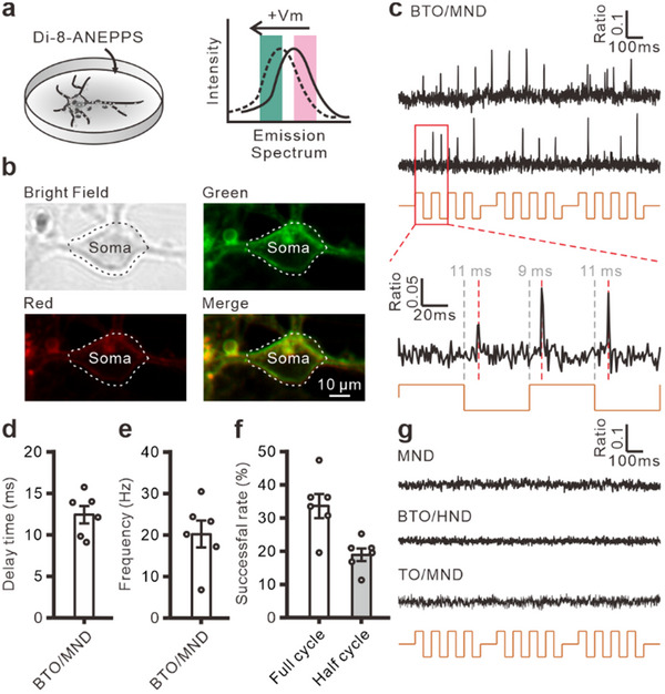
Calcium imaging captures response changes while voltage imaging tracks faster membrane events. Cultured neurons show voltage spikes during brief field pulses, with a mean control-to-peak delay of about 12.3 milliseconds.
Each field cycle changes direction twice, with responses possible at either alternation. Reported success rates are about one-third per full cycle and one-fifth per half cycle. Controls including magnetic discs alone show no detectable voltage response under the same brief-pulse conditions.
Source: Cheng CC et al. · Advanced healthcare materials · CC BY
Original figure and English caption ↗
The material pair accompanies greater amygdala activity markers
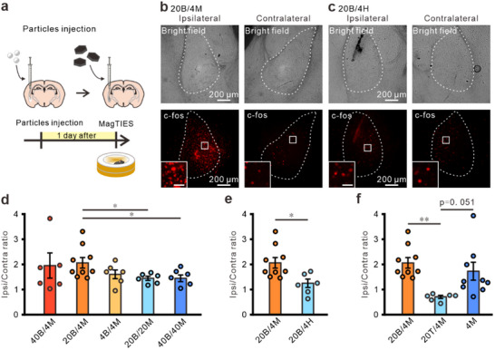
After local material delivery to the amygdala of male and female mice, the team applies an external alternating magnetic field. The BTO/MND combination produces a higher stimulated-to-opposite-side c-Fos ratio than a control with replacement magnetic particles. Its comparison with MND alone does not reach significance. Material locations are also close to regions displaying activity markers.
Changing amounts and ratios does not produce a simple more-is-better effect. One lower-MND-ratio combination has higher marker ratios than two equal-ratio combinations, while several BTO amounts at the lower ratio do not significantly differ.
Source: Cheng CC et al. · Advanced healthcare materials · CC BY
Original figure and English caption ↗
Calcium-signal outcomes vary with field strength
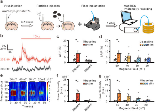
At 50 millitesla and 10 hertz, BTO/MND amygdala calcium fluorescence increases while controls show no clear change. At 40, 30 or 20 millitesla, upward trends do not reach significance in before/during comparisons.
The fluorescence spectrum gains power near 20 hertz under a 10-hertz field, with nonsignificant trends at lower strengths. The study then changes field frequency to test whether the spectral feature moves with stimulation timing.
Source: Cheng CC et al. · Advanced healthcare materials · CC BY
Original figure and English caption ↗
Fluorescence spectral enhancement follows field timing
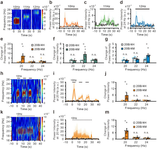
Fields at 10, 11 and 12 hertz yield relative spectral enhancement near 20, 22 and 24 hertz, without equivalent differences at adjacent comparison frequencies. The doubled relationship matches the two directional changes per field cycle.
Five- and one-second stimulation windows retain spectral enhancement associated with stimulation. Fast in vitro voltage events and in vivo fluorescence timing describe distinct temporal and frequency-related properties of the material pair.
Source: Cheng CC et al. · Advanced healthcare materials · CC BY
Original figure and English caption ↗
Nanoscale arrangement shapes neural modulation
Short-term cell-death assays and microglial/astrocytic markers three days after delivery show no evident increase. Long-term tissue effects and consequences for specific behavior remain questions for the approach.
MagTIES links the local arrangement of magnetic discs, piezoelectric particles and neural membranes with measurable responses. Component arrangement, rapid voltage events and stimulation-associated fluorescence spectra provide concrete experimental evidence for material-driven modulation.