Illuminating intracellular adenosine: a new sensor follows changes in sleep and seizures
HypnoS makes intracellular adenosine visible, revealing different dynamics in neurons and astrocytes and different effects of transporters on the two cell types.
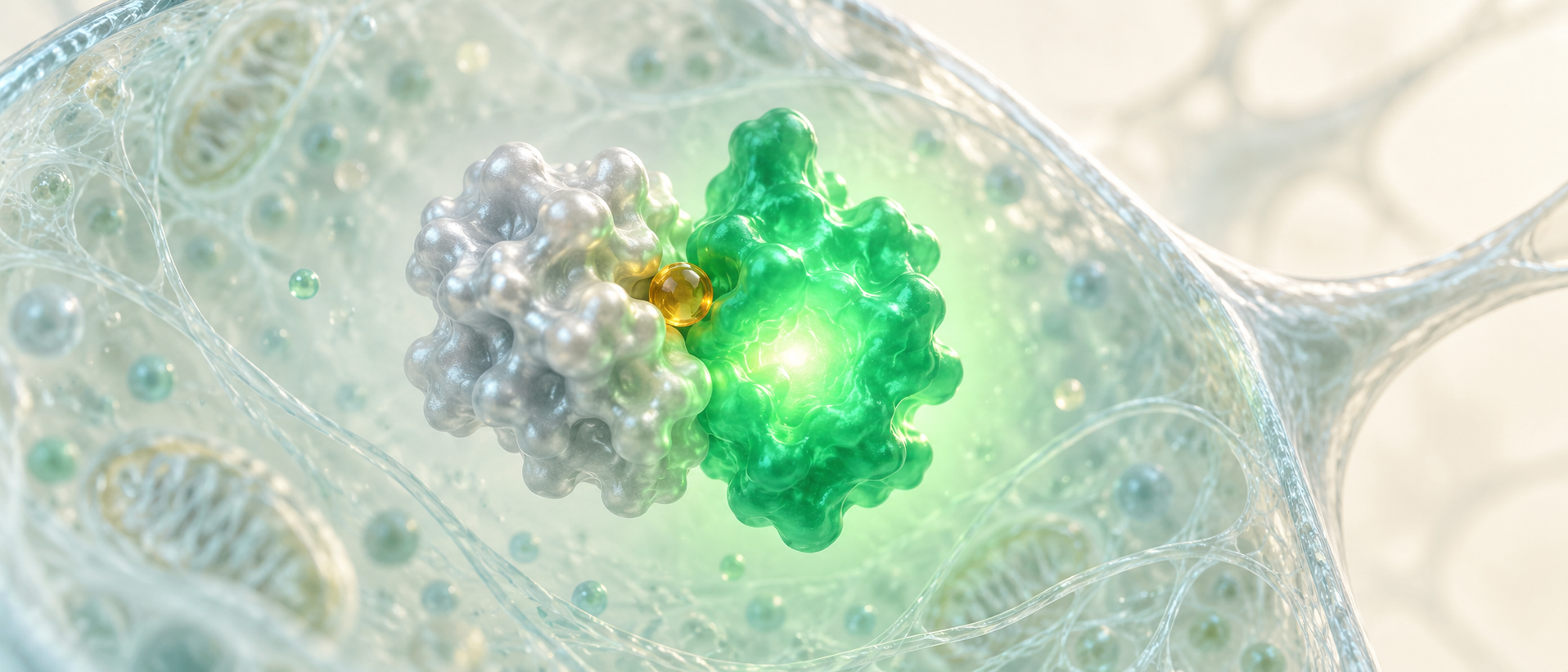
The same molecule can play different roles inside and outside a cell. Adenosine connects energy metabolism with intercellular communication, yet research has more readily tracked its extracellular changes. A study published on May 7, 2025 in Nature Communications develops the green fluorescent sensor HypnoS to look inside cells. It reveals adenosine waves following calcium waves in a seizure model, different signal-decay rates in neurons and astrocytes, and sleep-related intracellular dynamics more varied than a simple steady accumulation.
Cell identity and membrane transport shape adenosine dynamics: under the same brain state, intracellular signals in neurons and astrocytes can change in opposite directions.
Contents · 7 sections
An extracellular message can begin inside
Adenosine sits at the intersection of metabolism and communication. Extracellular adenosine can arise from extracellular ATP breakdown or from intracellular production followed by membrane transport. Measuring homogenized tissue mixes cell identity and molecular location, obscuring who produces or receives the signal and where changes begin.
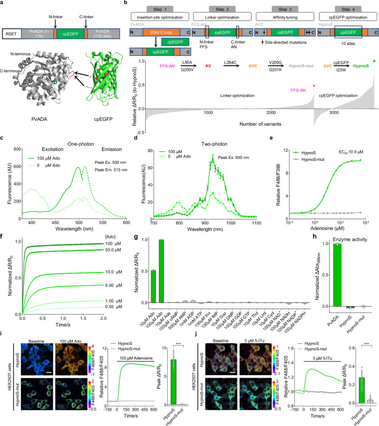
HypnoS links an adenosine-sensing protein to a green fluorescent protein, changing fluorescence when the molecule binds. Targeting the sensor to a particular cell type lets researchers observe intracellular adenosine in that population. Processes previously blended into tissue averages become distinct cellular stories.
Source: Wei Q et al. · Nature communications · CC BY-NC-ND
Original figure and English caption ↗
Fast molecular sensing, distinct cellular timing
In purified-protein tests, HypnoS shows a fluorescence-response increase exceeding ninefold, with subsecond binding and dissociation responses. Tested molecules including ATP, ADP, AMP and inosine do not evoke the same response. The sensor also lacks the adenosine-deaminase activity of its original sensing protein, so it does not directly catalyze adenosine breakdown as that enzyme does.
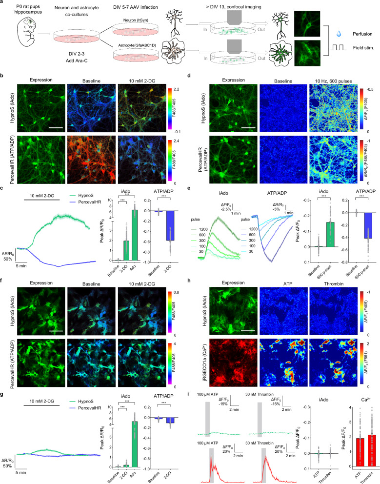
The sensor reacts quickly to molecules, while actual intracellular changes can last tens of seconds or longer. A fluorescent control that does not respond to adenosine helps identify molecule-associated changes. In cultured cells, intracellular adenosine can also be compared with changes in energy state.
Source: Wei Q et al. · Nature communications · CC BY-NC-ND
Original figure and English caption ↗
A calcium rise need not mean an adenosine rise
When cultured neuronal metabolism is perturbed or activity increases, intracellular adenosine rises as the ATP/ADP ratio falls. Astrocytic changes are smaller under corresponding conditions; some stimuli evoke clear calcium responses without a corresponding adenosine increase. The two molecules track different processes, and the two cell types respond differently metabolically.
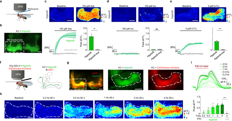
The link between activity and adenosine extends to mouse brain slices and living flies. Increasing activity in mushroom-body Kenyon cells raises intracellular adenosine signals, with stronger activation producing larger responses. Across cultures, tissue and intact animals, the sensor follows activity-associated changes.
Source: Wei Q et al. · Nature communications · CC BY-NC-ND
Original figure and English caption ↗
Adenosine waves follow calcium waves
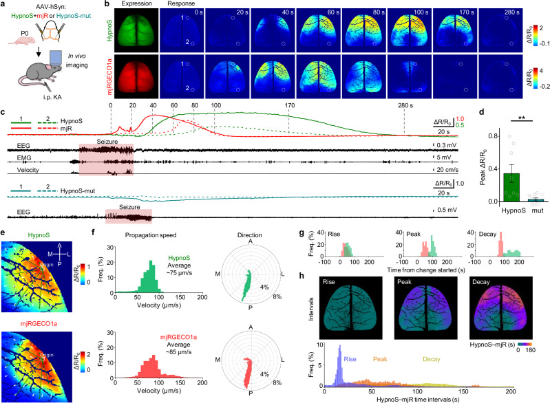
In a chemically induced mouse seizure model, cortical calcium and intracellular adenosine are recorded together. The waves begin at approximately the same location and propagate in similar directions, but the adenosine wave follows calcium by about twenty seconds and decays more slowly, making their temporal relationship visible.
Hippocampal recordings in freely moving mice also capture seizure-associated adenosine increases, appearing near seizure termination in EEG recordings. During successive seizures, the second response is smaller. Adenosine dynamics vary with events in their onset, delay and duration, rather than simply rising at a single fixed moment.
Source: Wei Q et al. · Nature communications · CC BY-NC-ND
Original figure and English caption ↗
Astrocytic signals recede faster
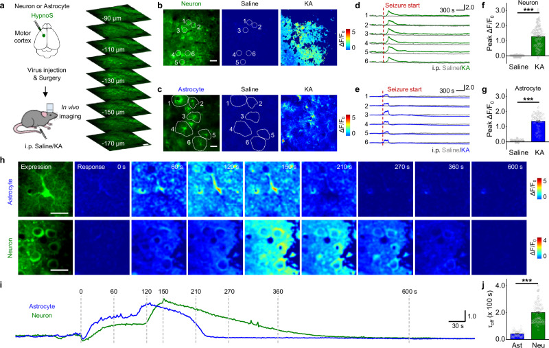
At single-cell resolution, both neurons and astrocytes show seizure-associated adenosine increases, but astrocytic signals decay faster. The same brain state does not produce identical dynamics in every cell: cell identity influences how long the intracellular signal persists.
Faster decay is consistent with differences in adenosine metabolism between the two cell types. It also illustrates why a tissue total cannot describe the full process: at one moment, signals have receded in some cells while remaining elevated in others, a timing difference that an average can conceal.
Source: Wei Q et al. · Nature communications · CC BY-NC-ND
Original figure and English caption ↗
Intracellular signals are higher in wakefulness and REM
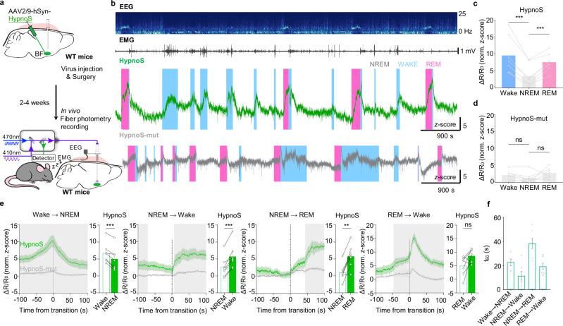
Adenosine is often linked to sleep pressure after wakefulness, but intracellular signals in the basal forebrain are more complex than high while awake and low while asleep. HypnoS recordings show higher signals during wakefulness and REM than during NREM, with changes unfolding over tens of seconds around state transitions.
Although REM is sleep, its molecular state differs from NREM. Intracellular adenosine dynamically tracks brain state, emphasizing the value of distinguishing internal metabolic signals from extracellular communication when asking how each participates in sleep.
Source: Wei Q et al. · Nature communications · CC BY-NC-ND
Original figure and English caption ↗
Reduced transport, opposite intracellular changes
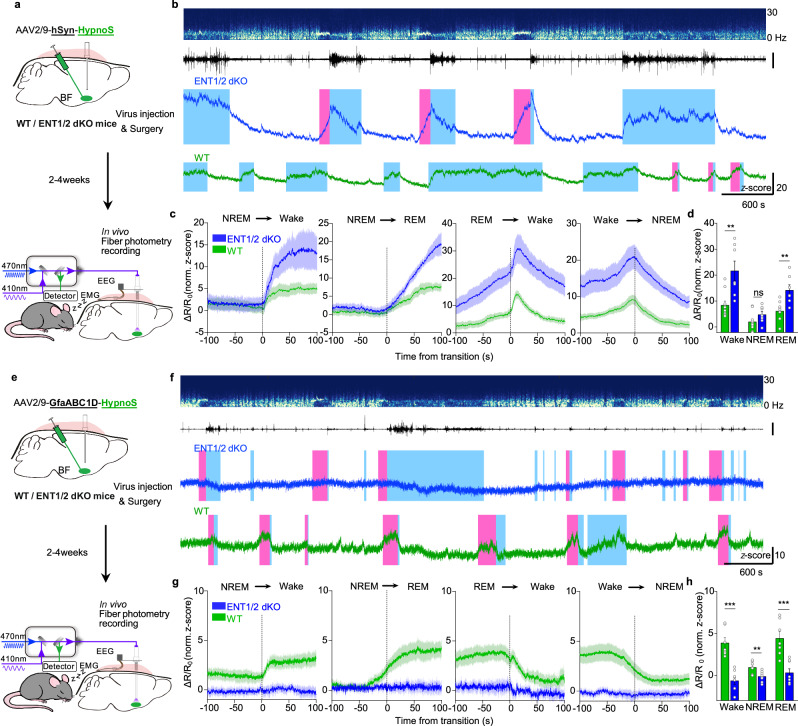
ENT1/2 are important adenosine membrane transporters. In mice with impaired transporter function involving the basal forebrain, neuronal intracellular signals increase during wakefulness and REM, with no clear NREM difference. Astrocytic signals decrease in all three states. The shared transporter perturbation produces different net effects in the two cell types.
Cultured neuronal experiments further show that reducing transport weakens responses to external adenosine while increasing intracellular accumulation during activity or metabolic perturbation. Membrane traffic and cellular metabolism jointly shape the molecule’s distribution. Fluorescence also depends on measurement conditions; HypnoS provides a spatially resolved window that turns an adenosine increase into a question of which cells and when.
Source: Wei Q et al. · Nature communications · CC BY-NC-ND
Original figure and English caption ↗