Why does sleep loss make touch more painful? A noradrenergic link from locus coeruleus to sensory cortex
A mouse study shows that sleep deprivation strengthens locus coeruleus noradrenergic input to primary somatosensory cortex, heightening neuronal excitability and delaying pain recovery.
Voluntary sleep loss driven by prolonged nighttime recreational activities—termed active sleep deprivation (ASD)—is pervasive in modern society. Although clinical and human neuroimaging studies have established that sleep deficiency lowers pain thresholds and exacerbates chronic pain, the specific neural circuits that integrate prolonged wakefulness signals to amplify cortical nociceptive processing have remained elusive. Published in Current Biology in May 2024, Zhi Zhang, Yan Jin, Di Wang, and colleagues at the University of Science and Technology of China, together with Yanli Luo at Renji Hospital of Shanghai Jiao Tong University School of Medicine, established a non-stressful novel-object ASD mouse model and delineated a direct monosynaptic pathway from locus coeruleus noradrenergic neurons to glutamatergic neurons in the hindlimb primary somatosensory cortex (LCNA→S1HLGlu). The authors demonstrate that 9 hours of ASD hyperactivates LCNA neurons and elevates noradrenaline release in the S1HL, driving S1HLGlu neuronal hyperexcitability to induce mechanical and thermal pain sensitization and delay recovery from inflammatory pain.
Non-stressful active sleep deprivation (ASD) hyperactivates brainstem locus coeruleus noradrenergic (LCNA) neurons projecting monosynaptically to layer II–V glutamatergic neurons in the hindlimb primary somatosensory cortex (S1HLGlu), sensitizing cortical responses to subthreshold stimuli to drive mechanical and thermal hyperalgesia and prolong inflammatory pain recovery.
Contents · 5 sections
Nine Hours of Non-stressful Active Sleep Deprivation Induces Pain Sensitization and Delays Inflammatory Pain Recovery
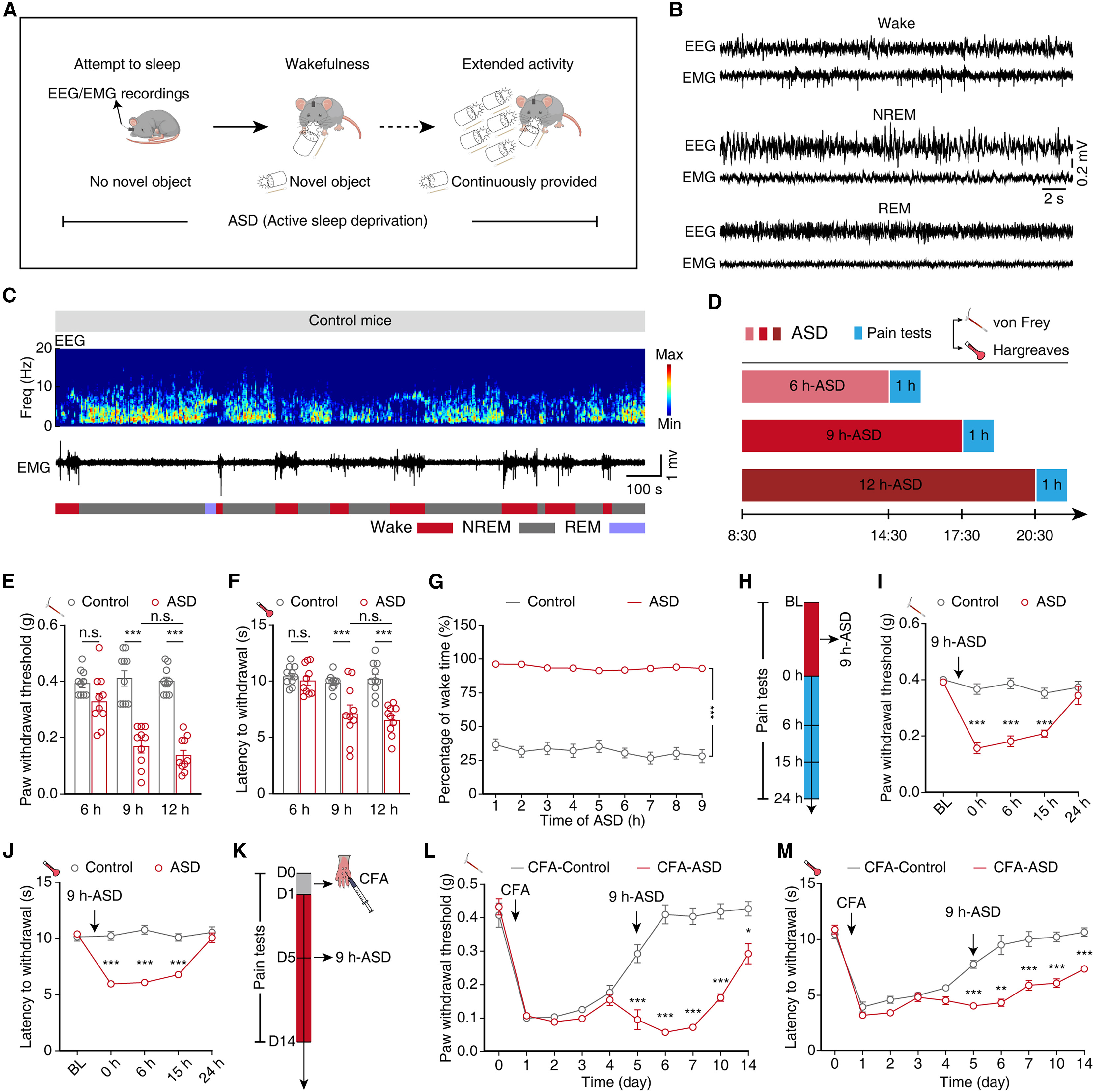
To model voluntary sleep loss caused by recreational activity without introducing physical stress, the researchers employed a non-stressful active sleep deprivation (ASD) paradigm. While continuously monitoring frontoparietal EEG and neck EMG starting at 08:30, experimenters introduced novel objects into the home cage whenever mice attempted to enter non-rapid eye movement (NREM) sleep, keeping them spontaneously awake without direct physical contact. Nociceptive testing with von Frey filaments and the Hargreaves radiant heat assay revealed that whereas 6 hours of ASD did not significantly alter pain thresholds, both 9 hours and 12 hours of acute ASD (as well as 6 hours of daily ASD for 5 consecutive days) significantly decreased mechanical and thermal pain thresholds to a comparable degree.
Time-course tracking after 9 hours of ASD showed that mechanical and thermal pain sensitization persisted at 0, 6, and 15 hours post-ASD before returning to baseline by 24 hours. Furthermore, subjecting mice with complete Freund's adjuvant (CFA)-induced hindpaw inflammatory pain to 9 hours of ASD on day 5 post-CFA markedly prolonged the recovery time required for mechanical and thermal thresholds to return to baseline, mirroring clinical observations in sleep-deprived pain patients.
Source: et al. · Current Biology · Original figure reproduced for academic literature review under fair scholarly commentary; copyright remains with the authors/publisher.
Original figure and English caption ↗
Active Sleep Deprivation Drives Hyperactivity of S1HL Glutamatergic Neurons
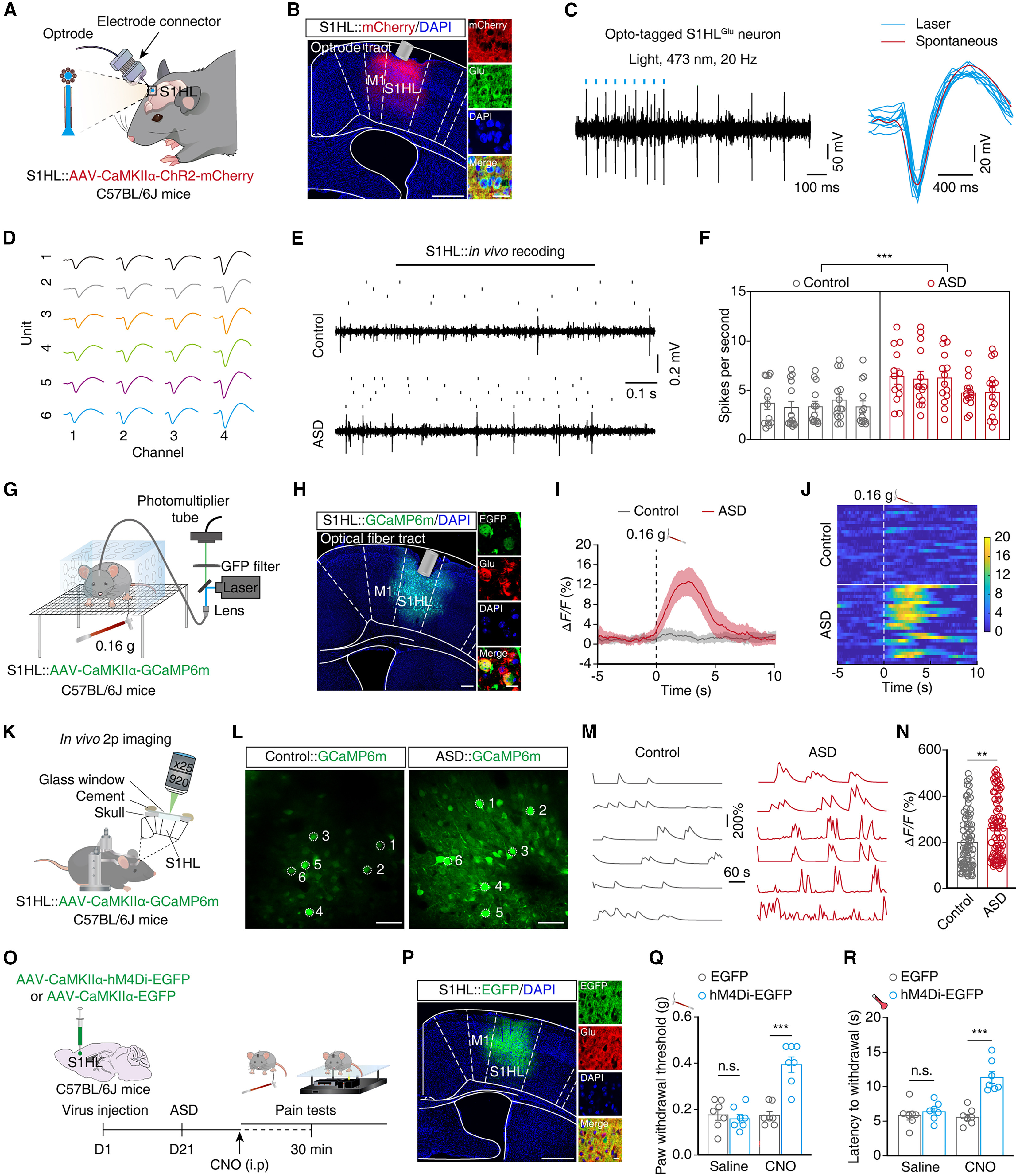
The primary somatosensory cortex (S1) is a critical cortical hub for processing nociceptive information. Immunofluorescence mapping after 9 hours of ASD revealed a marked elevation of c-Fos-positive neurons across layers II/III, IV, and V (but not layers I or VI) of the hindlimb S1 (S1HL), predominantly colocalizing with glutamatergic markers. Optogenetic tagging coupled with in vivo multi-channel tetrode recordings confirmed that spontaneous spike firing rates of S1HL glutamatergic (S1HLGlu) neurons were significantly elevated in awake ASD mice compared with controls.
To test whether S1HLGlu neurons become hypersensitive to subthreshold mechanical stimuli after ASD, the authors expressed AAV-CaMKIIα-GCaMP6m in the S1HL and performed in vivo fiber photometry (recording 470 nm GCaMP6m and 410 nm isosbestic control signals). Upon stimulation of the contralateral hindpaw with a subthreshold 0.16 g von Frey filament, awake ASD mice exhibited a rapid, robust surge in S1HLGlu calcium fluorescence (ΔF/F) compared with minimal responses in control mice. In vivo two-photon calcium imaging in awake head-fixed mice independently confirmed heightened spontaneous Ca2+ transients in individual S1HLGlu neurons after ASD, while chemogenetic inhibition of S1HLGlu neurons (AAV-CaMKIIα-hM4Di-EGFP plus CNO) rescued mechanical and thermal pain thresholds.
Source: et al. · Current Biology · Original figure reproduced for academic literature review under fair scholarly commentary; copyright remains with the authors/publisher.
Original figure and English caption ↗
Locus Coeruleus Noradrenergic Neurons Send Functional Monosynaptic Projections to S1HLGlu Neurons
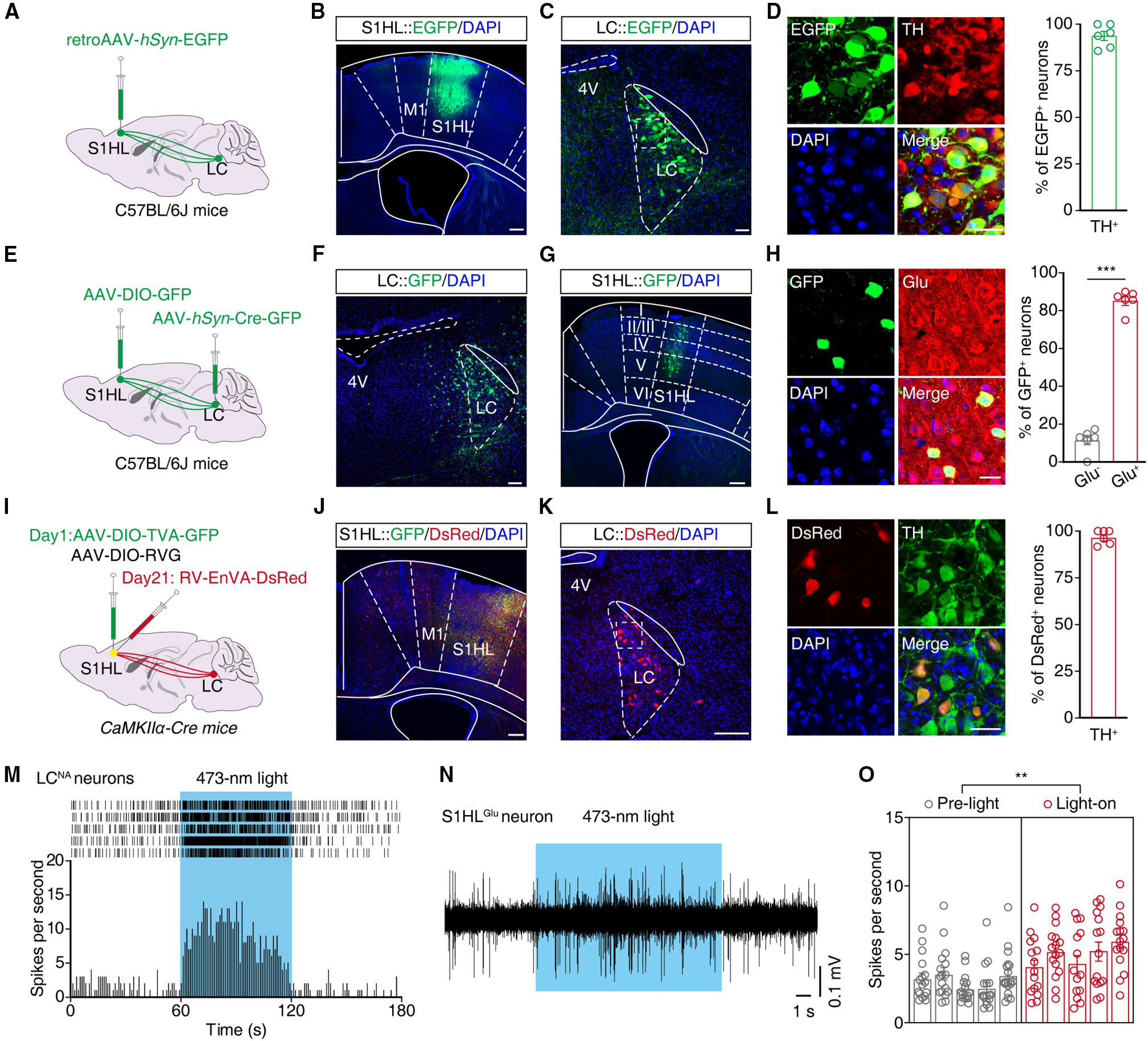
To identify the upstream sleep-wake nucleus driving S1HLGlu hyperactivity, the team injected retrograde retroAAV-hSyn-EGFP into the S1HL and found abundant EGFP-labeled neurons in the brainstem locus coeruleus (LC), ~90% of which colocalized with tyrosine hydroxylase (TH). Anterograde monosynaptic tracing (AAV2/1-hSyn-Cre-GFP in the LC and AAV-DIO-GFP in the ipsilateral S1HL) revealed GFP-positive postsynaptic neurons concentrated in layers II/III, IV, and V of the S1HL, with ~90% colocalizing with glutamate immunofluorescence. Complementary rabies-based trans-monosynaptic retrograde tracing from S1HLGlu starter cells in CaMKIIα-Cre mice confirmed direct monosynaptic inputs from TH-positive LC noradrenergic (LCNA) neurons.
To verify the functional impact of this LCNA→S1HLGlu pathway, the authors injected AAV-DIO-ChR2-EGFP into the LC of DBH-Cre mice, observing dense EGFP-positive noradrenergic fibers surrounding S1HLGlu neurons. In vivo multi-tetrode recordings in freely moving mice demonstrated that photostimulation (473 nm) of ChR2-expressing LCNA terminals within the S1HL significantly increased the firing rate of S1HLGlu neurons. Moreover, 9 hours of ASD markedly increased c-Fos expression in TH-positive LCNA neurons and elevated their spontaneous in vivo firing rate, whereas chemogenetic inhibition of LCNA neurons in DBH-Cre mice reversed ASD-induced pain sensitization.
Source: et al. · Current Biology · Original figure reproduced for academic literature review under fair scholarly commentary; copyright remains with the authors/publisher.
Original figure and English caption ↗
Inhibiting the LCNA→S1HLGlu Pathway Abolishes Cortical Calcium Hypersensitivity and Accelerates Inflammatory Pain Recovery
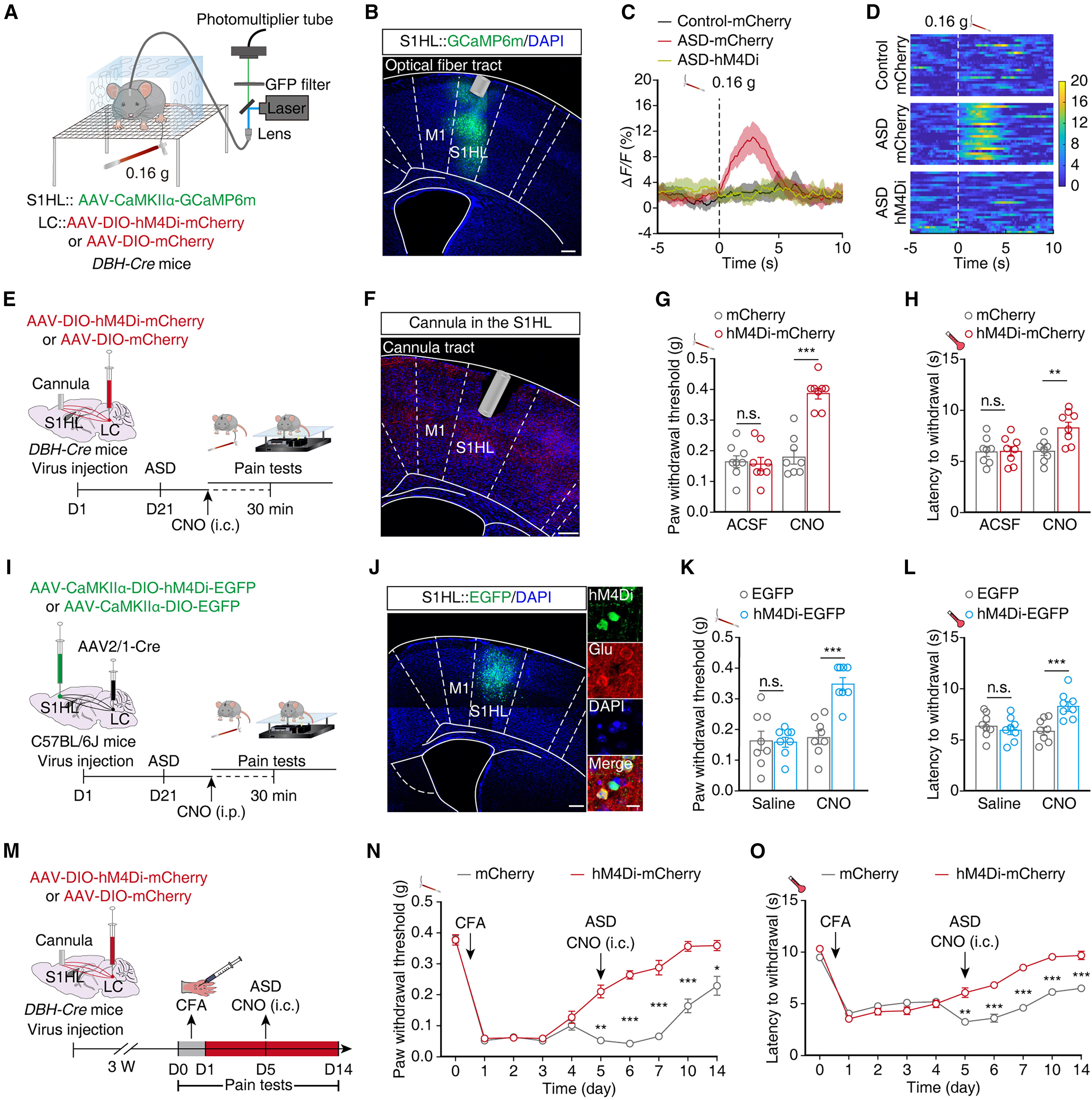
To test whether hyperactive LCNA inputs drive S1HLGlu hypersensitivity under ASD, the researchers combined chemogenetic inhibition of LCNA neurons (AAV-DIO-hM4Di-mCherry in the LC of DBH-Cre mice) with in vivo fiber photometry of S1HLGlu neurons (AAV-CaMKIIα-GCaMP6m in the S1HL). CNO-mediated suppression of LCNA neurons completely abolished the amplified S1HLGlu calcium transients elicited by 0.16 g von Frey stimulation of the contralateral hindpaw in ASD mice.
Behaviorally, terminal-specific chemogenetic inhibition (intra-S1HL CNO microinfusion) or optogenetic inhibition (eNpHR, 594 nm yellow light) of LCNA fibers in the S1HL—as well as postsynaptic inhibition of LC-innervated S1HLGlu neurons (via LC AAV2/1-Cre and S1HL AAV-CaMKIIα-DIO-hM4Di-EGFP)—significantly rescued mechanical and thermal pain thresholds in 9-hour ASD mice without affecting baseline nociception or sleep-wake architecture in naive mice. Crucially, intra-S1HL CNO infusion to inhibit LCNA terminals in CFA-ASD mice significantly shortened the time required for mechanical and thermal pain thresholds to recover to baseline.
Source: et al. · Current Biology · Original figure reproduced for academic literature review under fair scholarly commentary; copyright remains with the authors/publisher.
Original figure and English caption ↗
Activating the LCNA→S1HLGlu Pathway Elevates Cortical Noradrenaline Release and Mimics Pain Sensitization in Naive Mice
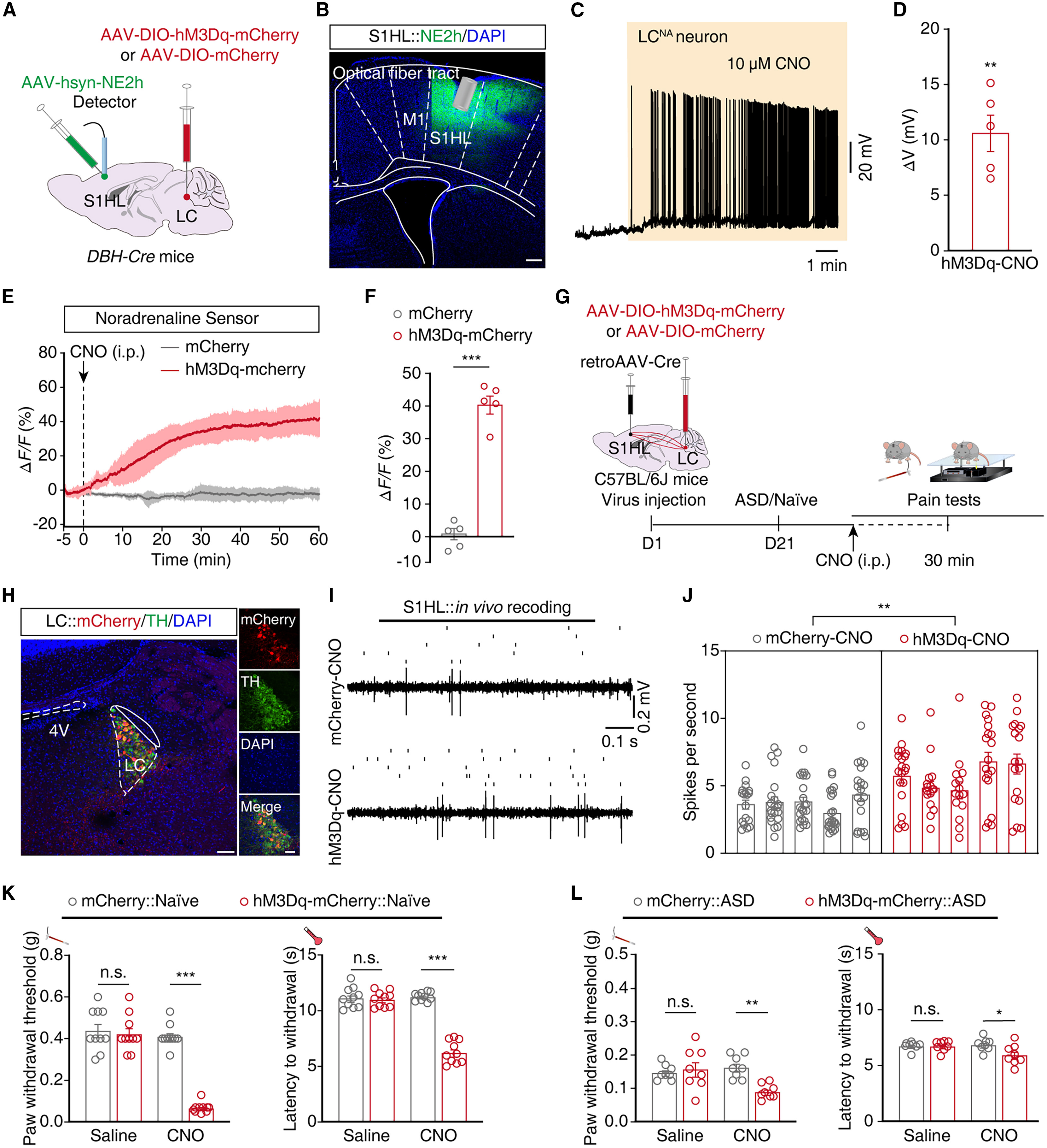
To directly monitor cortical noradrenaline dynamics in vivo, the authors expressed the genetically encoded noradrenaline fluorescent sensor NE2h (AAV-hSyn-NE2h) in the S1HL and excitatory hM3Dq (AAV-DIO-hM3Dq-mCherry) in the LC of DBH-Cre mice. In vivo fiber photometry demonstrated that chemogenetic activation of LCNA neurons triggered a robust, sustained increase in NE2h fluorescence (ΔF/F) within the S1HL.
To test whether selectively activating S1HL-projecting LCNA neurons suffices to induce pain sensitization in rested animals, the team injected retroAAV-hSyn-Cre into the S1HL and AAV-DIO-hM3Dq-mCherry into the ipsilateral LC of DBH-Cre mice. In awake naive mice, CNO administration significantly increased S1HLGlu firing rates and lowered both mechanical and thermal pain thresholds (and further aggravated hyperalgesia in ASD mice), without altering anxiety-like behaviors in the elevated plus maze or open-field test or changing sleep-wake percentages on EEG/EMG recordings. Thus, the LCNA→S1HLGlu pathway selectively gates sleep-loss-induced pain sensitization without perturbing general arousal or anxiety.
Source: et al. · Current Biology · Original figure reproduced for academic literature review under fair scholarly commentary; copyright remains with the authors/publisher.
Original figure and English caption ↗