After analgesia, greater sensitivity: a prefrontal circuit clue in rats
In male rats, fentanyl-associated hyperalgesia accompanies reduced prefrontal-to-accumbens-core activity. Recruiting this pathway improves mechanical and thermal responses.
Why can analgesia be followed by greater pain sensitivity in the same animal? A study published in Journal of Pain Research on March 20, 2024 follows changes after repeated acute fentanyl exposure in male rats. Initial analgesic effects gave way to greater mechanical and thermal sensitivity several hours later. The researchers examined connections from prelimbic medial prefrontal cortex (PL-mPFC) to nucleus accumbens core (NAc core). Reduced activity appeared at both ends of this pathway, and its recruitment altered stimulus responses.
Hyperalgesia involved reduced excitability of PL output and NAc-core recipient cells, with a lower evoked excitation-to-inhibition ratio. Pathway activation increased mechanical withdrawal thresholds and thermal withdrawal latencies, while inhibition increased sensitivity in control rats.
Contents · 6 sections
Analgesia precedes lower stimulus thresholds
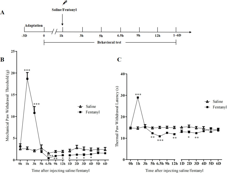
Early after fentanyl exposure, stronger mechanical stimulation was needed to elicit withdrawal, and thermal withdrawal occurred later. Both measures then decreased below control levels. This transition from analgesia to greater sensitivity represents the opioid-associated hyperalgesia examined here.
Increased sensitivity persisted for several days before gradually recovering. The study focused on the hyperalgesic phase several hours after exposure, when behavioral responses had changed, and examined which brain processes changed alongside them.
Source: Luo Q et al. · Journal of pain research · CC BY-NC
Original figure and English caption ↗
Prefrontal output cells become less excitable
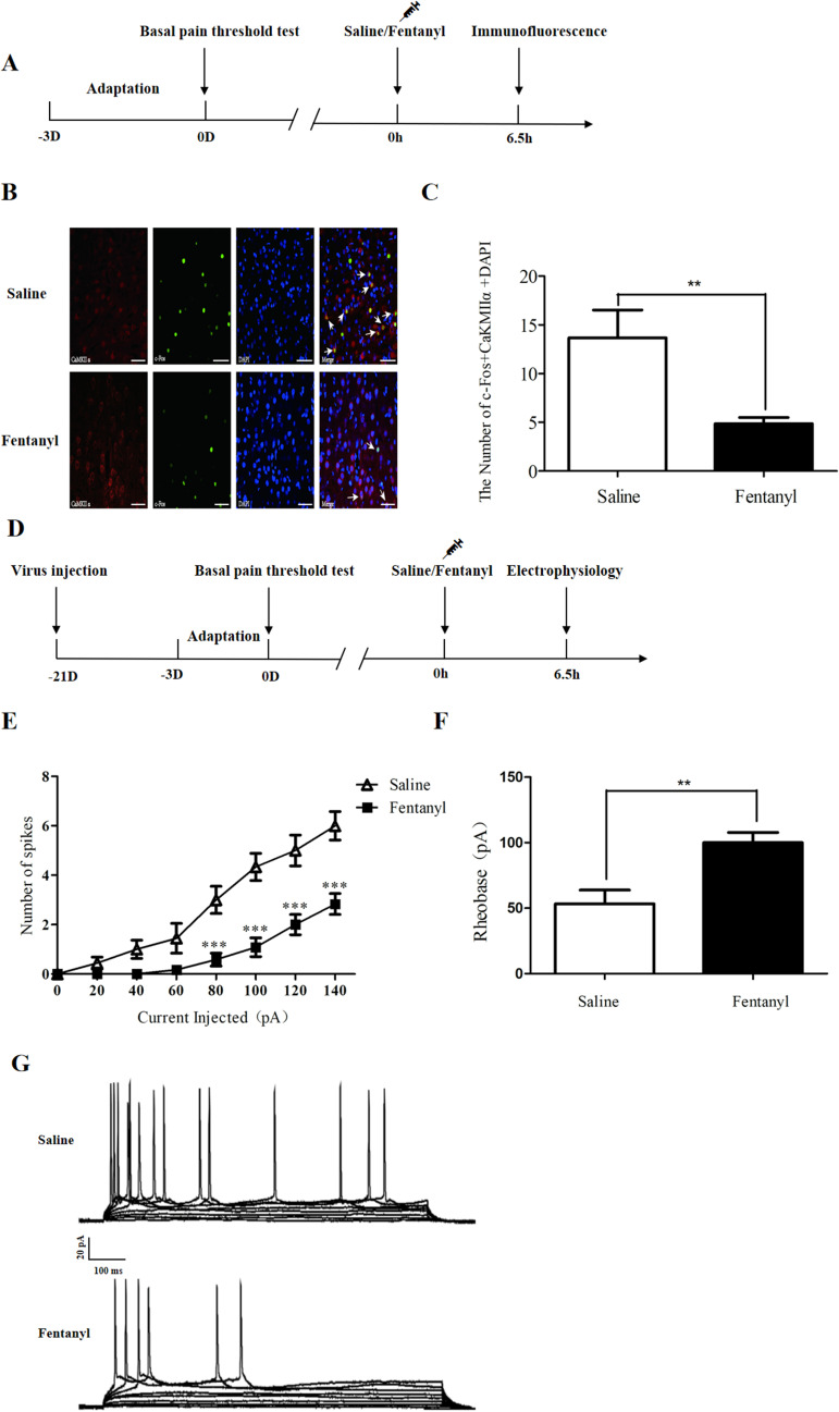
In PL-mPFC layer V, fewer cells co-expressed CaMKIIα and the activity marker c-Fos. Single-cell recordings showed fewer action potentials under comparable current injection, with larger currents needed to initiate firing in hyperalgesic rats.
Activity-marker and firing changes converge on reduced excitability in this output population. Prefrontal cortex participates in pain regulation through several connections; the study focused on its output to NAc core to examine changes downstream.
Source: Luo Q et al. · Journal of pain research · CC BY-NC
Original figure and English caption ↗
How does the prefrontal signal reach accumbens?
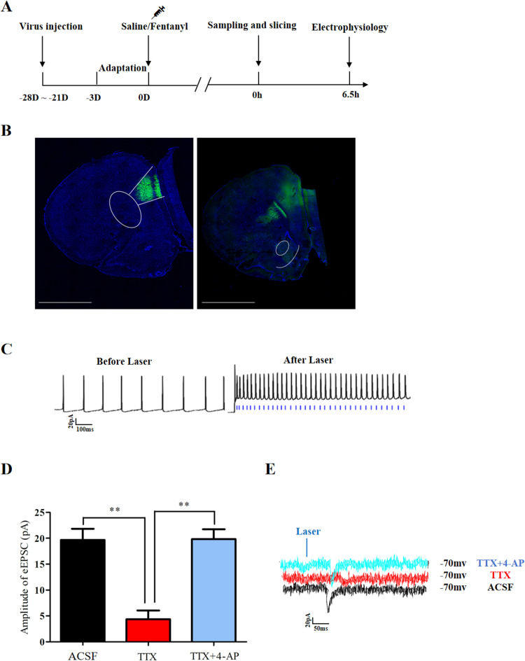
NAc core receives inputs from these prefrontal cells. PL fibers reached the region, and their recruitment elicited excitatory synaptic currents in recipient cells, placing changes at both ends within a traceable connection.
Current-blockade and recovery findings supported direct excitatory synaptic input from PL to NAc core. This glutamatergic signal enters cells that also integrate inhibitory inputs, so their firing reflects the combined state of multiple influences.
Accumbens is frequently studied in reward, but also participates in sensory and affective aspects of pain. Prefrontal–accumbens connections link cortical regulation with behavioral responses. This work focuses on the core as a specific recipient region; prefrontal signals reaching different locations may have different roles. Examining individual connections helps clarify regulation behind changes in broader regional activity.
Source: Luo Q et al. · Journal of pain research · CC BY-NC
Original figure and English caption ↗
The recipient cells’ input balance also changes
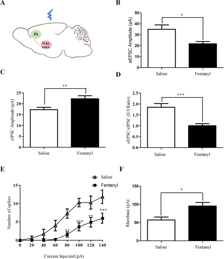
In NAc-core recipient cells from hyperalgesic rats, PL-terminal recruitment elicited smaller excitatory and larger inhibitory currents, lowering their excitation-to-inhibition ratio. The evoked input balance shifted toward inhibition relative to controls.
The cells also generated fewer action potentials under current injection and required larger currents to initiate firing. Weaker upstream excitability, altered downstream input balance and lower recipient-cell excitability form a set of functional changes in the hyperalgesic state.
Source: Luo Q et al. · Journal of pain research · CC BY-NC
Original figure and English caption ↗
Can recruiting the pathway improve responses?
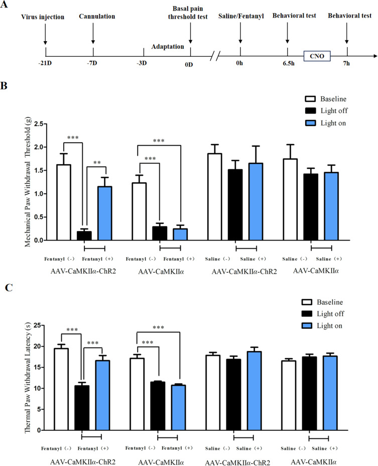
Recruiting PL terminals in NAc core increased mechanical withdrawal thresholds and prolonged thermal withdrawal latencies in hyperalgesic rats. Stronger mechanical stimulation was needed to elicit withdrawal, and thermal responses occurred less rapidly.
Light did not produce the corresponding improvement in controls lacking the relevant light-sensitive channel. These behavioral changes support a contribution from the reduced-activity pathway to pain regulation in this state. The findings concern responses during stimulation; sustained effects remain to be examined.
Source: Luo Q et al. · Journal of pain research · CC BY-NC
Original figure and English caption ↗
Suppressing the connection increases sensitivity in controls
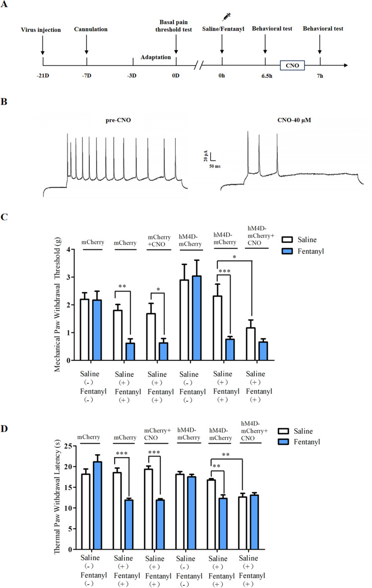
Suppressing PL-to-NAc-core inputs reduced mechanical thresholds and thermal withdrawal latencies in saline-treated controls. Rats already in the hyperalgesic state did not show a further significant decrease. The same manipulation yielded different observed outcomes in the two groups.
Improved responses with activation and greater sensitivity with inhibition in controls connect pathway activity with nociceptive behavior. The study identifies a regulatory component in a repeated acute-exposure model in male rats. The relevant NAc-core cell types, links to downstream pain-modulatory networks and corresponding functions in females remain open.
Source: Luo Q et al. · Journal of pain research · CC BY-NC
Original figure and English caption ↗