Why does pain sensitivity rise? A cortical clue from LCN2
Peripheral nerve injury also changes the cortex. In mice, increased anterior cingulate LCN2 accompanies excitatory-cell hyperactivity, while reducing the signal attenuates pain sensitization.
Pain can begin with a peripheral injury yet leave sensitivity that persists, making relatively mild contact evoke a stronger response. How does the brain contribute? A study published in Frontiers in Cellular Neuroscience on June 8, 2023 followed the anterior cingulate cortex, ACC, in a male mouse model of peripheral nerve injury. Lipocalin-2, or LCN2, increased alongside greater excitability in glutamatergic cells. Reducing LCN2 or suppressing relevant cellular activity attenuated mechanical and thermal sensitization in the model.
LCN2 links peripheral nerve injury, ACC glutamatergic excitability and pain sensitization. Both lowering expression before injury and locally neutralizing the protein after sensitization reduced pain-related responses, whereas adding LCN2 produced changes in the opposite direction.
Contents · 6 sections
After peripheral injury, ACC cells become more excitable
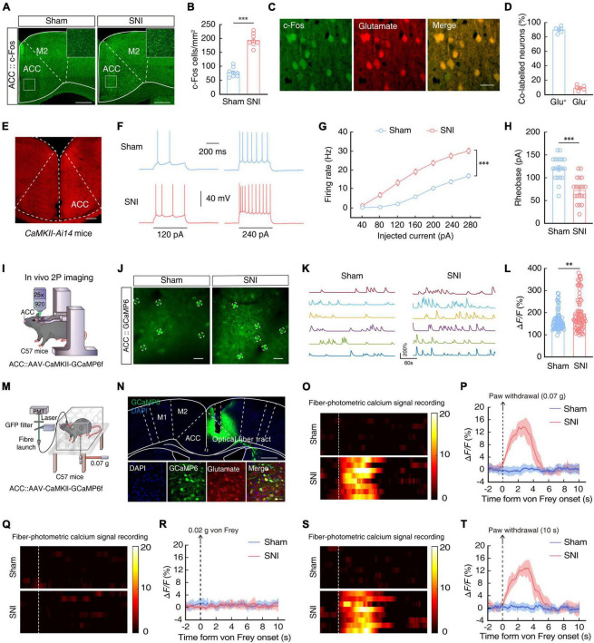
After nerve injury, mice showed a lower mechanical withdrawal threshold and shorter heat-withdrawal latency. ACC c-Fos labeling increased, while glutamatergic cells produced more spikes under matched current input and required less current to begin firing.
In vivo calcium signals distinguished spontaneous activity from evoked responses. Selected mild stimuli produced stronger behavioral and calcium responses after injury than in controls, whereas an even weaker condition elicited neither response in either group. Increased sensitivity therefore remained conditional rather than universal.
Source: Song XJ et al. · Frontiers in cellular neuroscience · CC BY
Original figure and English caption ↗
Lower cellular activity accompanies reduced sensitivity
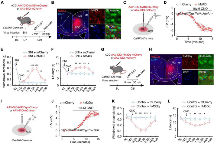
If heightened cortical activity merely accompanied pain, changing it might leave behavior unaffected. Suppressing ACC glutamatergic cells increased mechanical thresholds and heat-withdrawal latencies after injury. Conversely, activating these cells in uninjured mice increased mechanical and thermal sensitivity.
These complementary findings place the population within a functional sensitization pathway. The measured nociceptive behaviors do not cover every affective, cognitive or spontaneous aspect of chronic pain. They sharpen the next question: what makes these cells more excitable?
Source: Song XJ et al. · Frontiers in cellular neuroscience · CC BY
Original figure and English caption ↗
LCN2 is elevated before the two-week time point
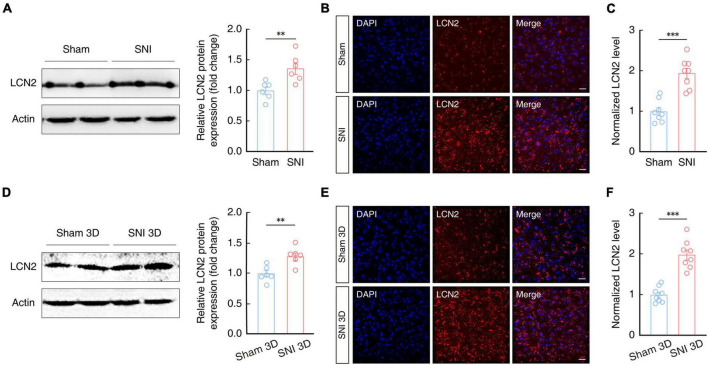
LCN2 participates in inflammation and neural plasticity. Its ACC protein levels were elevated two weeks after injury and were already higher at an earlier three-day time point. Changes at both stages identified a continuing lead for investigation.
Increased expression alone does not establish whether it is cause or consequence. Peripheral injury, cortical activity and inflammatory signaling can influence each other. Altering LCN2 and following both cells and behavior was therefore necessary to test its contribution.
Source: Song XJ et al. · Frontiers in cellular neuroscience · CC BY
Original figure and English caption ↗
The increase is concentrated in neuron-associated signals
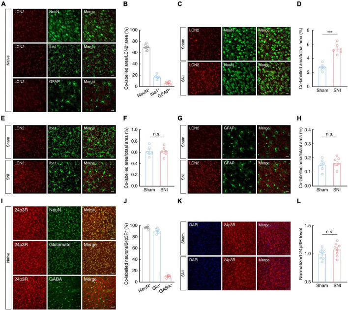
ACC LCN2 signals predominantly overlapped neuronal markers, with less overlap in microglia and astrocytes. After injury, neuron-associated colabeled area increased, whereas the corresponding glial measures did not change significantly, highlighting regional and cellular context.
The LCN2 receptor 24p3R also predominantly localized to neurons and overlapped substantially with glutamatergic labeling. Its protein level did not increase significantly after injury. Increased LCN2 therefore did not coincide with increased receptor abundance, and tissue colocalization did not directly establish the source of secretion.
Source: Song XJ et al. · Frontiers in cellular neuroscience · CC BY
Original figure and English caption ↗
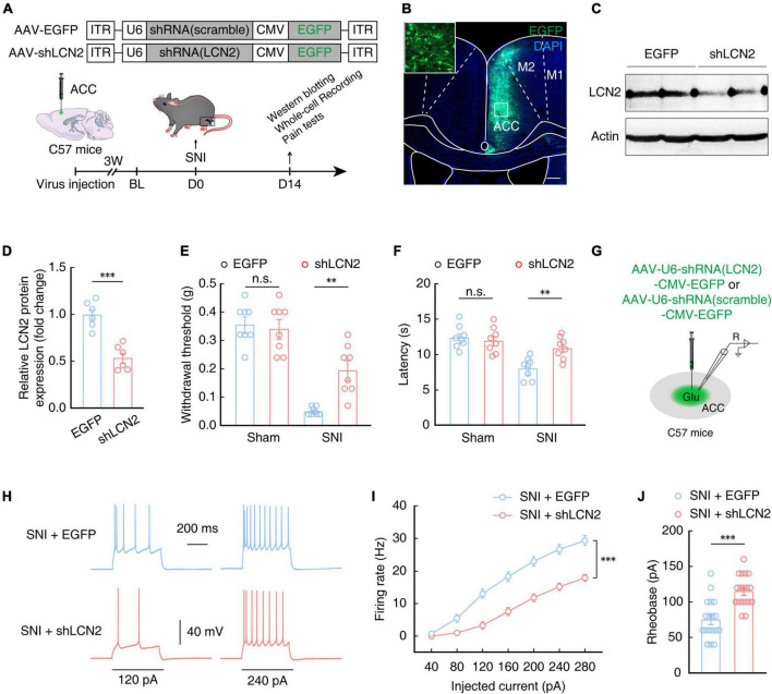
Lowering LCN2 before injury attenuates later sensitization
Lowering ACC LCN2 before injury increased subsequent mechanical thresholds and heat-withdrawal latencies in injured animals. Evoked firing decreased and the current required to initiate firing increased. Concordant cellular and behavioral changes extended the finding beyond expression alone.
Corresponding mechanical and thermal measures did not change significantly in sham controls. LCN2 therefore needs to be considered in the context of nerve injury; a role in pathological sensitivity does not establish general sensory suppression across all states.
This comparison addressed outcomes after a change made before injury. Whether an intervention could also affect established sensitization required a separate comparison with a different temporal sequence.
Source: Song XJ et al. · Frontiers in cellular neuroscience · CC BY
Original figure and English caption ↗
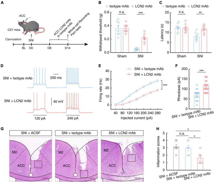
Neutralizing LCN2 also changes established sensitization
After sensitization had developed, local LCN2 neutralization likewise increased mechanical thresholds and heat-withdrawal latencies and reduced evoked firing. This complemented the earlier expression-reduction experiment, connecting LCN2 with persistent sensitization.
Adding LCN2 in uninjured mice produced changes in the opposite direction in sensitivity and excitability. Together these findings support an ACC contribution while leaving receptor necessity, downstream inflammatory signaling and relevance to human chronic pain unresolved. The result identifies a research target rather than an established clinical treatment.
Source: Song XJ et al. · Frontiers in cellular neuroscience · CC BY
Original figure and English caption ↗