How an oxytocin pathway changes avoidance after inflammatory pain
A male-mouse study links inflammatory pain with lower hypothalamic oxytocin-cell activity. Activating the population or local amygdala input improves anxiety-like measures, while local oxytocin-receptor blockade weakens improvement.
How long mice explore open space and whether they enter exposed arms change during inflammatory pain. Can a specific neural input regulate such avoidance? A study published online in CNS Neuroscience & Therapeutics on May 29, 2023 follows oxytocin cells in hypothalamic paraventricular nucleus, PVN, and their output to central amygdala, CeA. Activity falls in the inflammatory model, while enhancing oxytocin-related signals improves several anxiety-like measures. Oxytocin is a short peptide that can act in different regions through neuronal projections, with recipient location and receptors shaping its network effects. Population, projection and receptor experiments examine this input’s contribution to avoidance.
During inflammatory pain, activating PVN oxytocin cells or their CeA terminals increases exposed-arm and illuminated-area exploration and reduces marble burying. CeA receptor blockade weakens improvements from population activation. Without the inflammatory challenge, population or CeA-projecting-cell inhibition increases avoidance-like measures.
Contents · 7 sections
Less exploration and lower cell activity after inflammation
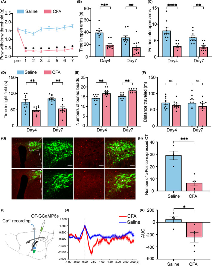
In the CFA inflammatory model, mechanical withdrawal thresholds fall. Days later, mice spend less time on exposed arms and in illuminated areas and bury more marbles, without a significant total-distance change. The study focuses on exploration and avoidance in the inflammatory setting.
PVN oxytocin-cell c-Fos activity labeling decreases, as do calcium signals during the elevated maze. These observations associate behavioral change with population activity without directly measuring oxytocin release. Subsequent interventions test the cells’ functional contribution.
Source: Li YJ et al. · CNS neuroscience & therapeutics · CC BY
Original figure and English caption ↗
Oxytocin supplementation shifts behavior toward exploration
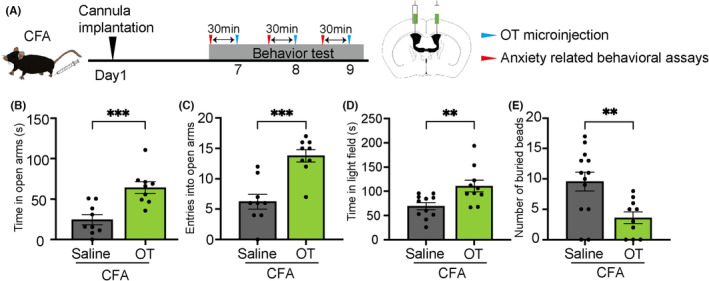
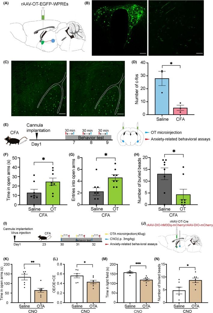
Ventricular oxytocin supplementation makes inflammatory-model mice spend longer on exposed arms, enter them more, explore illuminated areas more and bury fewer marbles. Measures across different settings consistently shift toward less avoidance-like behavior.
Mice without the inflammatory challenge lack the same significant anxiety-like changes, and tested novel-object recognition is not obviously altered. Because ventricular supplementation has broad reach, the team next asks whether the oxytocin-producing PVN population produces a similar direction of change.
Source: Li YJ et al. · CNS neuroscience & therapeutics · CC BY
Original figure and English caption ↗
Population activation improves several measures
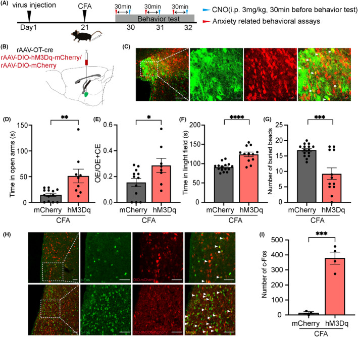
Activating PVN oxytocin cells in the inflammatory model increases exposed-arm time and entry proportion, illuminated-area time and reduces burying. Increased c-Fos labeling supports population recruitment, while behavior aligns with the supplementation results.
Oxytocin cells send axons to several regions, so activating the PVN population may engage multiple outputs. The experiment identifies a population-level contribution and motivates examining a particular output associated with the behaviors.
Source: Li YJ et al. · CNS neuroscience & therapeutics · CC BY
Original figure and English caption ↗
Inhibition alters avoidance without the inflammatory challenge
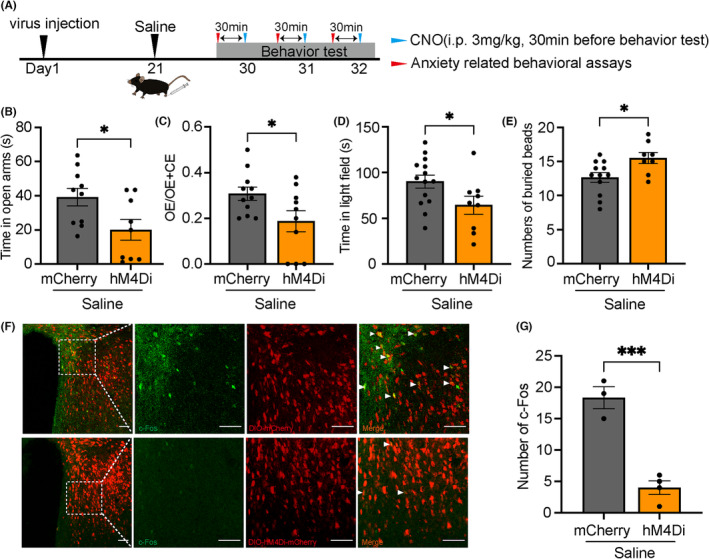
The opposite experiment uses the saline-control background. Inhibiting PVN oxytocin cells reduces exposed-arm time and entry proportion and illuminated-area time, increases burying and lowers PVN activity labeling. Reducing population activity itself alters the tested behaviors.
Activation and inhibition operate in different backgrounds: activation examines improvement following inflammation, while inhibition examines change without that challenge. Together they support a role for oxytocin cells in behavioral regulation while addressing different contextual questions.
Source: Li YJ et al. · CNS neuroscience & therapeutics · CC BY
Original figure and English caption ↗
Local amygdala signals and receptors converge
Tracing identifies PVN oxytocin-cell fibers in CeA, where overall activity labeling is lower in the inflammatory model. Local oxytocin supplementation increases exposed-arm time and entries and reduces burying, linking improvement to a specific downstream site.
Activating the PVN population while blocking CeA oxytocin receptors weakens improvements in several exploration and burying measures. Local supplementation and blockade provide complementary evidence for receptor involvement in the tested population effect.
Source: Li YJ et al. · CNS neuroscience & therapeutics · CC BY
Original figure and English caption ↗
Testing activation at the pathway terminals
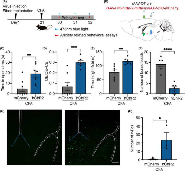
Activating PVN oxytocin-cell terminals in CeA also increases exposed-arm and illuminated-area exploration, reduces burying and raises CeA c-Fos labeling in the inflammatory model. Testing the output destination complements local supplementation and receptor-blockade results.
The observations concern local activity labeling and behavior. Recipient CeA cell types and how oxytocin release reshapes local excitation and inhibition are not resolved individually. The pathway’s behavioral contribution is evident while finer cellular mechanisms remain to be distinguished.
Source: Li YJ et al. · CNS neuroscience & therapeutics · CC BY
Original figure and English caption ↗
CeA-projecting cells also contribute in the unchallenged state
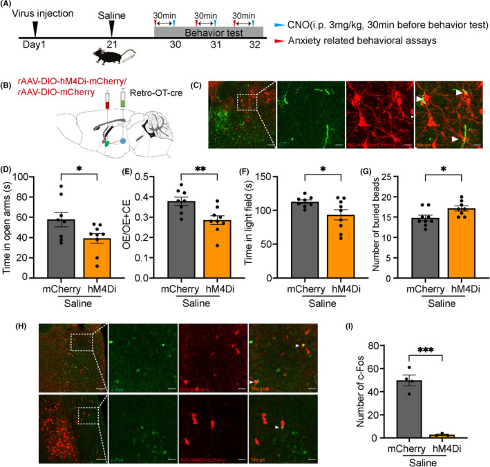
Labeling and inhibiting CeA-projecting PVN oxytocin cells in the saline background reduces exposed-arm and illuminated-area exploration and increases burying. This manipulation acts on PVN somata and may involve their other axonal branches, providing evidence at a different level from local CeA-terminal activation.
The study uses adult male mice and chiefly examines anxiety-like measures during inflammation and inhibition effects without it. It connects avoidance with oxytocin populations, CeA input and local receptors; whether those improvements also change inflammatory nociceptive thresholds is not answered across the interventions.
Source: Li YJ et al. · CNS neuroscience & therapeutics · CC BY
Original figure and English caption ↗