Why does pain linger? A reinforcing feedback loop in the mouse brain
Two inhibitory steps between the anterior cingulate cortex and ventral tegmental area form positive feedback linking persistent hypersensitivity with affect-related behavior in mice. Selected interventions show benefits beyond their end.
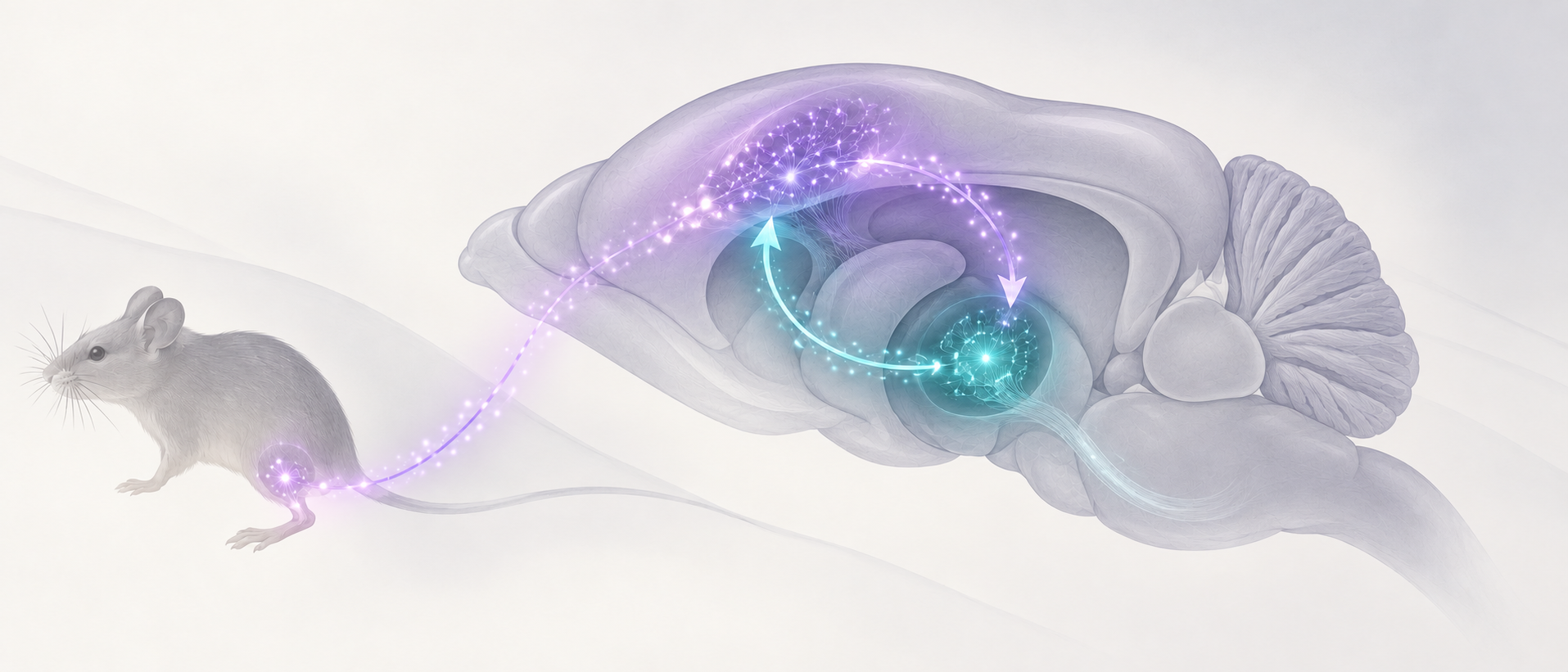
A light touch prompts a rapid paw withdrawal, while an open space invites cautious exploration near its edges. After nerve injury, pain and affect-related changes can resemble two connected gears. Could repeated brain responses build a circuit that keeps the state going? A study published in Nature Neuroscience on January 3, 2024 identifies a reciprocal connection between the anterior cingulate cortex and ventral tegmental area in mice. Its unusual feature is that two inhibitory steps can combine to reinforce sensitivity.
Excitatory cingulate output recruits local inhibition in the ventral tegmental area and reduces dopamine activity. Weaker dopamine return releases the original cortical output from restraint, linking persistent hypersensitivity and affect-related behavior to reinforcing feedback.
Contents · 6 sections
Two brain regions move in opposite directions as pain persists
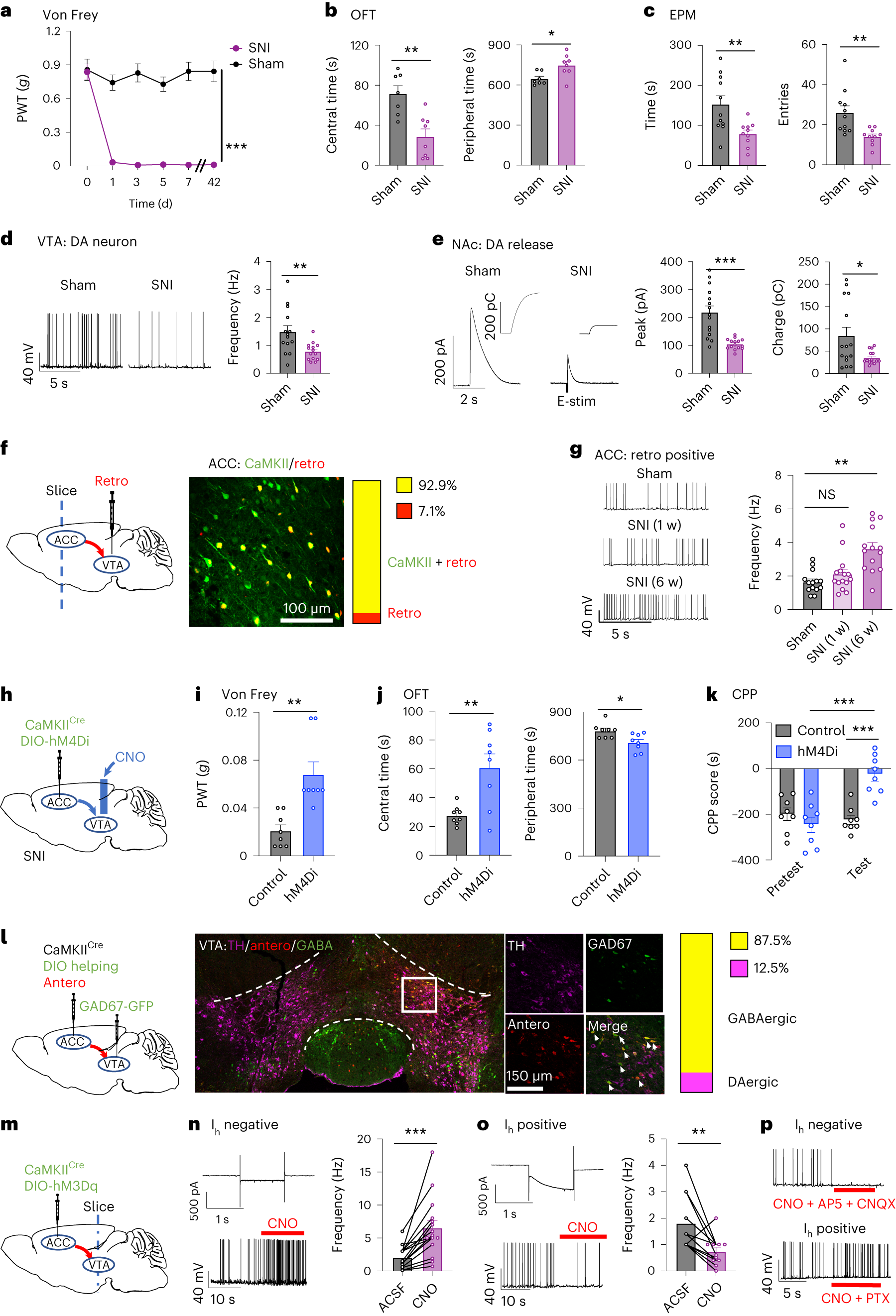
The story begins with nerve-injured mice. Lower paw-withdrawal thresholds mean that lighter pressure evokes a response, while exploration in the open field and elevated plus maze also changes at six weeks. The researchers ask whether persistent sensitivity and affect-related behavior share a circuit background.
Excitatory anterior cingulate cortex (ACC) neurons projecting to the ventral tegmental area (VTA) fire more spontaneously at six weeks; the one-week comparison is not significant. Meanwhile, VTA dopamine-cell firing and dopamine release in the nucleus accumbens decrease. The opposing changes suggest something more specific than a uniform rise in brain activity.
Source: Qian Song et al. · Nature Neuroscience ·
Original figure and English caption ↗
Dopamine returning to cortex restrains the outgoing signal
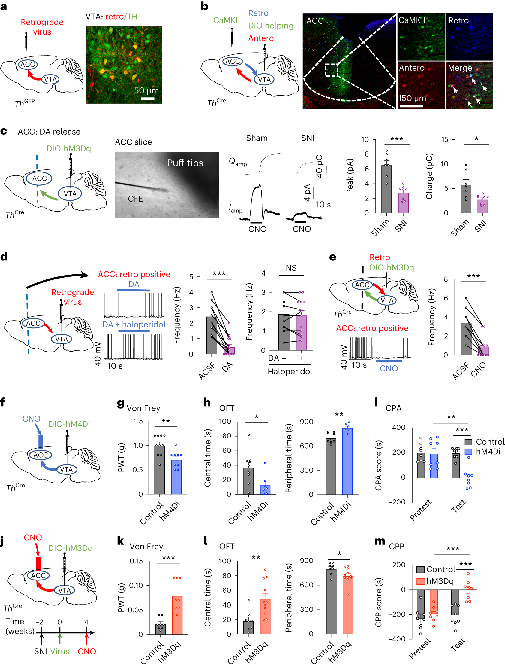
Dopamine does not simply leave the VTA and continue forward. VTA dopamine neurons also send signals back to the ACC, reaching the excitatory cells that originally project to the VTA. The outgoing and returning pathways therefore meet: information leaving cortex can influence its own starting point.
Dopamine reduces firing in the recorded ACC projection cells, and D2-related pharmacological blockade interferes with this effect. Dopamine release reaching the ACC falls after nerve injury. Activating local dopamine terminals in injured mice raises withdrawal thresholds, changes open-field exploration and produces preference for an intervention-paired context. Suppressing this return in normal mice produces oppositely directed sensory and avoidance changes.
Source: Qian Song et al. · Nature Neuroscience ·
Original figure and English caption ↗
How can two inhibitory steps reinforce a loop?
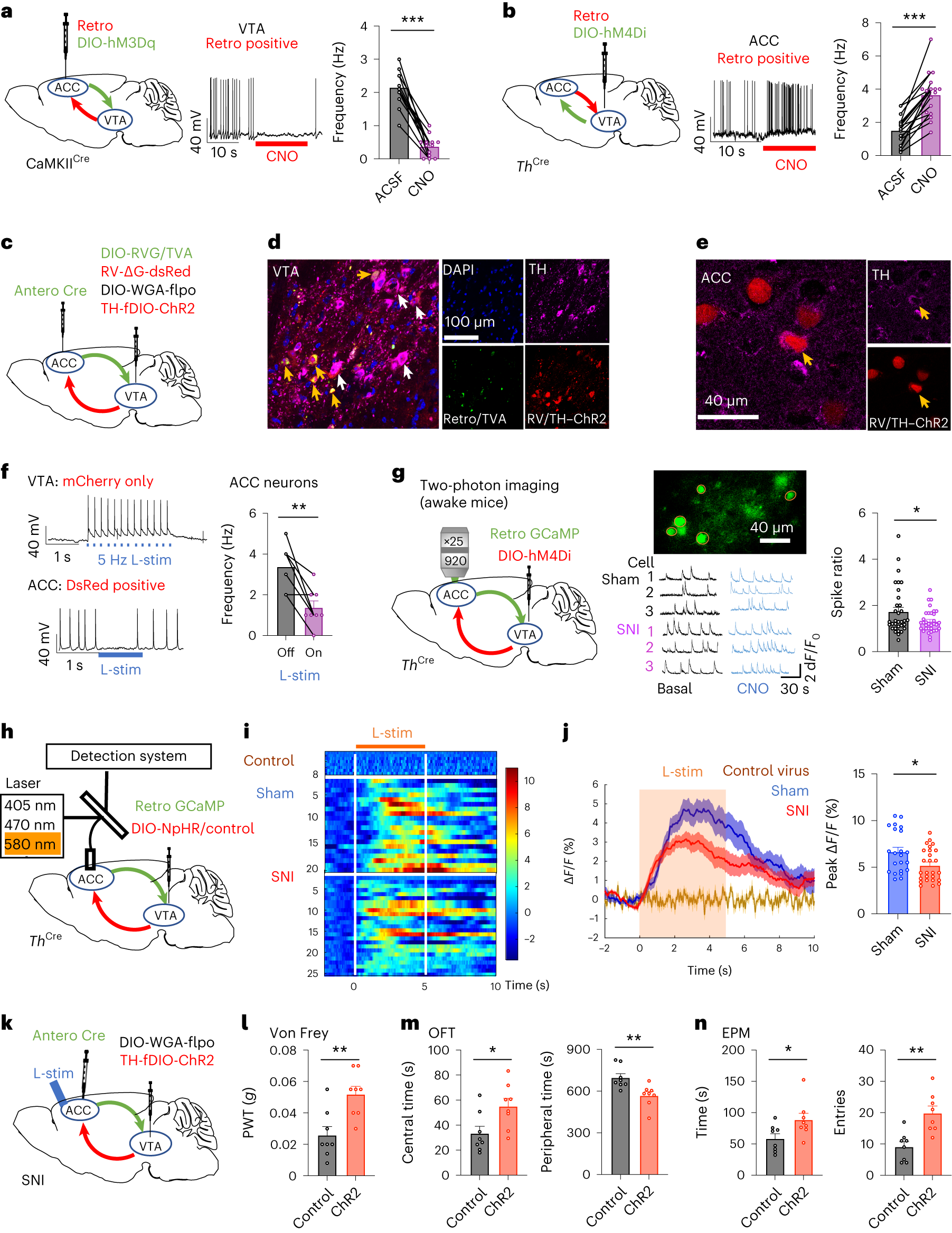
It matters which cells each brake acts on. Excitatory ACC output recruits local VTA GABA cells, which suppress dopamine neurons. Those dopamine neurons normally restrain the original ACC output. Stronger cortical output can therefore weaken dopamine return, and weaker return releases cortical output from restraint.
This is positive feedback: after a change travels around the circuit, it comes back to reinforce itself. Tracing and recordings support connections between the steps. In awake animals, suppressing VTA dopamine terminals increases calcium signals in the relevant ACC cells, while activating terminals within the loop attenuates sensory and avoidance responses after nerve injury. The loop has anatomical, functional and behavioral support.
Source: Qian Song et al. · Nature Neuroscience ·
Original figure and English caption ↗
Repeatedly driving the loop can leave changes behind
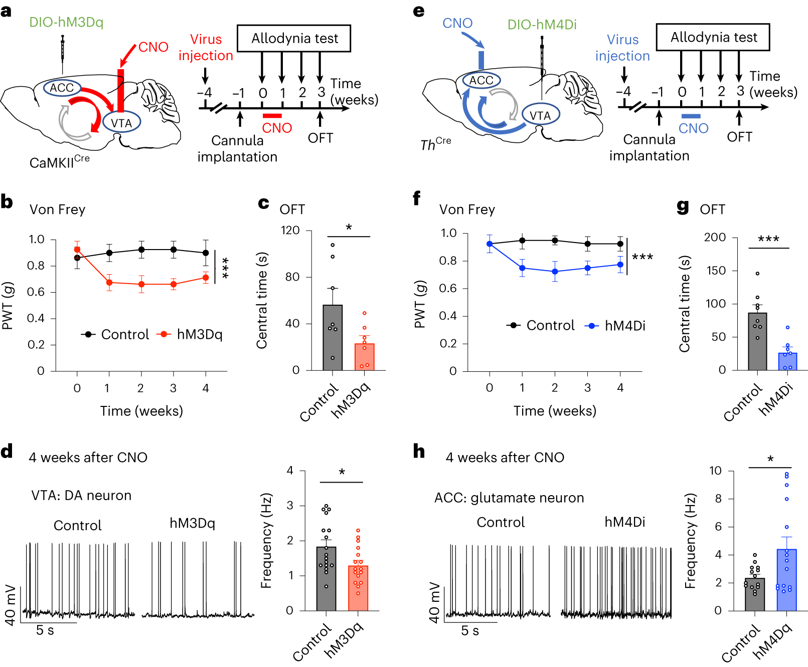
If the circuit simply followed pain, changing it might produce only a brief response. The team repeatedly enhances ACC-to-VTA output or suppresses VTA-to-ACC dopamine return in normal mice. These interventions drive the loop at different points, and both are followed by lower withdrawal thresholds and altered exploration.
Some changes persist for weeks after repeated manipulation ends, together with changes in spontaneous cellular firing. This links the circuit more closely to a lasting state. The observations concern specific mouse interventions and weeks of measured effects, rather than establishing that all pain-related memories become permanently fixed.
Source: Qian Song et al. · Nature Neuroscience ·
Original figure and English caption ↗
Sensitivity can persist after the peripheral ligation is removed
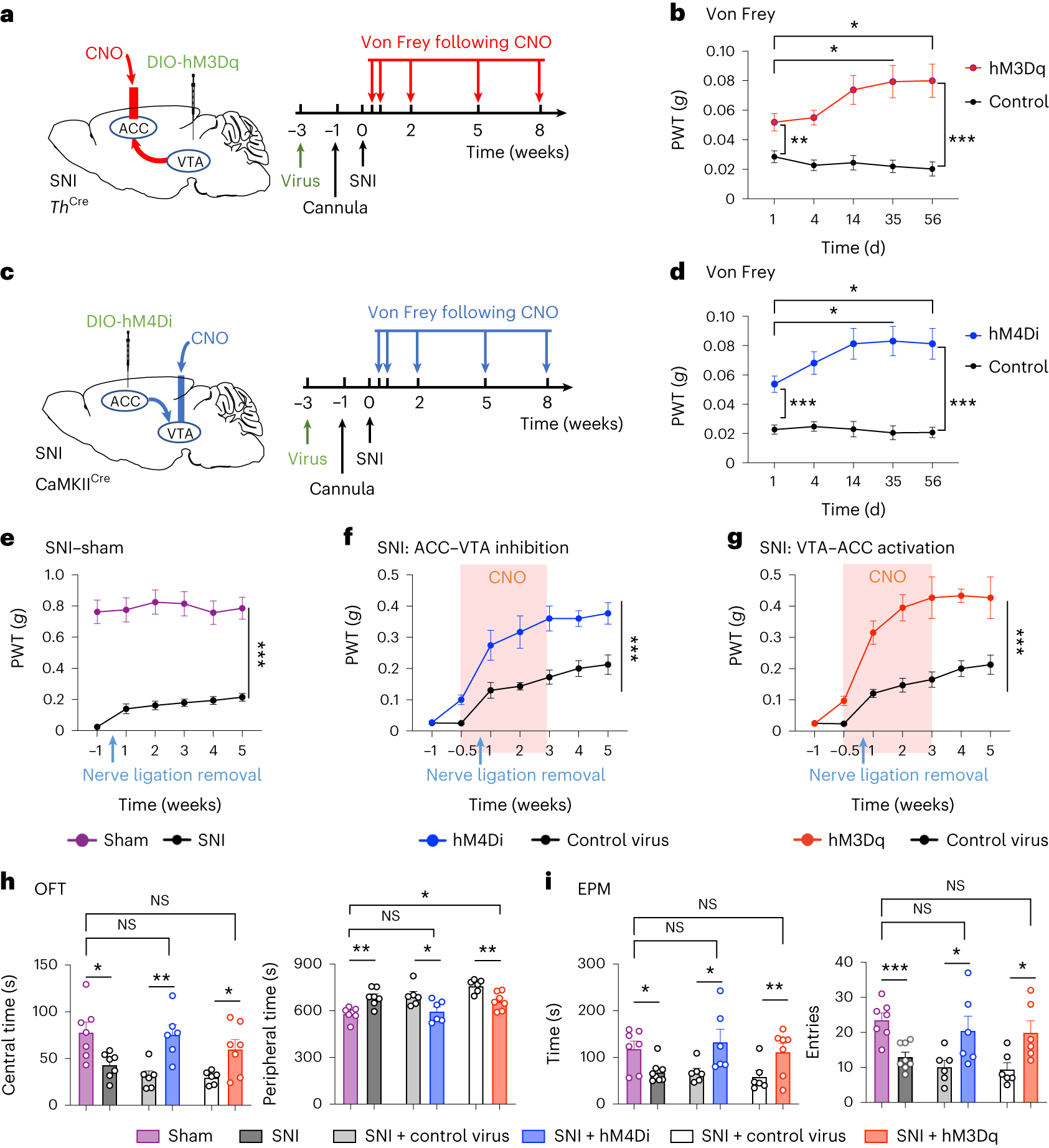
In nerve-injured mice, sustained enhancement of dopamine return to ACC or inhibition of ACC output to VTA improves mechanical hypersensitivity. A further condition removes the original nerve ligation. Withdrawal thresholds do not immediately return to sham levels, allowing peripheral treatment and the persistent brain-associated state to be examined separately.
Under this condition, repeated circuit intervention for two weeks produces threshold improvements that persist at later observation points after intervention ends. Open-field and elevated-plus-maze behavior also shifts toward improvement three weeks later. The thresholds are not uniformly restored to sham levels, but the findings show that the feedback-associated state remains adjustable after sensitivity has developed.
Source: Qian Song et al. · Nature Neuroscience ·
Original figure and English caption ↗
The same receptor family plays different roles at different sites
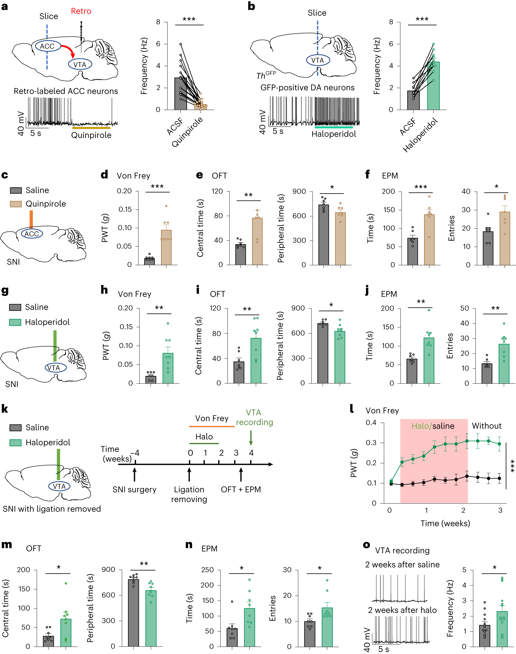
D2-related findings add a further twist. A D2 agonist applied locally in ACC reduces firing of relevant cortical cells and improves sensory and exploration responses in injured mice. A D2 antagonist applied locally in VTA increases dopamine-cell firing and also shifts behavior toward improvement.
Opposite pharmacological operations at different circuit locations can thus weaken the same feedback process. In the ligation-removal condition, some benefits of repeated local VTA intervention also continue after its end. These local mouse results identify adjustable circuit nodes; translation to treatment in people requires further evidence about dosage, targeting and long-term effects.
Source: Qian Song et al. · Nature Neuroscience ·
Original figure and English caption ↗