How can excitatory prefrontal input reduce hyperalgesia?
In male rats with fentanyl-associated hyperalgesia, infralimbic inputs to the laterocapsular central amygdala change. Pathway recruitment improves stimulus responses, suggesting a compensatory contribution.
Why can an analgesic be followed by greater stimulus sensitivity? Opioid-associated hyperalgesia raises a further question: does the brain recruit mechanisms that counter excessive nociceptive responses? A study published in Molecular Pain on February 7, 2024 examines infralimbic cortex (IL) inputs to the laterocapsular central amygdala (CeLC) in male rats. Hyperalgesia accompanied increased activity-marker expression in right CeLC and altered synaptic input. Recruiting IL terminals further improved mechanical and thermal responses, raising the question of how an excitatory connection can contribute to a brake.
IL provides direct excitation to CeLC and can recruit indirect inhibition. Hyperalgesia involves a higher evoked inhibition-to-excitation ratio; recruitment improves responses and suppression worsens them, supporting a possible compensatory contribution.
Contents · 6 sections
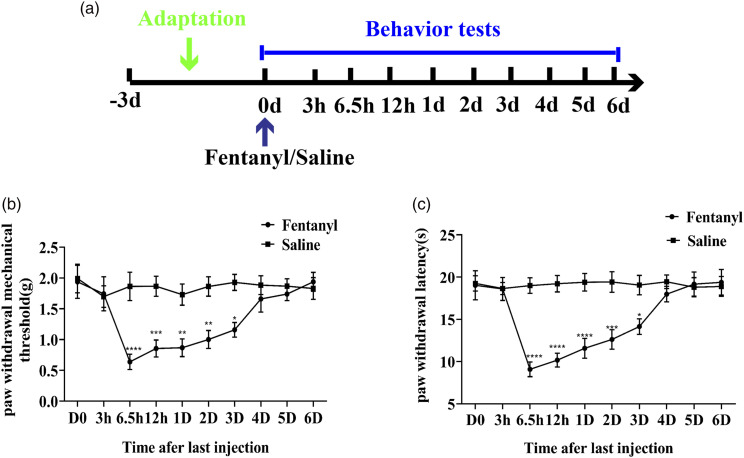
Stimulus responses change for several days
Repeated acute fentanyl exposure lowered mechanical withdrawal thresholds and shortened thermal withdrawal latencies. Weaker mechanical stimulation elicited withdrawal, and thermal withdrawal occurred sooner. Increased sensitivity appeared several hours after exposure and gradually recovered over subsequent days.
These two stimulus measures define the setting for the study. Examining a prominent hyperalgesic phase then allows assessment of accompanying neural changes. The findings describe rats after repeated acute exposure, providing a specific model of opioid-associated hyperalgesia.
Source: Cui LL et al. · Molecular pain · CC BY-NC
Original figure and English caption ↗
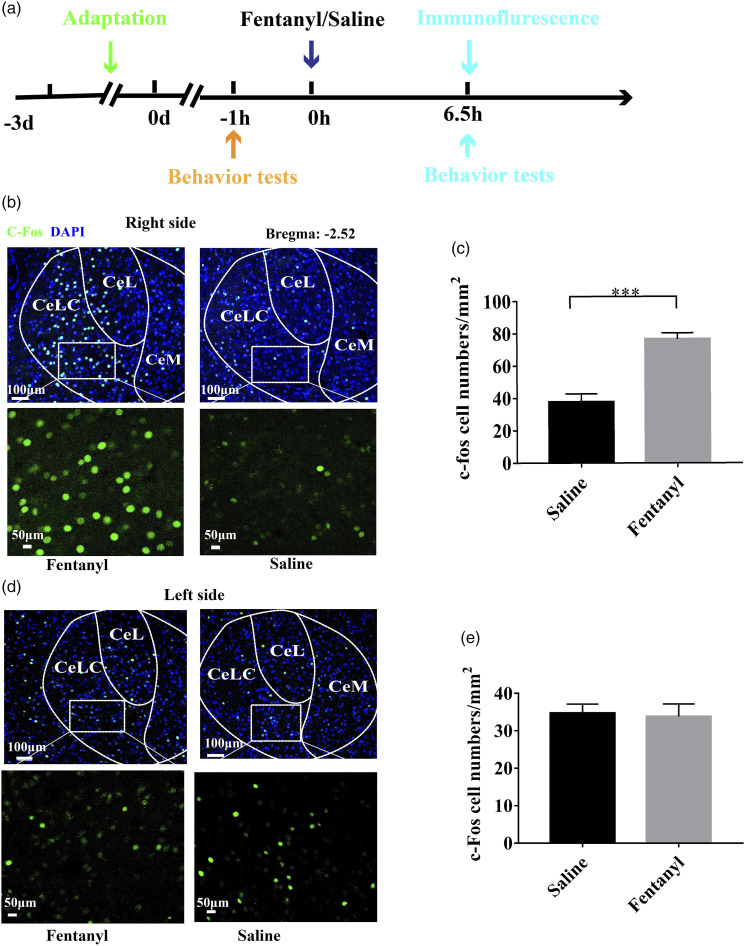
Activity labeling increases on the right
At the prominent hyperalgesic phase, more cells expressed c-Fos in right CeLC. This activity-related marker directs attention to the right amygdala node. Left CeLC did not show a significant difference, revealing a lateralized pattern.
The amygdala participates in several dimensions of pain, with roles varying among subregions. This study focuses on CeLC and examines prefrontal input as a regulatory clue. Increased activity labeling provides a starting point for linking connectivity with behavioral responses.
Source: Cui LL et al. · Molecular pain · CC BY-NC
Original figure and English caption ↗
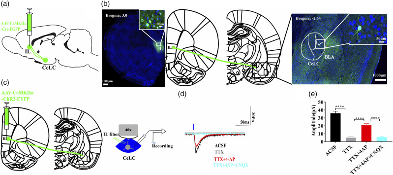
Where does the prefrontal excitatory signal arrive?
IL is a medial prefrontal subregion. Its projection fibers reached CeLC, and recruiting them elicited excitatory synaptic currents in recipient cells. Blockade and recovery findings further support direct glutamatergic input between the regions.
The connection provides a traceable bridge between the regions. Excitatory prefrontal output need not make every downstream cell more active, because it enters local networks and interacts with other excitatory and inhibitory components. Both direct responses and local interactions matter.
Source: Cui LL et al. · Molecular pain · CC BY-NC
Original figure and English caption ↗
Excitatory input also recruits delayed inhibition
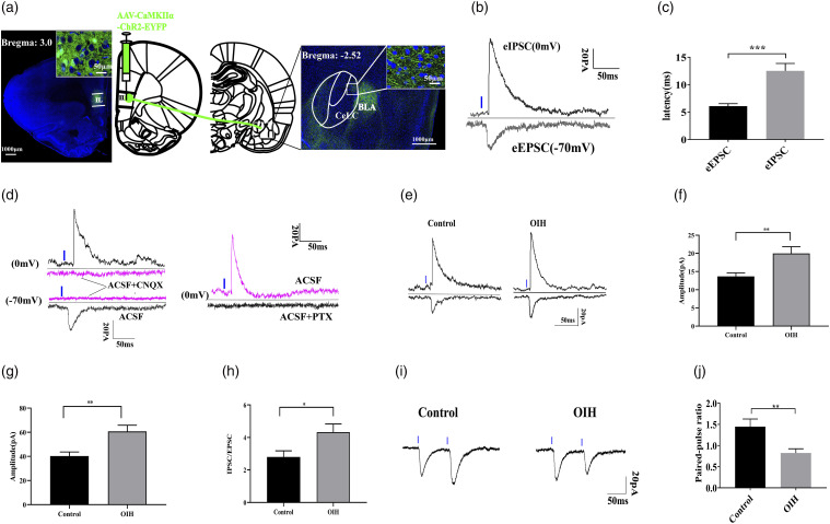
Recruiting IL input elicited both excitatory currents and inhibitory currents with later onset. Sensitivity to glutamate- and GABA-receptor-related blockade supports indirect inhibition recruited through the local network.
In hyperalgesic rats, input-evoked inhibitory currents increased and the inhibition-to-excitation ratio rose. A lower paired-pulse ratio also suggests altered presynaptic release regulation. The input thus shows component-specific remodeling rather than a simple uniform decrease.
The measured current ratio describes an evoked input balance but does not by itself establish whether the pathway alleviates nociceptive responses. Changes may contribute to compensation or aggravation. Behavioral effects of recruitment and suppression help distinguish these possibilities.
Source: Cui LL et al. · Molecular pain · CC BY-NC
Original figure and English caption ↗
Stronger input improves withdrawal responses
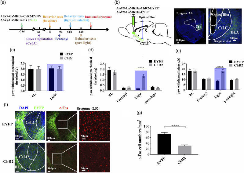
Recruiting IL terminals in CeLC increased mechanical withdrawal thresholds and prolonged thermal withdrawal latencies in hyperalgesic rats. Responses returned toward the hypersensitive state after light ended, linking improvement to pathway recruitment during stimulation.
CeLC c-Fos labeling also decreased. In rats without fentanyl exposure, activation did not significantly change mechanical thresholds. Effects depend on the background state, supporting a contribution to limiting responses during hyperalgesia; sustained effects remain to be examined.
Source: Cui LL et al. · Molecular pain · CC BY-NC
Original figure and English caption ↗
Suppressing the input worsens hyperalgesic responses
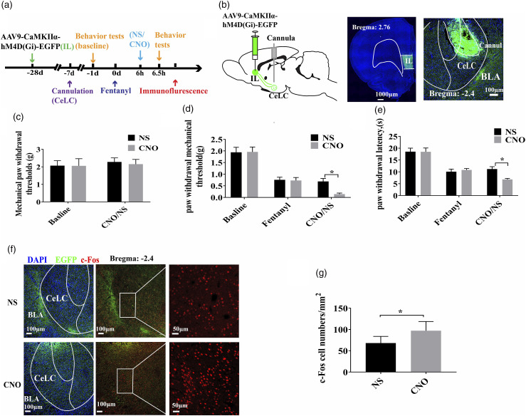
Suppressing IL input in CeLC further lowered mechanical thresholds and shortened thermal withdrawal latencies in hyperalgesic rats while increasing CeLC c-Fos labeling. Mechanical thresholds in unexposed rats again did not significantly change.
Improvement with recruitment and worsening with suppression support a compensatory interpretation: stronger cortical input during hyperalgesia may help constrain downstream responses. The relevant CeLC cell types, functions at other times or in other pain models, and corresponding patterns in females remain to be clarified.
Source: Cui LL et al. · Molecular pain · CC BY-NC
Original figure and English caption ↗