Why does hypersensitivity last longer after the same incision? A cortical stress pathway
A mouse study finds stronger orbitofrontal input to anterior insula after preoperative stress, linking dexmedetomidine effects to α2-receptor signaling at this connection.
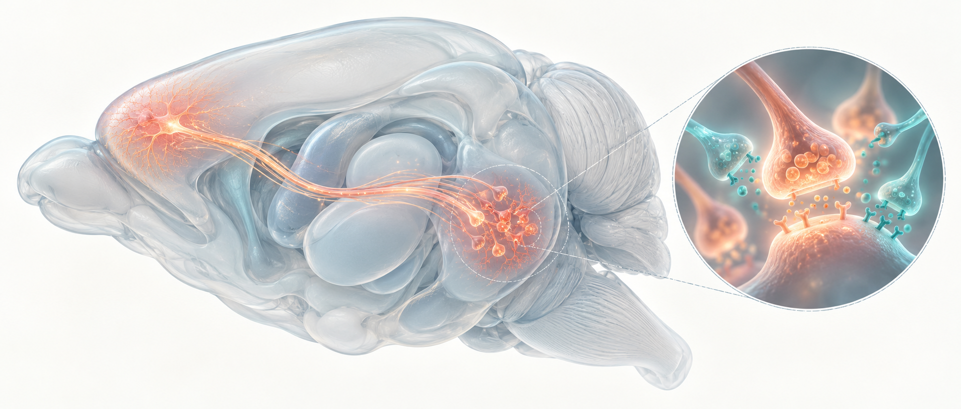
Why does the same incision cause more persistent hypersensitivity in previously stressed mice? Combining preoperative stress with a hind-paw incision revealed changes even in the unincised paw. Researchers followed excessive anterior-insular excitation to orbitofrontal input and examined how dexmedetomidine modifies the connection.
Stress plus incision increases anterior-insular excitation and orbitofrontal input. Pathway inhibition reduces hypersensitivity, while dexmedetomidine lowers excessive transmission through an α2-related mechanism.
Contents · 6 sections
The unincised paw also becomes more sensitive
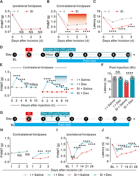
After modified single prolonged stress, mice received a right-paw incision. Compared with incision alone, the combined group had lower mechanical thresholds in the unincised paw and greater thermal sensitivity bilaterally, with altered anxiety-related exploration but comparable distance.
Early incised-paw thresholds were very low in both groups without a significant difference. Fentanyl dose-response tests supported the authors’ floor-effect interpretation. The unincised paw became the main readout in later experiments to track generalized stress-related sensitization.
A later effect remains after the acute threshold rise
Systemic dexmedetomidine initially raised mechanical thresholds in both groups. At around six hours, incision-only mice returned toward baseline while the combined group retained improvement, motivating later behavioral and neural comparisons. Brief treatment over the first two postoperative days accelerated subsequent recovery, followed for four weeks.
The early effect faded in incision-only mice but remained later in the combined group, revealing different state-dependent trajectories. Early short-course treatment also changed later recovery. The researchers then examined cellular and synaptic activity behind the sustained difference.
Source: Rong H et al. · MedComm · CC BY
Original figure and English caption ↗
Two anterior-insular cell types respond differently
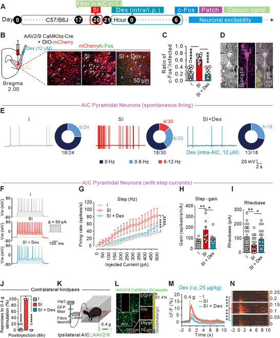
Imaging and activity labeling highlighted anterior insula. Activating its glutamatergic cells lowered mechanical and thermal thresholds, inhibition reduced model hypersensitivity, and GABAergic activation also improved readouts. The cell types could move behavior in different directions.
Glutamatergic cells showed increased spontaneous and evoked excitability and larger calcium responses to low-force stimulation, reduced by dexmedetomidine. GABAergic excitability and calcium measures did not show the same group differences, directing attention to excitatory input.
Source: Rong H et al. · MedComm · CC BY
Original figure and English caption ↗
More frequent excitatory events point upstream
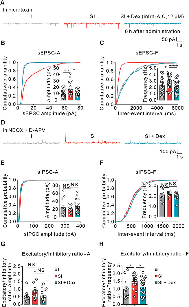
Excitatory synaptic events increased in the combined model, whereas inhibitory amplitude and frequency did not significantly change. The excitatory/inhibitory frequency ratio changed without a corresponding significant amplitude-ratio change. Drug treatment reduced excessive excitation and restored the relevant ratio.
Frequency changes stood out while amplitude ratios remained comparable, drawing attention to event occurrence. Connectivity imaging and projection resources suggested orbitofrontal cortex as a candidate, leading the team to test a defined source of enhanced drive.
Source: Rong H et al. · MedComm · CC BY
Original figure and English caption ↗
Orbitofrontal terminals link circuit effects with α2 signaling
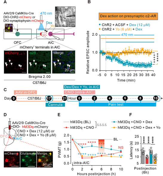
Tracing and slice recordings supported direct glutamatergic input. Terminal inhibition reduced model hypersensitivity, whereas activation sensitized otherwise untreated mice. Bulk orbitofrontal imaging lacked the same marked change despite recruitment of the insula-projecting population.
α2 labeling was associated with these terminals. Dexmedetomidine reduced optically evoked excitatory currents, opposed by yohimbine, which also blocked local drug-related behavioral improvement. Anatomy, currents and behavior supported an α2-related presynaptic contribution.
Source: Rong H et al. · MedComm · CC BY
Original figure and English caption ↗
How stress amplifies input becomes a tractable question
The same incision in different preoperative states led to a cortical-input question. Insular excitability and orbitofrontal drive changed together, with drug effects linked to receptor-related modulation. Both acute responses and later network states emerged as relevant.
The evidence comes from a male-mouse stress/incision model. Other inputs, α2 subtypes and locations, and the sequence of intrinsic and synaptic changes remain unresolved. More precise genetic localization and state comparisons can clarify the pathway’s role.