Why does chronic stress heighten pain sensitivity? Clues from an amygdala-to-brainstem pathway
A mouse study links chronic restraint stress to a central amygdala–locus coeruleus–periaqueductal gray pathway that shapes stress-induced pain sensitivity.
Chronic stress frequently precipitates both mood disorders and heightened pain sensitivity, yet whether stress-induced allodynia and depression-like behaviors are governed by shared or distinct neural circuits remains poorly understood. Recently, a collaborative team led by Prof. Qian Meng at Anhui Medical University, Prof. Zhi Zhang at the University of Science and Technology of China, and Dr. Zi-Hua Song at the National Innovation Institute of Defense Technology (with Jing Wei, An Liu, and Yu Cao as co-first authors) published a research article titled "A central amygdala-locus coeruleus pathway mediating pain under chronic stress" in *Cell Reports*. Using trans-monosynaptic viral tracing, slice electrophysiology, in vivo fiber photometry, and bidirectional chemogenetic and optogenetic manipulations, the authors demonstrate that chronic restraint stress (CRS) hyperactivates central amygdala GABAergic neurons (GABACeA) that monosynaptically inhibit locus coeruleus noradrenergic neurons (NELC), thereby reducing excitatory noradrenergic drive onto lateral periaqueductal gray glutamatergic neurons (GluLPAG) to selectively mediate stress-induced pain without altering depression-like behaviors.
Chronic restraint stress hyperactivates GABACeA neurons to monosynaptically suppress NELC neurons and downstream GluLPAG neurons, selectively driving stress-induced allodynia and pain aversion independently of depression-like behaviors and acute inflammatory pain.
Contents · 5 sections
Chronic Stress Suppresses Locus Coeruleus Noradrenergic Neurons to Drive Allodynia
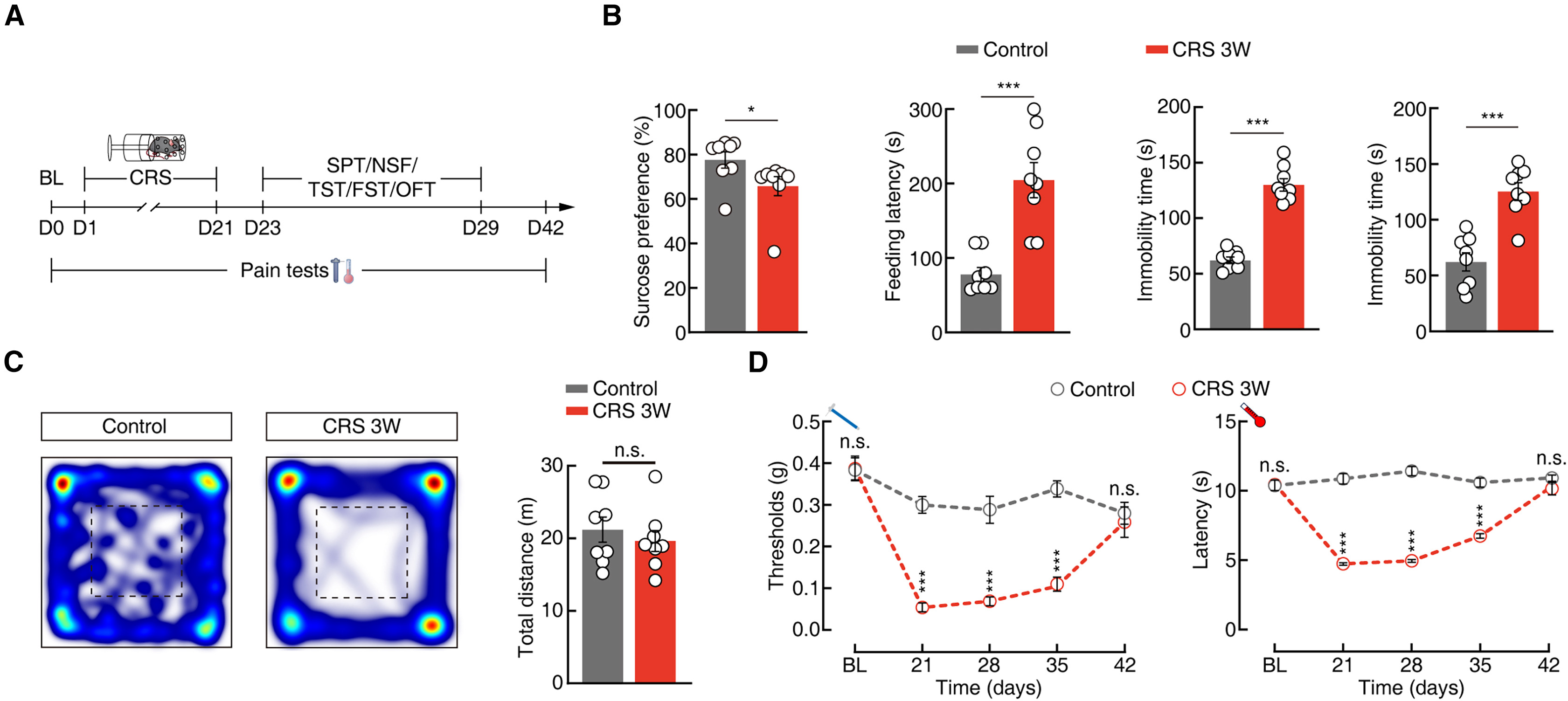
To model chronic stress-induced pain, the authors subjected male and female C57BL/6J mice to chronic restraint stress for 6 h daily over 21 consecutive days (CRS 3W; Figure 1A). CRS 3W mice developed robust depression-like behaviors—decreased sucrose preference in the SPT, prolonged feeding latency in the NSF test, and increased immobility in the TST and FST—without changes in open-field locomotion (Figures 1B and 1C), alongside persistent mechanical allodynia (von Frey test) and thermal hyperalgesia (Hargreaves test) starting from week 2 (Figure 1D). Whole-cell patch-clamp recordings in acute brain slices revealed that locus coeruleus noradrenergic (NELC) neurons exhibited a significantly reduced firing rate in CRS 3W mice compared with unstressed controls.
To test whether reduced NELC activity causally mediates CRS-induced pain, the authors bilaterally injected AAV-DBH-hM3Dq-EYFP or AAV-DBH-hM4Di-EYFP into the LC. Chemogenetic activation of NELC neurons with CNO (5 mg/kg) in CRS 3W mice significantly alleviated mechanical and thermal allodynia and produced conditioned place preference (CPP) for the CNO-paired chamber, indicating relief of ongoing pain aversion. Conversely, chemogenetic inhibition of NELC neurons in naive mice induced mechanical and thermal hypersensitivity as well as conditioned place aversion. Strikingly, neither activating nor inhibiting NELC neurons altered depression-like behaviors in the NSF, TST, or FST assays, demonstrating that NELC hypoactivity selectively governs the nociceptive and aversive dimensions of chronic stress.
Source: Jing Wei et al. · Cell Reports · © Elsevier Inc.
Original figure and English caption ↗
Anatomical and Synaptic Dissection of the Monosynaptic GABACeA → NELC Pathway
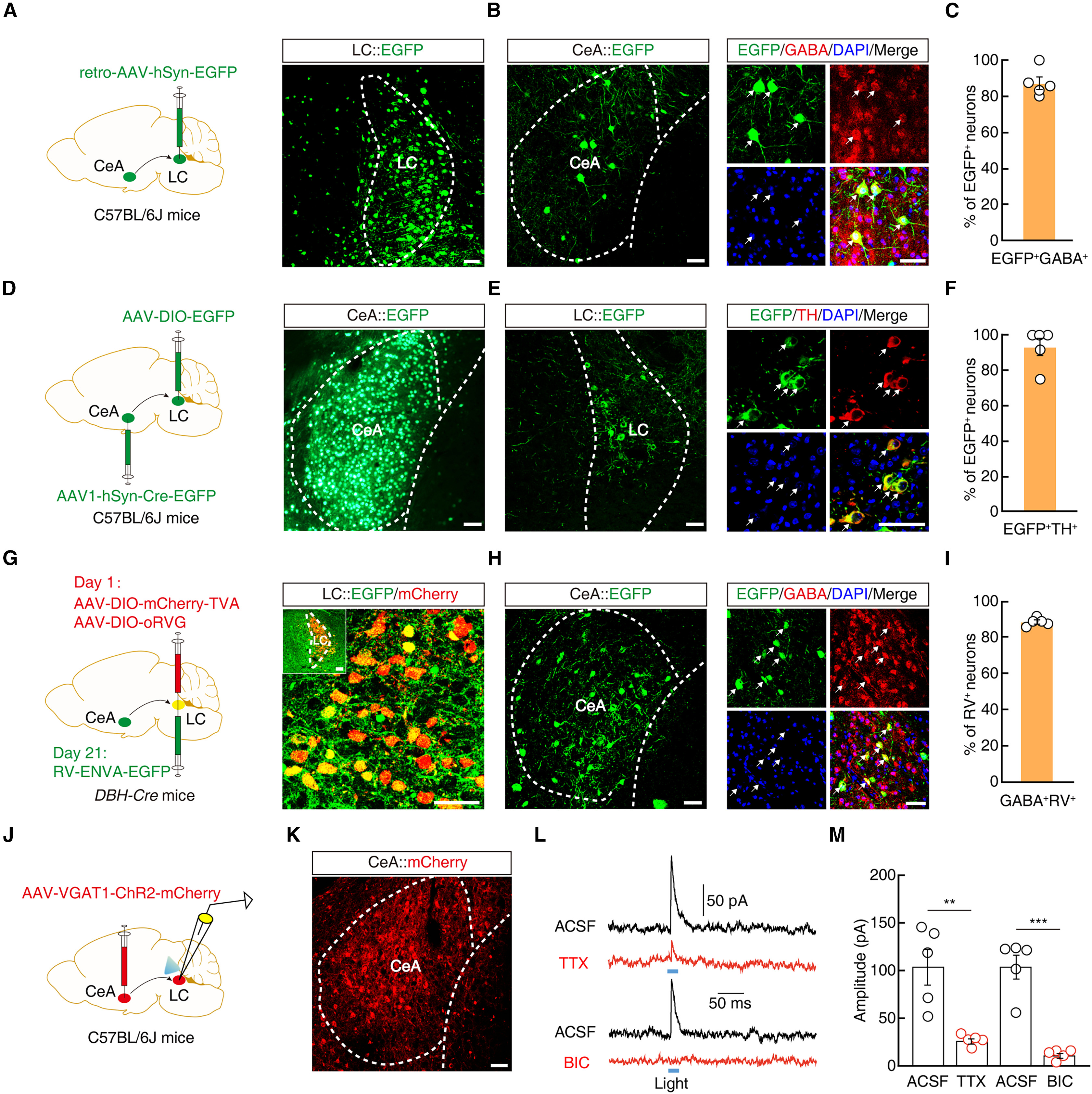
To identify the upstream source suppressing NELC neurons, the authors mapped brain-wide inputs to the LC using three complementary viral tracing strategies (Figures 2A–2I). First, retrograde tracing with retro-AAV-hSyn-EGFP injected into the LC labeled a dense population of central amygdala (CeA) neurons, ~90% of which co-localized with GABA (Figures 2A–2C). Second, anterograde trans-monosynaptic tracing with AAV1-hSyn-Cre-EGFP in the CeA and AAV-DIO-EGFP in the ipsilateral LC showed that ~90% of postsynaptic LC neurons were TH+ noradrenergic neurons (Figures 2D–2F). Third, cell-type-specific monosynaptic rabies tracing in DBH-Cre mice (AAV-DIO-mCherry-TVA, AAV-DIO-oRVG, and RV-ENVA-EGFP in the LC) confirmed that ~90% of rabies-labeled CeA input neurons were GABAergic (Figures 2G–2I).
To functionally characterize these projections, the authors injected AAV-VGAT1-ChR2-mCherry into the CeA and performed whole-cell patch-clamp recordings from NELC neurons in acute LC slices (Figures 2J and 2K). Blue light (473 nm) photostimulation of ChR2-expressing CeA terminals in the LC reliably evoked robust inhibitory postsynaptic currents (IPSCs) that were sensitive to TTX and completely abolished by the GABAA receptor antagonist bicuculline (BIC; Figures 2L and 2M), establishing a direct monosynaptic GABAergic inhibitory projection from the CeA to NELC neurons (GABACeA → NELC).
Source: Jing Wei et al. · Cell Reports · © Elsevier Inc.
Original figure and English caption ↗
CRS Hyperactivates the GABACeA → NELC Pathway to Bidirectionally Gate Stress-Induced Pain
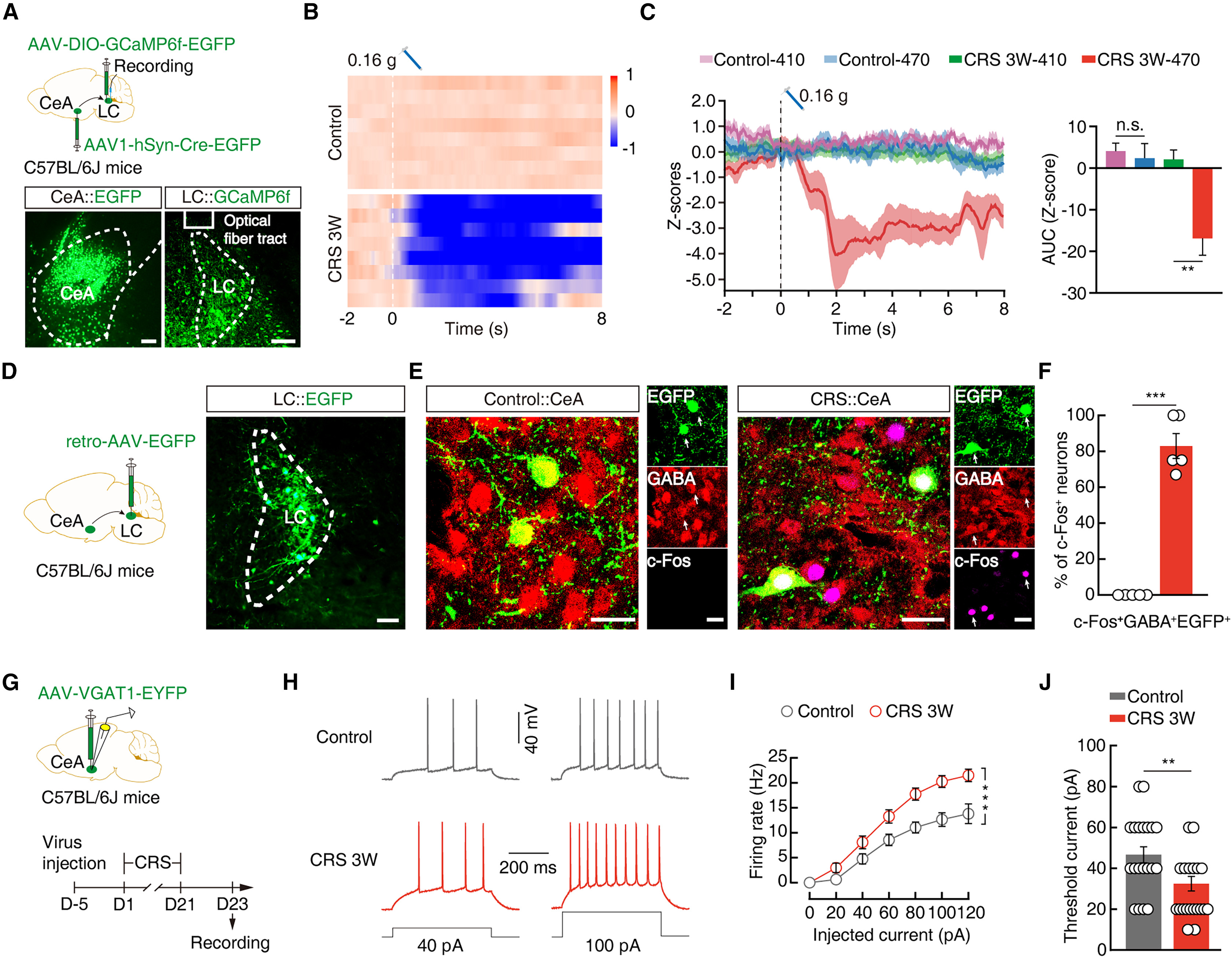
To monitor how chronic stress alters activity along this pathway in vivo, the authors injected AAV1-hSyn-Cre-EGFP into the CeA and Cre-dependent AAV-DIO-GCaMP6f into the LC for fiber photometry recordings in freely behaving mice (Figures 3A and 3B). Upon subthreshold (0.16 g) von Frey filament stimulation of the hind paw, CeA-recipient LC neurons in CRS 3W mice exhibited a rapid, pronounced decrease in GCaMP6f Ca2+ signals (Z-score and ΔF/F) compared with control mice (Figures 3C–3E). Consistently, retrograde labeling combined with c-Fos immunostaining and slice patch-clamp recordings demonstrated that CRS 3W markedly increased c-Fos expression in LC-projecting CeA neurons (~80% GABA+; Figures 3F–3I) and significantly elevated the firing rate while lowering the rheobase of GABACeA neurons (Figures 3J–3M).
Pathway-specific chemogenetic manipulations (retro-AAV-Cre-EGFP in the LC plus AAV-DIO-hM4Di/hM3Dq-mCherry in the CeA) and terminal optogenetic manipulations in Vgat-Cre mice (AAV-DIO-eNpHR3.0-EYFP or AAV-DIO-ChR2-mCherry in the CeA with optical fibers implanted in the LC) confirmed the causal necessity and sufficiency of this circuit (Figures 4A–4P). Inhibiting LC-projecting GABACeA somata or their axon terminals in the LC robustly reversed mechanical and thermal allodynia and induced CPP in both male and female CRS 3W mice (Figures 4A–4D and 4I–4L), whereas activating this pathway in naive mice triggered allodynia and place aversion (Figures 4E–4H and 4M–4P). Importantly, manipulating the GABACeA → NELC circuit did not affect depression-like behaviors or complete Freund's adjuvant (CFA 2D)-induced acute inflammatory pain, highlighting its etiological specificity for chronic stress-induced pain.
Source: Jing Wei et al. · Cell Reports · © Elsevier Inc.
Original figure and English caption ↗
Downstream NELC → GluLPAG Noradrenergic Projections Mediate Antinociceptive Output
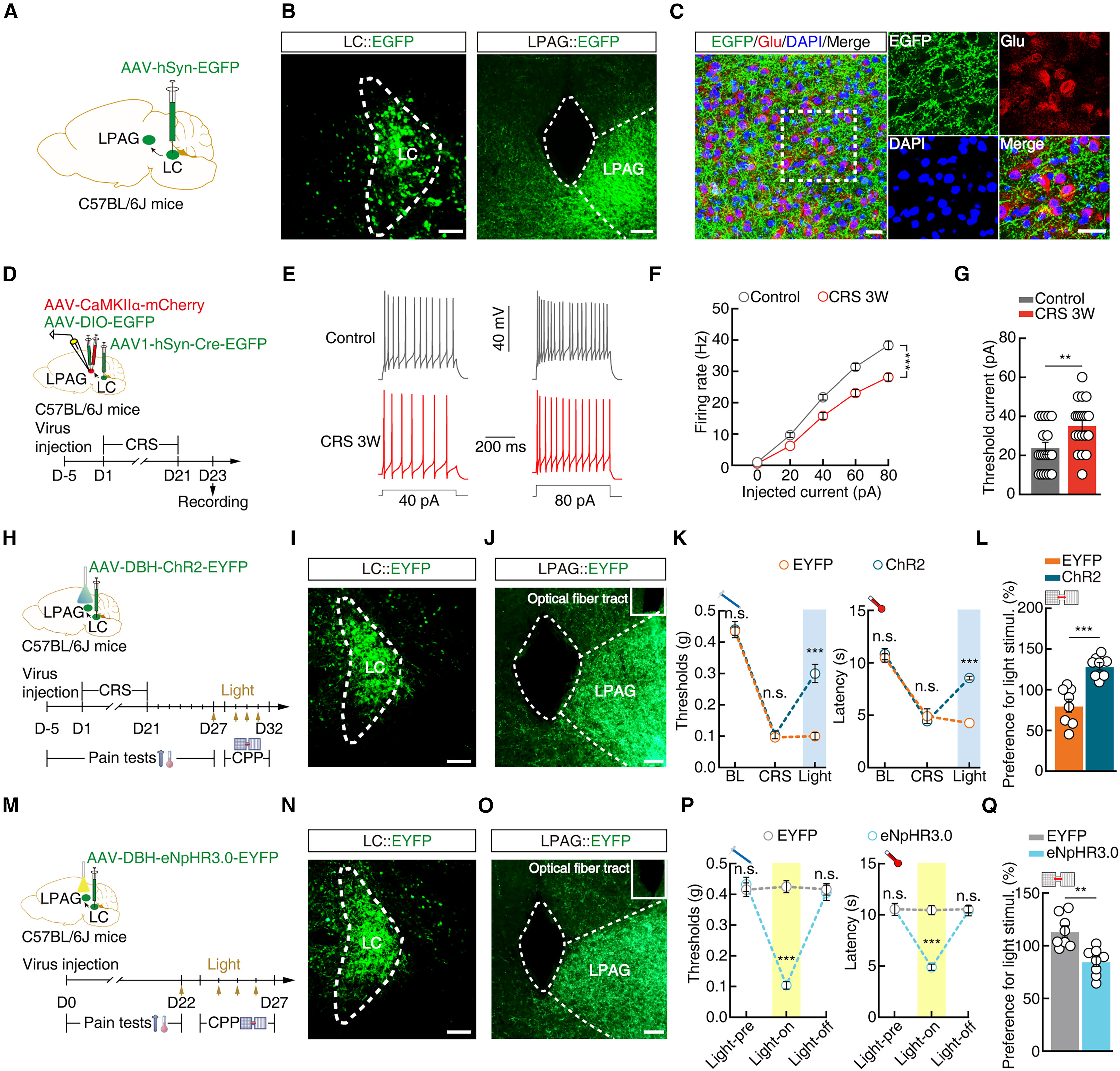
To determine how NELC hypoactivity is relayed downstream to amplify nociception, the authors traced LC efferents and identified dense projections to the lateral periaqueductal gray (LPAG), a key hub for descending pain modulation. Anterograde trans-monosynaptic tracing (AAV1-hSyn-Cre-EGFP in the LC and AAV-DIO-EGFP in the LPAG) showed that ~85% of LC-recipient LPAG neurons were glutamatergic (GluLPAG; Figures 5A–5D). In CRS 3W mice, LC-recipient GluLPAG neurons (co-labeled by AAV-CaMKIIα-mCherry and trans-synaptic EGFP) displayed a significantly decreased firing rate and increased rheobase (Figures 5E–5H). In acute LPAG slices from DBH-Cre mice, optogenetic stimulation of ChR2-expressing NELC terminals evoked inward currents that were blocked by the adrenoceptor antagonist carvedilol, confirming excitatory noradrenergic transmission onto GluLPAG neurons.
Optogenetic behavioral manipulations further established the functional role of this downstream projection: activating ChR2-expressing NELC terminals in the LPAG (AAV-DBH-ChR2-EYFP in the LC, optical fiber in the LPAG) significantly alleviated mechanical and thermal allodynia and produced CPP in CRS 3W mice (Figures 5I–5L), whereas photoinhibiting eNpHR3.0-expressing NELC terminals in the LPAG of naive mice induced mechanical and thermal hypersensitivity and place aversion (Figures 5M–5P) without affecting depression-like behaviors.
Source: Jing Wei et al. · Cell Reports · © Elsevier Inc.
Original figure and English caption ↗
Closing the Loop: Dissection of the Disynaptic GABACeA → NELC → GluLPAG Circuit
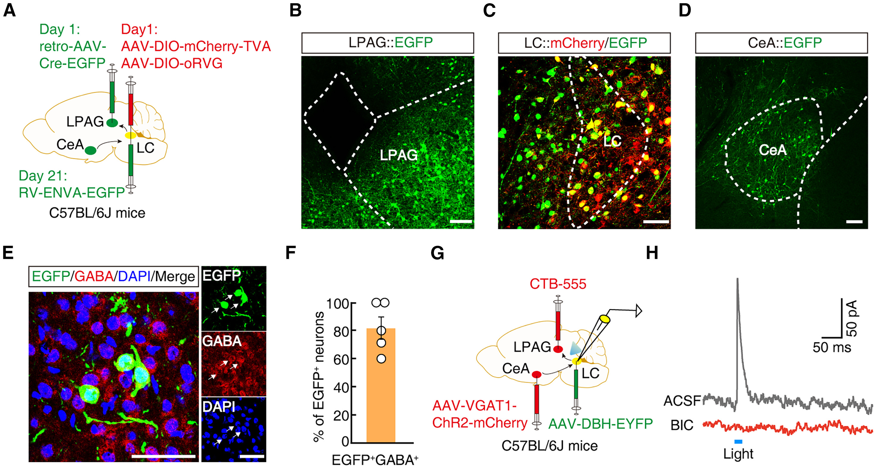
To directly verify that GABACeA, NELC, and GluLPAG form a continuous disynaptic pathway, the authors performed a triple-viral tracing experiment (Figure 6A): retro-AAV-Cre-EGFP was injected into the LPAG while Cre-dependent rabies helper viruses (AAV-DIO-mCherry-TVA and AAV-DIO-oRVG) were co-injected into the ipsilateral LC, followed 3 weeks later by RV-ENVA-EGFP in the LC (Figures 6B and 6C). Rabies virus monosynaptically traced from LPAG-projecting LC starter neurons labeled a substantial population of upstream neurons in the CeA, ~80% of which were immunopositive for GABA (Figures 6D–6F).
Finally, the authors injected AAV-VGAT1-ChR2-mCherry into the CeA, AAV-DBH-EYFP into the LC, and the retrograde tracer CTB-555 into the LPAG (Figure 6G). Whole-cell patch-clamp recordings from dual-labeled (EYFP+ and CTB-555+) LPAG-projecting NELC neurons in acute LC slices demonstrated that blue light stimulation of ChR2-expressing GABACeA terminals evoked robust, bicuculline-sensitive IPSCs (Figure 6H). Collectively, these findings establish a complete disynaptic GABACeA → NELC → GluLPAG circuit mechanism explaining how chronic stress selectively gates nociceptive hypersensitivity and pain aversion independently of depression-like behaviors.
Source: Jing Wei et al. · Cell Reports · © Elsevier Inc.
Original figure and English caption ↗