How can the vagus nerve soften pain? A brainstem signal is reshaped
A mouse brainstem population links withdrawal, avoidance and dopamine dips. Vagus nerve stimulation recruits local inhibition to dampen pain-related responses.
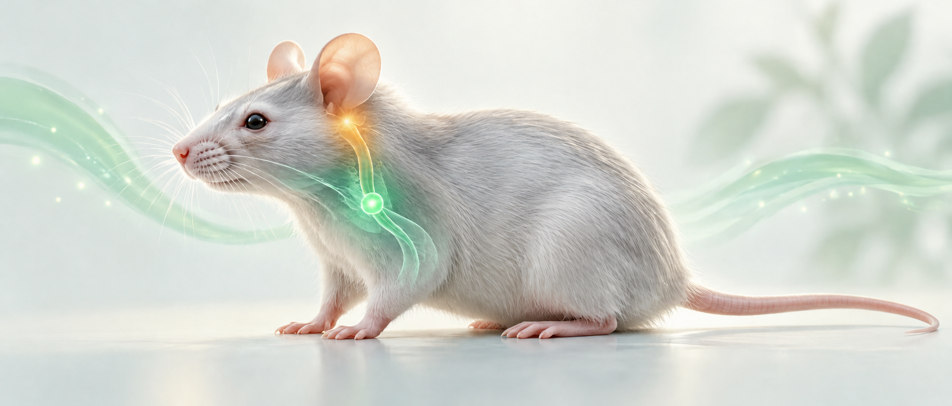
Pain brings rapid defensive movements and avoidance of the place associated with harm. How does one noxious event acquire both a bodily response and an aversive quality? And how can the vagus nerve, a major link between viscera and brain, influence this process? A study published in Nature Neuroscience on June 3, 2026 focuses on the caudal nucleus of the solitary tract in the mouse brainstem. A population projecting to the periaqueductal gray responds to noxious inputs and acquires responses to predictive cues during learning. Vagus nerve stimulation recruits nearby inhibitory neurons to dampen selected pain-related signals.
PAG-projecting neurons in the caudal solitary tract nucleus form a node for vagal modulation of mechanical nociception and pain-related aversion. Local inhibition accompanies reduced noxious-evoked activity and smaller accumbal dopamine dips.
Contents · 7 sections
A brainstem population recruits more than withdrawal
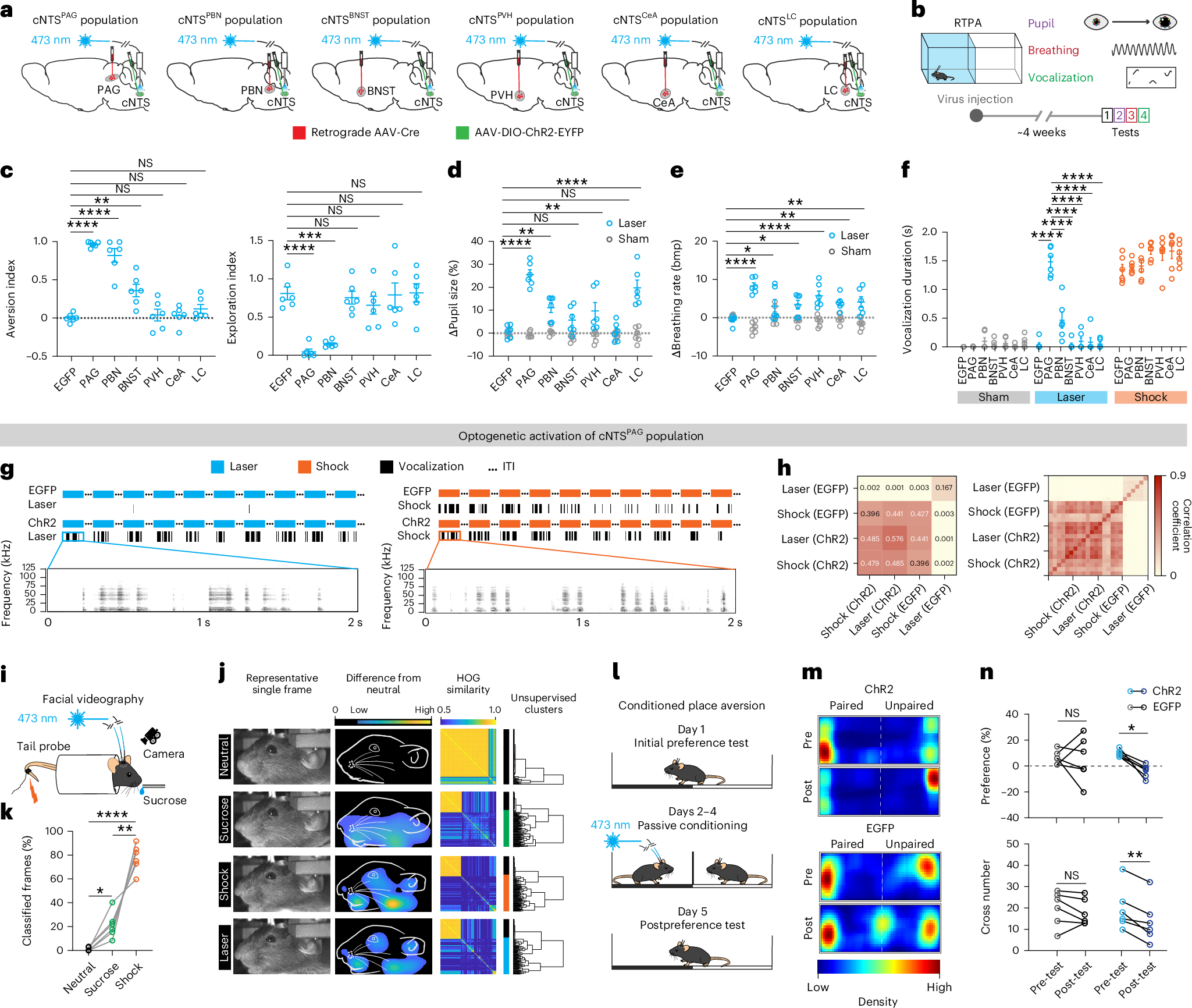
The caudal solitary tract nucleus is often discussed in relation to visceral signals, but it also receives somatic noxious information. Comparing six output-defined populations revealed that activation of PAG-projecting neurons produced place avoidance alongside changes in pupils, breathing and vocalization. Facial responses also resembled those evoked by tail shock.
Place avoidance alone can reflect different forms of discomfort. Comparisons of vocal and facial patterns further connected this population to pain-related responses. Pairing stimulation with a context also changed subsequent chamber preference, linking the brainstem population to defensive behavior, aversion and experience-dependent responses.
Source: Yuan Tang et al. · Nature Neuroscience ·
Original figure and English caption ↗
Pinch and heat remain distinguishable in population activity
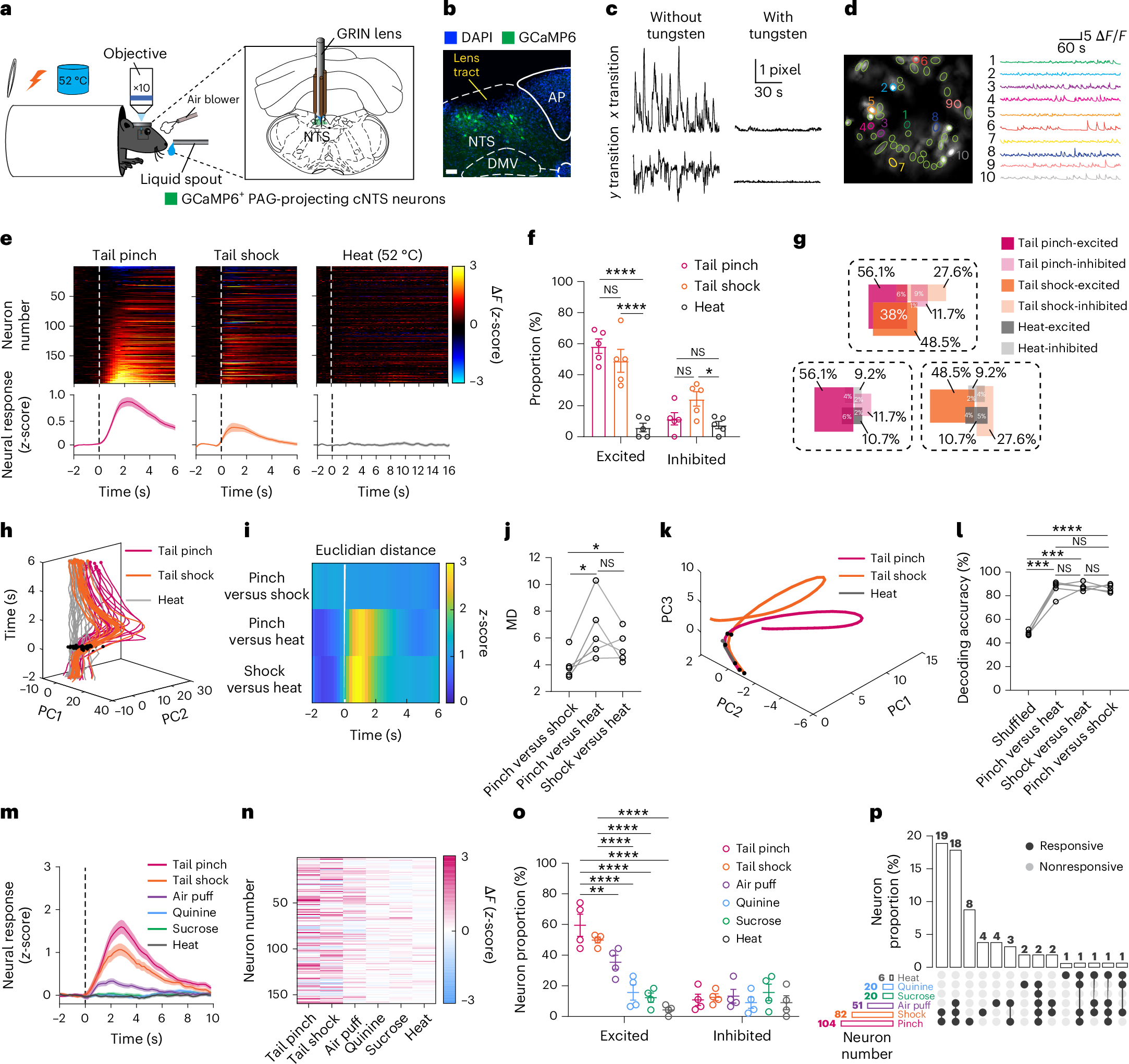
A uniform alarm would carry little information about the incoming event. Recordings instead showed different neuronal response combinations for tail pinch, tail shock and heat. Some neurons increased their activity while others decreased it, allowing the population to retain distinctions among noxious inputs.
Responses to air puff, bitter and sweet solutions were also heterogeneous, with overlap among responsive groups. This organization combines stimulus discrimination with behavioral relevance rather than acting as a switch for every event. It also provides a basis for asking how vagal stimulation changes the population pattern.
Source: Yuan Tang et al. · Nature Neuroscience ·
Original figure and English caption ↗
A predictive sound begins to recruit the population
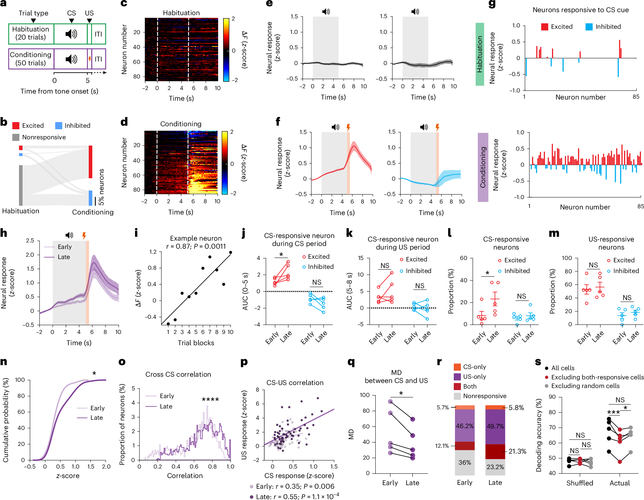
Pain-related responses can begin before harm itself. Pairing a sound with a later tail shock strengthened cue-evoked activity in a subset of neurons as training progressed. Population responses to the cue and the noxious event also became more similar.
Cue responses increased between early and late training, while the analyzed shock responses did not significantly change. Repeated sound presentation without shock did not produce the same population response. The brainstem population thus incorporates pairing experience into anticipatory activity, extending its role beyond immediate sensory relay.
Source: Yuan Tang et al. · Nature Neuroscience ·
Original figure and English caption ↗
Spinal inputs define a functionally selective route
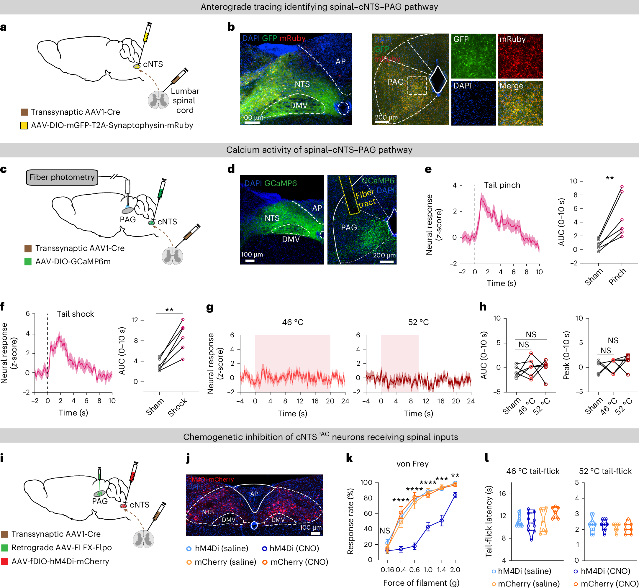
The study traced cNTS neurons receiving spinal input and projecting to PAG. This route responded to pinch and shock. Inhibiting the input-defined population reduced paw withdrawal at several mechanical stimulus strengths, while changes at the weakest tested level were not significant.
Thermal responses showed a different pattern. Tail-flick latency in hot water and measured hot-plate responses did not significantly change, and locomotor measures were also unaffected. This separation indicates modality-selective involvement of the spinal-input-defined population rather than equivalent control over every noxious input.
Source: Yuan Tang et al. · Nature Neuroscience ·
Original figure and English caption ↗
Vagal stimulation dampens selected responses
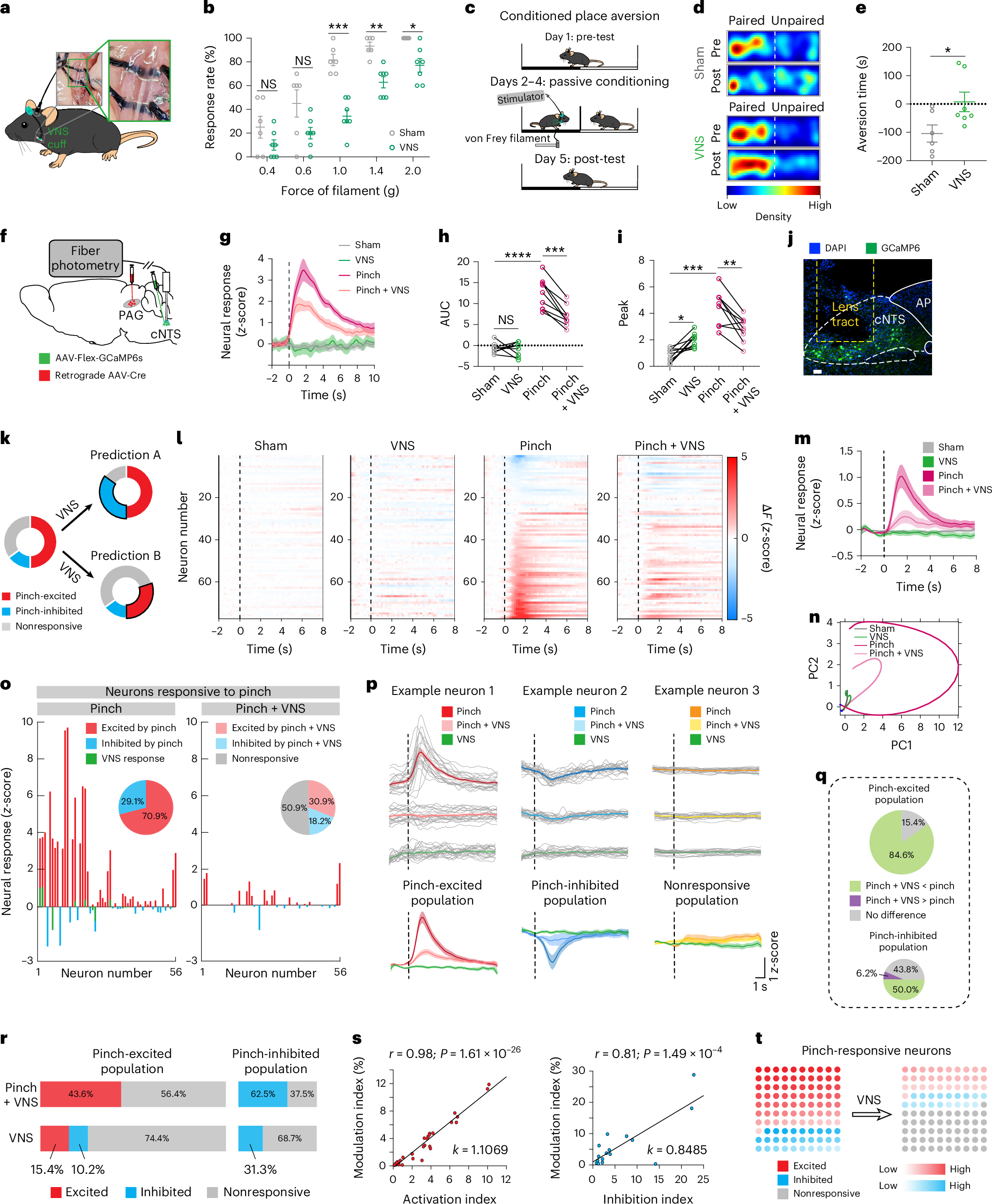
Vagus nerve stimulation reduced withdrawal to stronger mechanical stimuli and weakened aversion to a context paired with mechanical stimulation. Concurrent recordings showed smaller pinch-evoked cNTS responses. At single-cell resolution, many pinch-excited neurons showed attenuated responses.
Modulation was heterogeneous rather than a uniform shutdown. Responses persisted in some neurons, and pinch-inhibited cells were also modulated to different extents. Vagal stimulation reshaped the population pattern. Under the experimental parameters, rotarod performance and measured open-space exploration did not significantly change.
Source: Yuan Tang et al. · Nature Neuroscience ·
Original figure and English caption ↗
Nearby inhibitory neurons help apply the brake
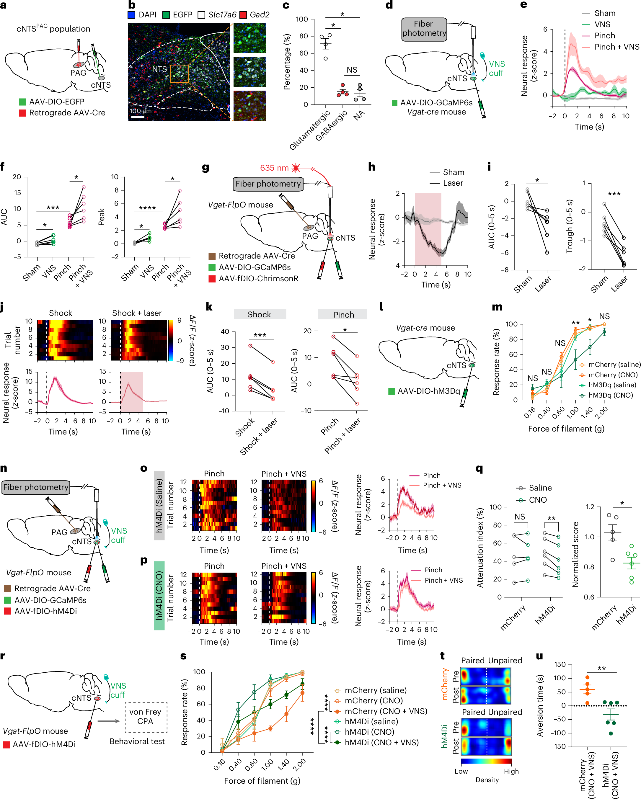
The mechanism points to local GABAergic neurons. Vagal stimulation increased their activity, and activating them suppressed the PAG-projecting population and attenuated noxious-evoked responses. Activating local inhibitory neurons also reduced responses to selected mechanical stimulus strengths.
Conversely, inhibiting these GABAergic neurons weakened vagal attenuation of pinch-evoked activity and reduced its effects on mechanical responses and associated place aversion. Together, these manipulations place local inhibition within the mechanism by which vagal stimulation modulates the noxious-response pathway.
Source: Yuan Tang et al. · Nature Neuroscience ·
Original figure and English caption ↗
Brainstem modulation reaches pain-associated dopamine dips
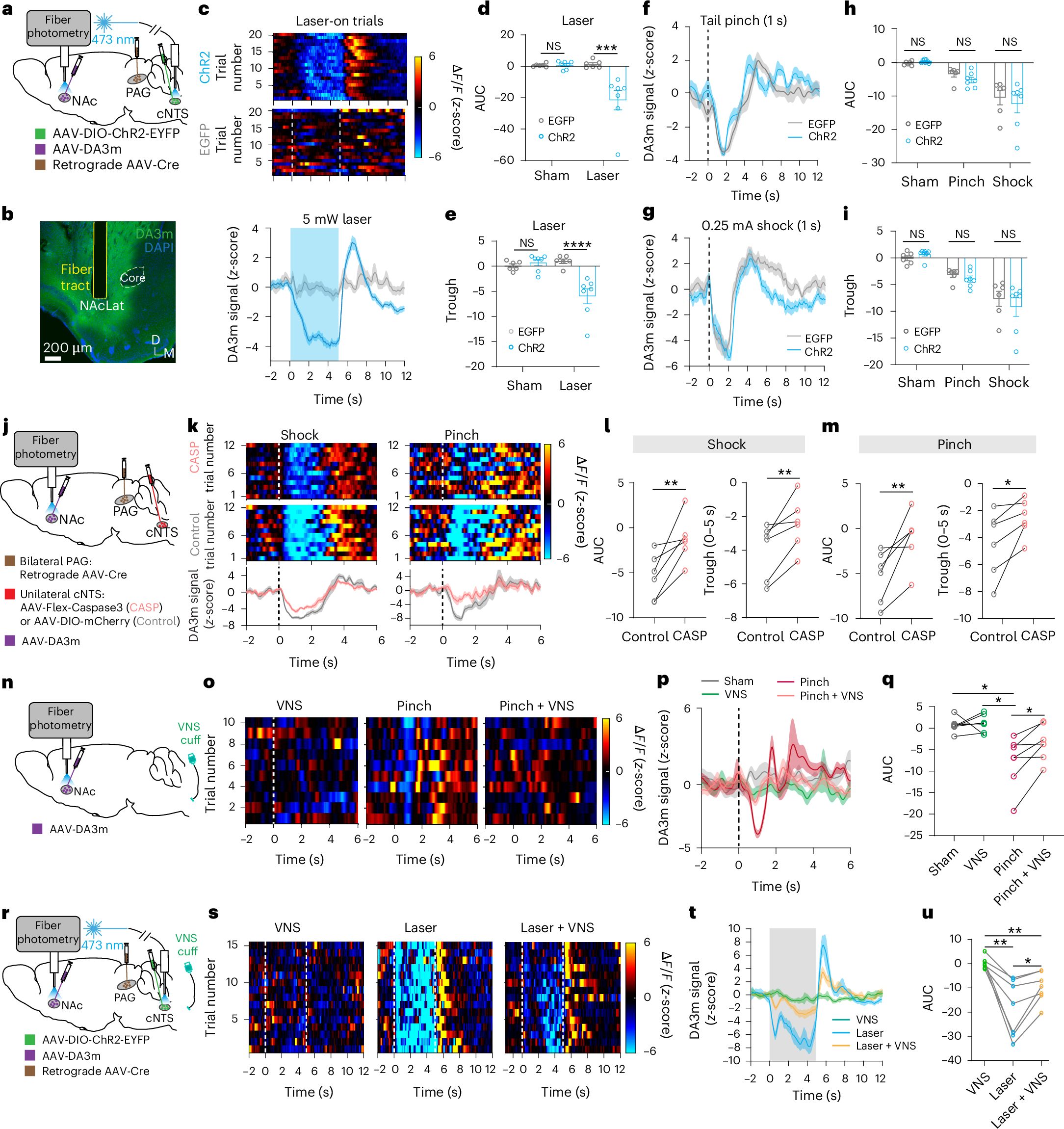
Noxious inputs also alter signals associated with motivation and aversion. Pinch, shock and activation of PAG-projecting cNTS neurons accompanied dopamine dips in the lateral accumbal shell. Unilateral removal of this cNTS population reduced noxious-evoked dopamine dips in the accumbal hemisphere on the ablated side, connecting the population to downstream signal changes.
Vagal stimulation attenuated dopamine dips evoked by pinch and by activation of the brainstem population. Stimulating its terminals in VTA produced dopamine decreases and place avoidance, whereas the CeA comparison did not show the same significant effects. These mouse findings connect somatic input, local inhibition and downstream dopamine changes, identifying a circuit-level basis for more targeted investigation of vagal pain modulation.
Source: Yuan Tang et al. · Nature Neuroscience ·
Original figure and English caption ↗