Why does a thalamic pathway alter hypersensitivity without changing avoidance?
In male nitroglycerin-model mice, posterior paraventricular thalamic–amygdala populations contribute to mechanical hypersensitivity, with different cellular dependence for tested anxiety-like behavior.
Greater mechanical responsiveness and reduced open-area exploration can coexist in one pain model. Are they driven by the same circuit? A study in The Journal of Headache and Pain follows posterior paraventricular thalamus, or pPVT, input to central amygdala, or CeA, in repeatedly nitroglycerin-treated male mice. Broad CeA GABA-cell inhibition improves hypersensitivity and some anxiety-like measures. Restricting manipulation to thalamic-input-related populations improves mechanical responses without significant anxiety-like changes.
Inhibiting pPVT glutamatergic cells, CeA-projecting pPVT cells or pPVT-recipient CeA GABA cells improves paw and periorbital hypersensitivity without significant improvement in tested anxiety-like measures.
Contents · 6 sections
Sensitivity and exploration do not change every measure
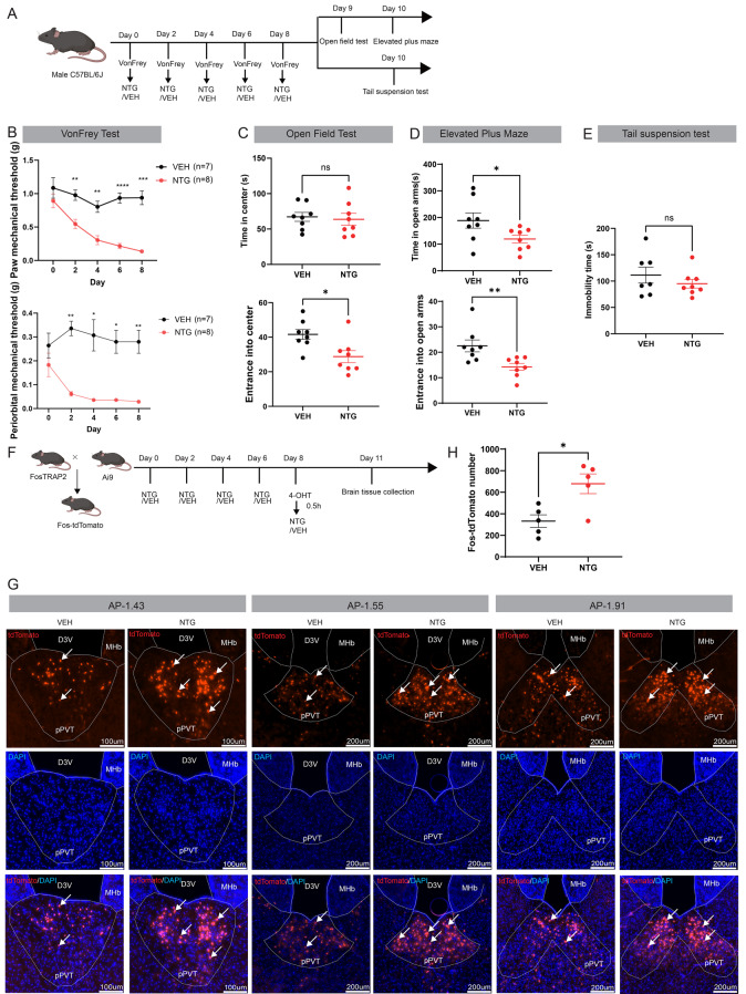
Repeated NTG lowers male-mouse paw and periorbital thresholds. Open-field center entries decrease without significant center-duration changes; elevated-plus-maze open-arm duration and entries fall, while tail-suspension immobility does not significantly rise. Hypersensitivity accompanies some, but not all, affect-related changes.
Because PVT has substantial baseline activity, temporally controlled activity labeling compares model and control animals. More labeled pPVT cells indicate greater recruitment during the selected window. This midline thalamic area becomes the starting point for examining hypersensitivity.
Source: Na W et al. · The journal of headache and pain · CC BY-NC-ND
Original figure and English caption ↗
Lower pPVT activity improves hypersensitivity
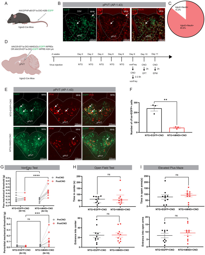
Labeling identifies a predominantly Vglut2 glutamatergic pPVT population. Inhibiting it after model establishment improves paw and periorbital thresholds without significant open-field or plus-maze improvement. The effect centers on mechanical responses rather than every associated behavior.
The result narrows the question. pPVT contributes to hypersensitivity but has many outputs, potentially serving distinct functions at different targets. Tracing and downstream activity comparisons identify which target stands out in the model.
Both periorbital and paw thresholds improve, extending the sensory effect beyond one facial measurement site. Open-space choices do not significantly recover, making the distinction between coexisting sensory and exploration measures clearer.
Source: Na W et al. · The journal of headache and pain · CC BY-NC-ND
Original figure and English caption ↗
Lateral and capsular CeA stand out
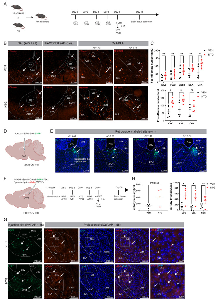
pPVT projects to accumbens, bed nucleus, interstitial nucleus of the posterior anterior-commissure limb, and basolateral and central amygdala. Activity labeling increases in CeA but not significantly in the other tested regions. Capsular CeC and lateral CeL changes are significant, unlike medial CeM.
Tracing activity-tagged pPVT cells finds stronger terminal fluorescence in CeC and CeL. The combined CeA increase is a nonsignificant trend, with no significant CeM change. Regional analysis places connectivity and activity clues in the same two subdivisions.
Source: Na W et al. · The journal of headache and pain · CC BY-NC-ND
Original figure and English caption ↗
Broader CeA inhibition affects both dimensions
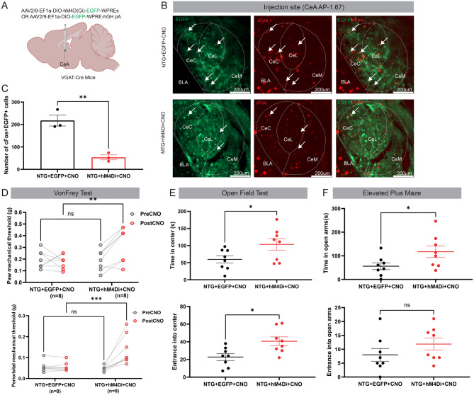
CeA is predominantly GABAergic. Inhibiting its broader VGAT-labeled population after modeling improves thresholds, increases open-field center duration and entries, and increases open-arm time. The open-arm entry trend is nonsignificant. Broader inhibition changes both sensory and some exploration measures.
This differs from pPVT inhibition. CeA receives diverse inputs and contains varied populations. Broadly suppressing its dominant cell class may therefore differ from suppressing cells associated with one input. The next tests restrict manipulation to the thalamic–amygdala relationship.
Source: Na W et al. · The journal of headache and pain · CC BY-NC-ND
Original figure and English caption ↗
Thalamic-terminal stimulation raises GABA-population calcium
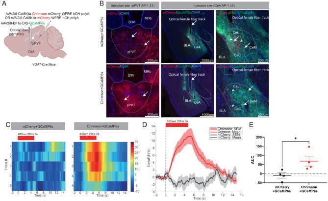
The local CeA inhibitory network makes net responses difficult to predict from glutamatergic input alone. Stimulating pPVT terminals within CeA raises GABA-cell calcium and response area, demonstrating recruitment of the downstream population.
Tracing identifies the route and responses show its influence on recipients. Together they motivate behavioral tests at each end: CeA-projecting pPVT cells and pPVT-recipient CeA GABA cells.
Source: Na W et al. · The journal of headache and pain · CC BY-NC-ND
Original figure and English caption ↗
Mechanical effects persist with route-defined targeting
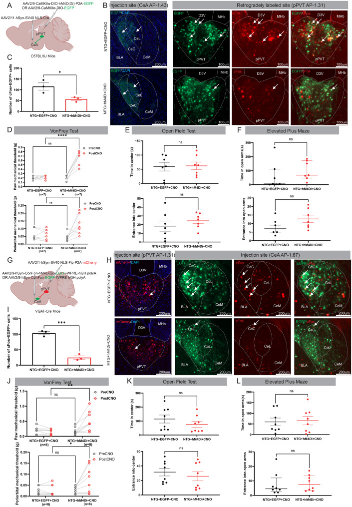
Inhibiting CeA-projecting glutamatergic-associated pPVT cells improves paw and periorbital hypersensitivity without significant exploration changes. Inhibiting pPVT-recipient CeA GABA cells yields the same dissociation: higher thresholds without significant anxiety-like improvement. Both ends give aligned outcomes.
In male NTG-model mice, hypersensitivity and avoidance coexist with different cellular dependence. Broader CeA inhibition affects both dimensions, while thalamic-input-associated populations primarily affect mechanical responses. Function depends on selected inputs and cells within active regions.
Source: Na W et al. · The journal of headache and pain · CC BY-NC-ND
Original figure and English caption ↗