Why can bright light ease scratching? Tracing a visual pathway from retina to parabrachial nucleus
A mouse study traces a pathway from retinal ganglion cells through the visual thalamus to the lateral parabrachial nucleus, explaining how bright light dampens itch-related activity.
Bright light treatment is a non-invasive physical intervention known to modulate mood, spatial memory, sleep, and pain perception, and pilot clinical observations have suggested its potential to relieve pruritus. Yet how ambient bright light signals are routed from the visual system to central itch-processing nuclei to suppress scratching behaviors has remained unknown. Published in Cell Reports in June 2024, Chaoran Ren, Qian Tao, Lu Huang, and colleagues at the First Affiliated Hospital and GHM Institute of CNS Regeneration of Jinan University delineate a three-node retina–ventral lateral geniculate nucleus/intergeniculate leaflet (vLGN/IGL)–lateral parabrachial nucleus (LPBN) circuit in mice. The authors demonstrate that ON-type SMI-32-positive M4 melanopsin-expressing retinal ganglion cells (mRGCs) directly innervate GABAergic neurons in the vLGN/IGL, which in turn send monosynaptic inhibitory projections to CaMKIIα-positive excitatory neurons in the LPBN. Exposure to bright light (≥3,000 lux) activates this retina–vLGN/IGL–LPBN pathway to dampen pruritogen-evoked LPBN neuronal excitation, alleviating both histamine-dependent and histamine-independent acute scratching as well as itch-associated conditioned place aversion.
Bright light exposure (≥3,000 lux) activates ON-type SMI-32-positive M4 retinal ganglion cells that innervate vLGN/IGL GABAergic neurons, which monosynaptically inhibit LPBN CaMKIIα-positive neurons to attenuate pruritogen-induced LPBN excitation and reduce acute scratching and itch-related aversion.
Contents · 6 sections
vLGN/IGL GABAergic Neurons Monosynaptically Inhibit LPBN CaMKIIα+ Excitatory Neurons
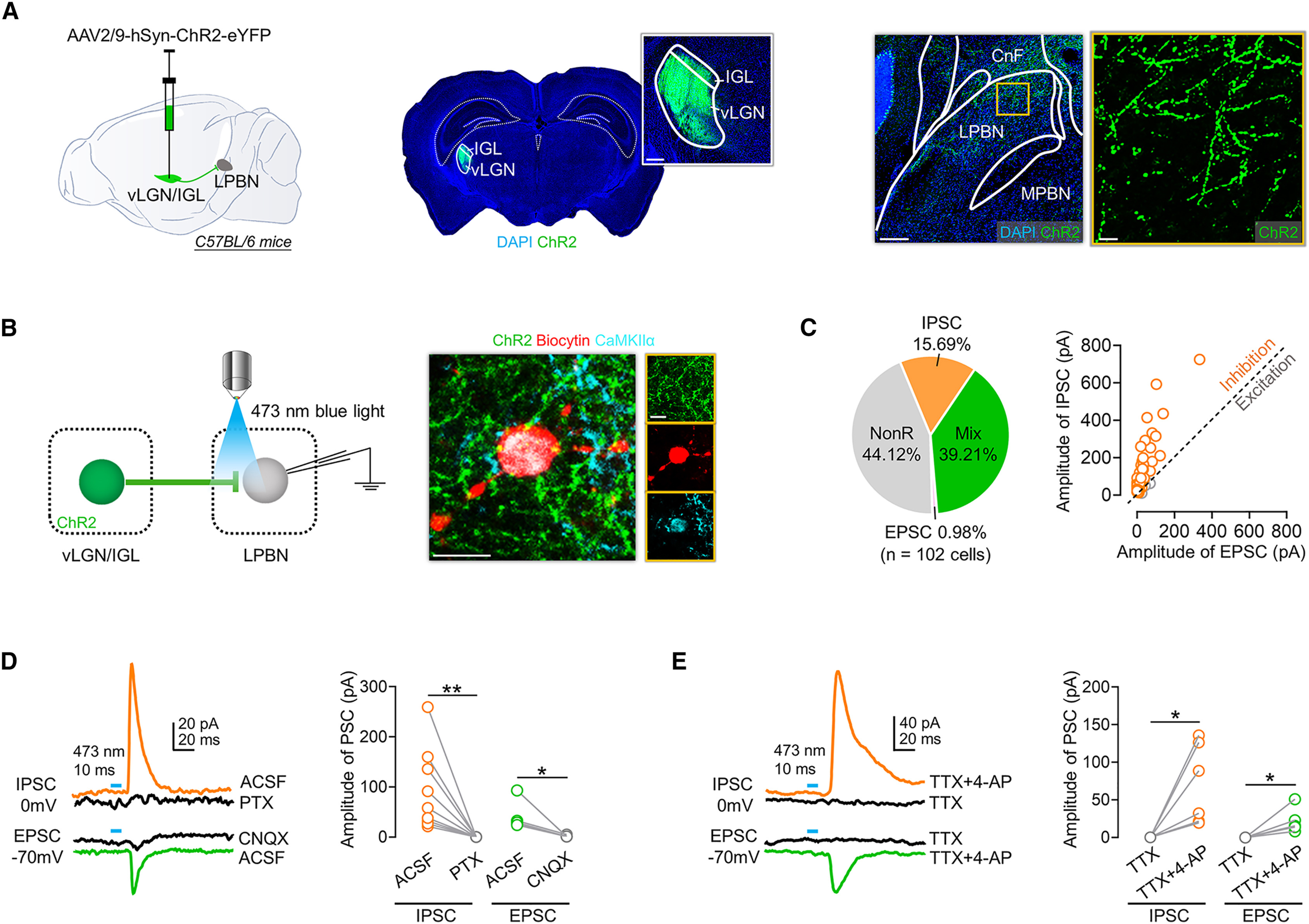
The lateral parabrachial nucleus (LPBN) is a primary supraspinal hub that receives spinal itch signals and broadcasts pruritic information to forebrain and brainstem targets. Using anterograde and retrograde viral tracing in C57BL/6 mice, the authors first mapped the anatomical organization of the vLGN/IGL-to-LPBN pathway. Injecting AAV2/9-hSyn-ChR2-eYFP into the vLGN/IGL revealed dense axonal projections in the LPBN and sparse fibers in the medial PBN (MPBN). Retrograde labeling via angled rAAV2/2-Retro-Cre injection into the LPBN (avoiding the cuneiform nucleus) combined with AAV2/9-DIO-eYFP in the vLGN/IGL showed that LPBN-projecting neurons resided mainly in the medial and internal vLGN/IGL, with 97.38% immunopositive for GABA. Conversely, anterograde trans-monosynaptic tagging with AAV2/1-Cre in the vLGN/IGL and AAV2/9-DIO-mCherry in the LPBN demonstrated that postsynaptic neurons were concentrated in the dorsal LPBN (dLPBN), with 96.05% expressing CaMKIIα and projecting downstream to itch-modulatory regions including the central medial thalamus (CM), central amygdala (CeA), lateral/ventrolateral periaqueductal gray (l/vlPAG), and rostral ventromedial medulla (RVM).
Whole-cell patch-clamp recordings in acute LPBN slices defined the physiological properties of this projection. Optogenetic activation (473 nm) of ChR2-expressing vLGN/IGL terminals across 102 recorded LPBN neurons evoked purely inhibitory postsynaptic currents (IPSCs) in 15.69% of cells and mixed IPSCs/EPSCs in 39.21% of cells, with IPSC amplitudes markedly exceeding EPSC amplitudes. Evoked IPSCs and EPSCs were blocked by the GABAA receptor antagonist picrotoxin (PTX) and the AMPA/kainate antagonist CNQX, respectively, and were abolished by tetrodotoxin (TTX) but restored by TTX plus 4-aminopyridine (4-AP), confirming direct monosynaptic predominantly inhibitory transmission from vLGN/IGL GABAergic neurons onto LPBN CaMKIIα+ neurons.
Source: et al. · Cell Reports · Original figure reproduced for academic literature review under fair scholarly commentary; copyright remains with the authors/publisher.
Original figure and English caption ↗
Activating the vLGN/IGL–LPBN Pathway Dampens Pruritogen-Evoked LPBN Calcium Transients and Alleviates Itch Behaviors
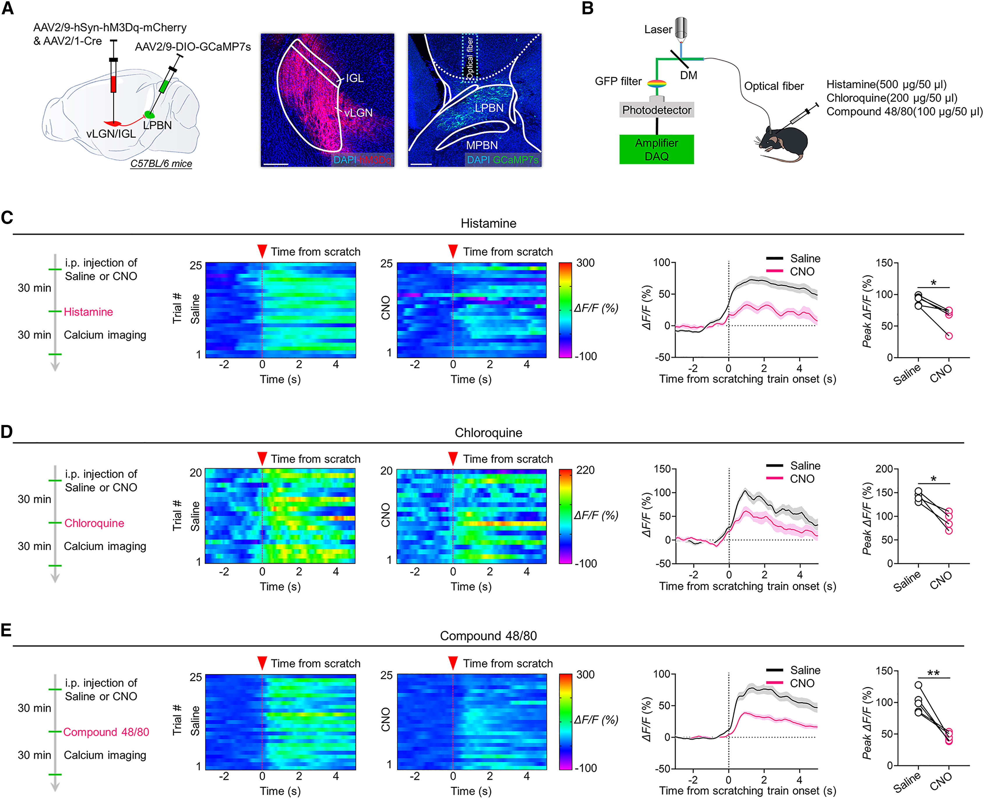
To test in vivo whether vLGN/IGL activation dampens pruritogen-evoked excitation of LPBN neurons, the team co-injected anterograde trans-synaptic AAV2/1-Cre and AAV2/9-hSyn-hM3Dq-mCherry into the vLGN/IGL alongside Cre-dependent AAV2/9-DIO-GCaMP7s in the LPBN, monitoring postsynaptic calcium signals via fiber photometry (470 nm signal and 410 nm isosbestic reference). Intradermal nape injection of histamine, chloroquine, or compound 48/80 triggered robust Ca2+ signal increases (ΔF/F) in vLGN/IGL-recipient LPBN neurons time-locked to scratching onset, whereas chemogenetic activation of vLGN/IGL neurons with intraperitoneal CNO (1 mg/kg) significantly attenuated these pruritogen-evoked calcium surges across all three itch models.
Behavioral manipulations confirmed pathway-specific antipruritic efficacy: bilateral optogenetic stimulation of vLGN/IGL-to-LPBN terminals significantly reduced histamine-induced scratching bouts without altering anxiety-like behaviors in the open-field or elevated plus maze tests. By contrast, optogenetic activation of vLGN/IGL-to-l/vlPAG terminals—previously shown to mediate bright-light analgesia—did not affect histamine-induced scratching, revealing distinct downstream pathways for pain versus itch modulation. Moreover, projection-specific chemogenetic activation of vLGN/IGL-to-LPBN projections (via intra-LPBN CNO infusion) markedly reduced scratching bouts and duration evoked by histamine, chloroquine, or compound 48/80, and significantly attenuated histamine-induced conditioned place aversion (CPA), demonstrating relief of both sensory scratching and itch-related negative affect.
Source: et al. · Cell Reports · Original figure reproduced for academic literature review under fair scholarly commentary; copyright remains with the authors/publisher.
Original figure and English caption ↗
Retinal M4-Type Melanopsin Ganglion Cells Disynaptically Innervate the LPBN and Suppress Itch Behaviors
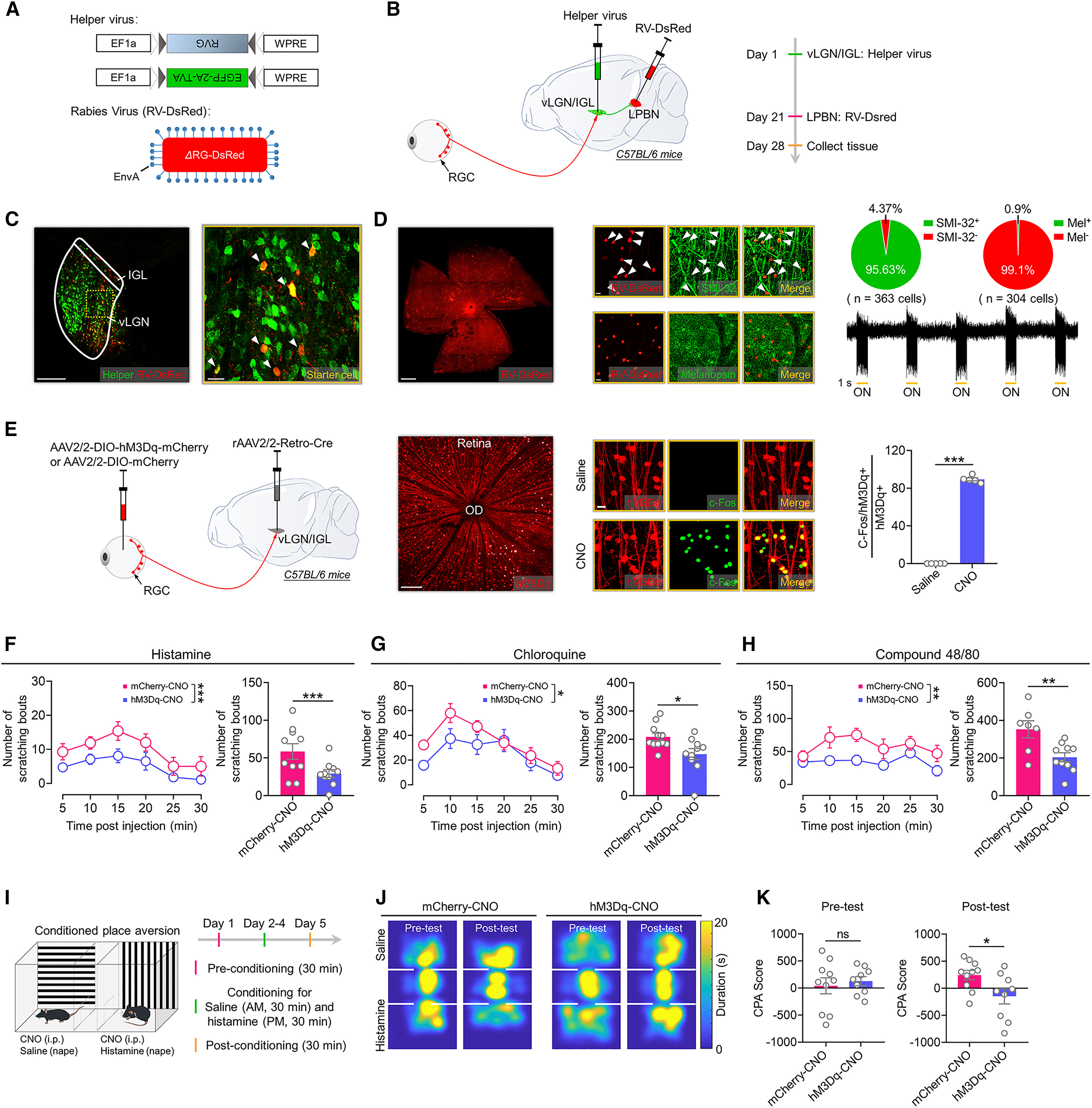
To identify the specific retinal ganglion cell (RGC) subtype innervating LPBN-projecting vLGN/IGL neurons, the authors performed modified rabies-virus-based disynaptic retrograde tracing by injecting helper virus (TVA and RVG) into the vLGN/IGL followed 3 weeks later by SAD-ΔG-DsRed (EnvA) into the LPBN. Doubly infected vLGN/IGL starter cells trans-synaptically labeled 617 ± 28 RGCs per retina. Whole-mount retinal immunostaining and extracellular recordings revealed that 95.63% of rabies-labeled RGCs were immunopositive for SMI-32 and all recorded cells (32/32) exhibited sustained ON-type depolarization to light flashes. Disynaptic tracing in Opn4-Cre mice further confirmed that ~81.3% of rabies-labeled RGCs co-labeled with melanopsin-expressing RGCs (mRGCs), matching the morphological and physiological profile of M4-type mRGCs.
Because RGCs release excitatory glutamate onto vLGN/IGL targets, the researchers tested whether selectively activating vLGN/IGL-projecting RGCs suppresses itch. Delivering rAAV2/2-Retro-Cre into the bilateral vLGN/IGL and AAV2/2-DIO-hM3Dq-mCherry intraocularly into both eyes enabled selective chemogenetic activation of vLGN/IGL-projecting RGCs upon intraperitoneal CNO administration. Activating these RGCs significantly reduced scratching bouts and duration induced by histamine, chloroquine, or compound 48/80 and attenuated histamine-induced CPA, establishing a functional retina–vLGN/IGL–LPBN antipruritic circuit.
Source: et al. · Cell Reports · Original figure reproduced for academic literature review under fair scholarly commentary; copyright remains with the authors/publisher.
Original figure and English caption ↗
Bright Light Exposure (3,000 Lux) Suppresses LPBN Neuronal Excitation and Relieves Acute Pruritus
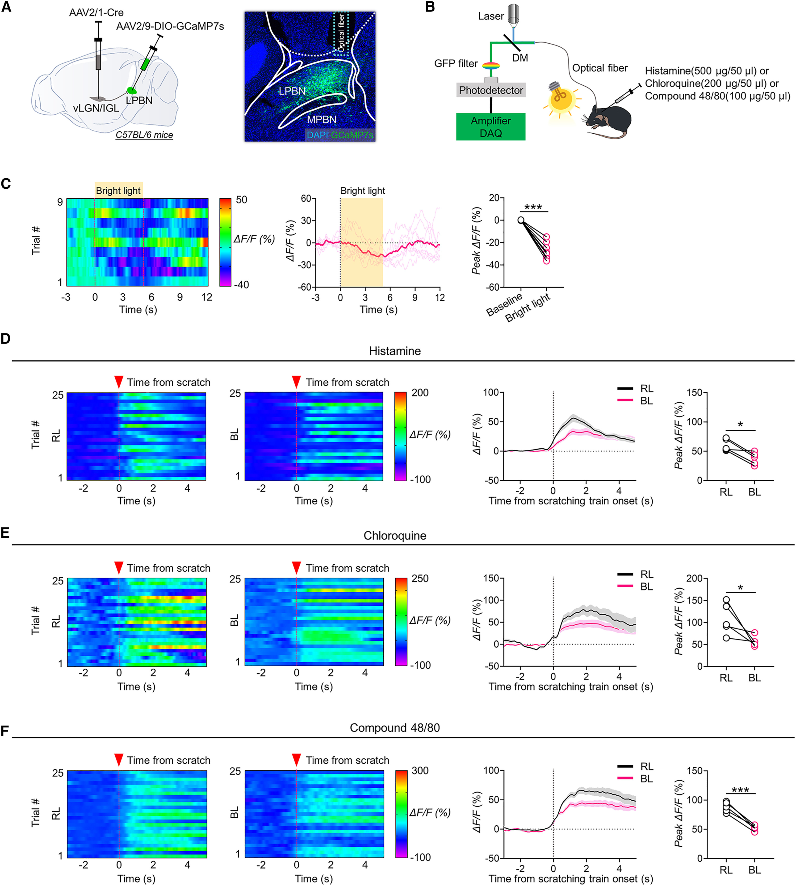
Given that ON-type RGCs drive the vLGN/IGL–LPBN pathway, the team next examined whether ambient bright light exposure suppresses LPBN neuronal activity and itch behavior. In vivo fiber photometry in vLGN/IGL-recipient LPBN neurons expressing GCaMP7s revealed that exposure to UV-free bright light (3,000 lux), compared with ambient room light (200 lux), significantly lowered baseline Ca2+ signal amplitude and markedly blunted scratching-associated Ca2+ transients evoked by histamine, chloroquine, or compound 48/80 within the same animals.
Testing graded light intensities (0, 200, 1,000, 3,000, and 5,000 lux) against histamine-induced scratching revealed an intensity-dependent antipruritic effect requiring a minimum threshold of 3,000 lux for significant relief. Exposure to 3,000 lux bright light also significantly reduced scratching bouts and duration triggered by chloroquine or compound 48/80 and attenuated histamine-induced CPA without affecting anxiety-like behaviors in the open-field or elevated plus maze tests. Notably, acute 3,000 lux exposure did not alleviate chronic pruritus induced by 4 weeks of repeated 1-fluoro-2,4-dinitrobenzene (DNFB) sensitization, suggesting potential differences in LPBN neuronal subpopulations or circuit plasticity between acute and chronic itch.
Source: et al. · Cell Reports · Original figure reproduced for academic literature review under fair scholarly commentary; copyright remains with the authors/publisher.
Original figure and English caption ↗
Stepwise Inhibition of the Retina–vLGN/IGL–LPBN Pathway Abolishes the Antipruritic Effects of Bright Light
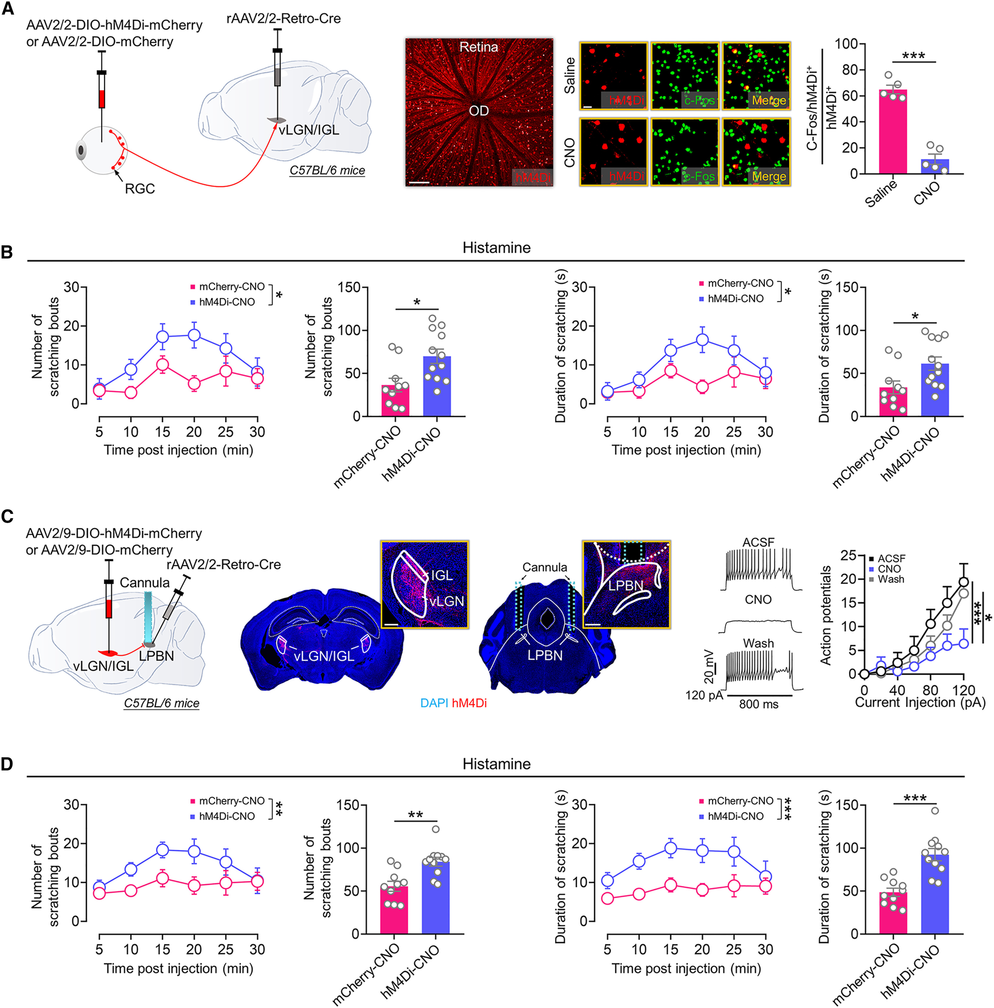
To rigorously test whether the retina–vLGN/IGL–LPBN circuit is required for bright-light antipruritic action, the authors performed stepwise loss-of-function manipulations across all three nodes during 3,000 lux exposure. First, chemogenetic inhibition of vLGN/IGL-projecting RGCs (via bilateral vLGN/IGL rAAV2/2-Retro-Cre and intraocular AAV2/2-DIO-hM4Di-mCherry plus i.p. CNO) suppressed bright-light-induced RGC c-Fos expression and abolished the antipruritic effect of 3,000 lux bright light on histamine-induced scratching, without altering baseline scratching under 200 lux room light.
Second, selective chemogenetic inhibition of vLGN/IGL-to-LPBN terminals (via bilateral LPBN rAAV2/2-Retro-Cre, vLGN/IGL AAV2/9-DIO-hM4Di-mCherry, and intra-LPBN CNO infusion) likewise negated bright-light-induced reductions in histamine scratching. Third, chemogenetically activating vLGN/IGL-recipient LPBN postsynaptic neurons (via vLGN/IGL AAV2/1-Cre and LPBN AAV2/9-DIO-hM3Dq-mCherry) during 3,000 lux exposure counteracted upstream inhibition and impaired the antipruritic effect of bright light. Together, these node-by-node manipulations prove that activation of the retina–vLGN/IGL–LPBN pathway is indispensable for the antipruritic effects of bright light treatment.
Source: et al. · Cell Reports · Original figure reproduced for academic literature review under fair scholarly commentary; copyright remains with the authors/publisher.
Original figure and English caption ↗
Parallel Non-Image-Forming Visual Circuits for Multisensory Modulation
Viewed alongside prior work on bright-light analgesia, antidepressant action, and sleep regulation, this study reveals a modular circuit logic within the non-image-forming visual system. While ON-type SMI-32+ M4 melanopsin RGCs funnel ambient bright light signals into the vLGN/IGL GABAergic hub, divergent downstream projections segregate sensory control: the vLGN/IGL-to-l/vlPAG pathway mediates antinociception without affecting histamine itch, whereas the newly defined vLGN/IGL-to-LPBN pathway directly silences excitatory LPBN neurons to suppress acute itch and itch-related aversion.
Because both the visual thalamus and parabrachial nucleus are evolutionarily conserved across mammals, elucidating the retina–vLGN/IGL–LPBN circuit expands our understanding of non-image-forming visual physiology and provides a neurobiological foundation for developing non-invasive phototherapy protocols to manage acute pruritus and its affective comorbidities.