The urge to scratch and to avoid: where does the lateral habenula fit?
Itch drives scratching and an aversive experience. A mouse study links excitatory lateral habenula cells to both, with inhibition changing scratching and place avoidance.
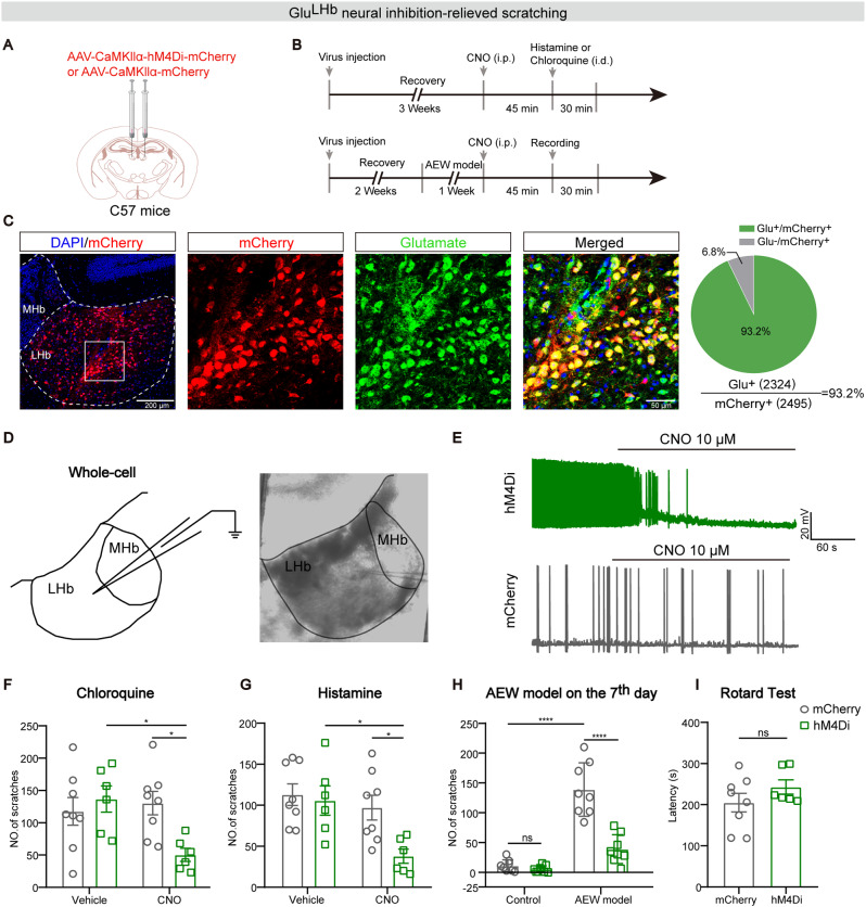
Itch is more than a skin signal: it drives scratching and can make an associated situation worth avoiding. How do sensation, action and aversion meet in the brain? A study published in Molecular Brain on June 27, 2023 examined the lateral habenula, a region associated with negative experiences. Scratching during acute and dry-skin-associated itch coincided with higher population activity. Inhibiting the relevant cells reduced scratching, while the usual within-group place-avoidance change was no longer detected.
Glutamatergic lateral habenula cells contribute to itch-related scratching and aversive learning in mice. Population recordings, excitability and inhibition results provide complementary evidence, while the cellular organization of sensory and affective components remains unresolved.
Contents · 6 sections
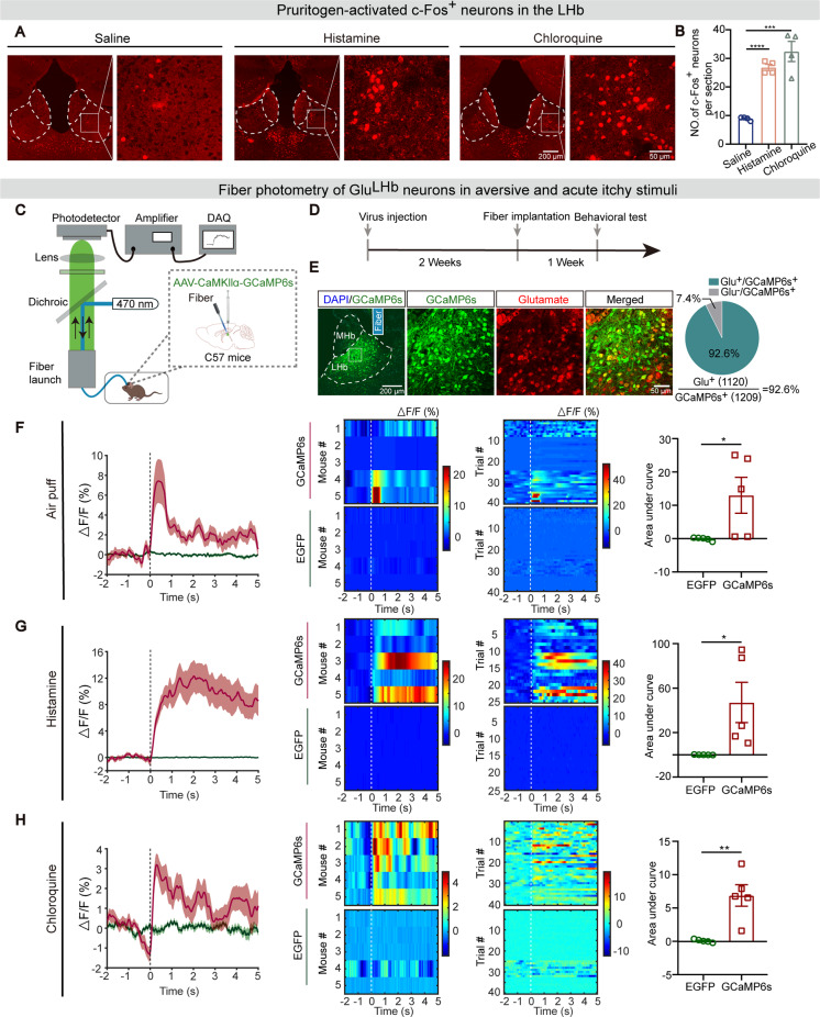
Two forms of acute itch accompany rising population signals
Both histamine-dependent and nonhistaminergic acute itch increased an activity-related marker in the lateral habenula. Behavior-aligned calcium recordings showed a brief rise in the relevant glutamatergic population around scratching onset, without the same response in fluorescence controls.
The temporal link places the lateral habenula within itch processing. Yet a population signal coincident with scratching does not separately quantify sensation, movement and aversion. Responses to another aversive stimulus also show that activity is not exclusive to itch.
Source: Chen R et al. · Molecular brain · CC BY
Original figure and English caption ↗
Dry-skin-associated itch accompanies altered neural responses
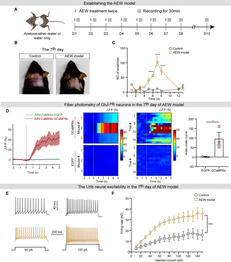
In the dry-skin-associated model, scratching increased, peaked and later declined during observation. At a prominent scratching stage, lateral habenula calcium signals rose around scratching, extending the association beyond a single acute event.
In slices, relevant neurons from model mice fired more in response to comparable injected current, supporting increased excitability. This stronger input response complements behavior-linked recordings without implying constant overactivity in every lateral habenula cell.
Source: Chen R et al. · Molecular brain · CC BY
Original figure and English caption ↗
Lowering cellular activity reduces scratching
Inhibition reduced scratching in both acute-itch conditions and spontaneous scratching in the dry-skin model, without the same change in marker controls. This extends the evidence beyond coincident activity to a functional influence on behavior.
Rotarod performance did not significantly change, arguing against a simple loss of motor ability. Scratching nevertheless remains a behavioral measure rather than a direct scale of subjective itch. Place-choice tests then addressed the aversive dimension from another angle.
Source: Chen R et al. · Molecular brain · CC BY
Original figure and English caption ↗
Would a mouse return to an itch-associated place?
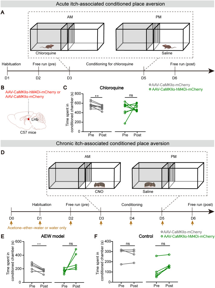
After itch was paired with a particular place, controls spent less time there. Inhibited mice did not show the same significant pre/post decline, a pattern observed in acute and dry-skin-associated conditions and consistent with a contribution to aversive association formation.
In controls without dry-skin itch, inhibition alone did not produce significant place preference or avoidance, limiting a simple added-reward explanation. Place choice still combines learning and experience rather than directly measuring a human emotional rating.
Source: Chen R et al. · Molecular brain · CC BY
Original figure and English caption ↗
A connection map is not yet a complete itch pathway
Anatomical labeling identifies several connected regions, including anterior cingulate and medial prefrontal cortex and lateral hypothalamus. These are candidate sources, not individually established itch-transmission pathways in this study. How sensory and aversive information meet requires more detailed functional evidence.
Understanding itch includes both action and aversion
The study places the lateral habenula at an intersection of itch-related scratching and aversive behavior. Its evidence comes from adult male mice and specific models, without defining the cellular subgroups for sensation versus affect or establishing human treatment efficacy. Those remain central questions for further work.
Timing also matters: inhibition accompanied the formation of the place–itch association. The findings therefore primarily concern acquisition of that aversive association. Whether later intervention would similarly influence an established, repeatedly recalled memory remains a separate open question.