Male mice become sensitive again in a pain-associated setting
Context-associated hypersensitivity involves hippocampus, free testosterone and a hypothalamic–midbrain connection. Females lack the same contextual change, but circuit activation can increase their sensitivity too.
Can returning to a place where pain occurred make the body sensitive again? A 2025 Nature Communications mouse study observes a context-associated response: males returning to a setting previously paired with abdominal discomfort show enhanced hindpaw mechanical and thermal sensitivity, whereas females lack the same clear change. The study follows hippocampus, free testosterone and a hypothalamic–midbrain connection to examine how environmental cues relate to sensory responses.
The tested contextual hypersensitivity accompanies increased excitatory mPOA activity and free testosterone in males. Reducing androgen receptors in these cells or weakening the connection to vlPAG alleviates context-associated sensitivity.
Contents · 7 sections
Similar initial responses, different next-day sensitivity
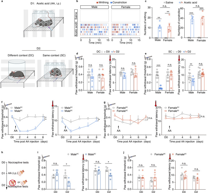
Mice experience abdominal stimulation in a particular setting, initially showing similar writhing and constriction in both sexes. On returning the next day, males have lower mechanical withdrawal thresholds and shorter thermal withdrawal latencies. A different setting, or female mice, does not produce the same clear change.
The male hypersensitivity persists for several days, and associated auditory cues produce a similar direction. The effect involves previous experience and returning cues rather than the environment making every animal more sensitive; tested anxiety-like behavior does not clearly change.
Source: Zhang M et al. · Nature communications · CC BY-NC-ND
Original figure and English caption ↗
The same setting recruits different population responses
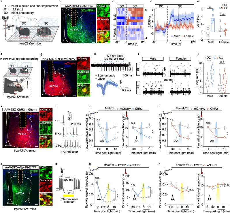
The medial preoptic area (mPOA) participates in behaviors influenced by hormonal state. Returning males show increased excitatory-population calcium and in-vivo firing, without the same clear female change. The earlier brief response to abdominal stimulation occurs in both sexes and subsides within hours.
Activating these cells increases sensitivity and induces place avoidance in both sexes in the different-context condition. Inhibition alleviates male contextual hypersensitivity but has no significant effect in females lacking the phenotype. Cellular capacity and natural contextual recruitment are distinct.
Source: Zhang M et al. · Nature communications · CC BY-NC-ND
Original figure and English caption ↗
Hormonal state changes the conditions for a contextual response
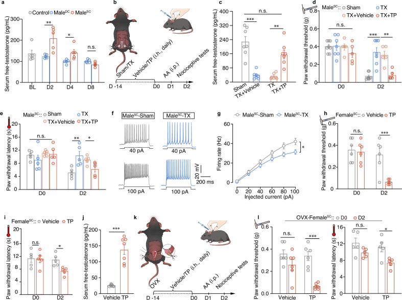
Free testosterone rises during male contextual hypersensitivity and later declines, paralleling cellular activity. This unbound fraction can engage receptor signaling, making the temporal relationship a candidate for further testing.
Reducing male testosterone accompanies weaker contextual hypersensitivity and lower mPOA activity; an analogue restores sensitivity. Supplemented females also develop mechanical sensitivity, whereas ovariectomy alone does not produce the same contextual change.
Source: Zhang M et al. · Nature communications · CC BY-NC-ND
Original figure and English caption ↗
Previous experience relates to hormonal and cellular responses
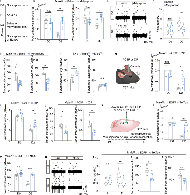
Dorsal hippocampal CA1 (dCA1) participates in contextual processing. Reducing its synaptic transmission alleviates male mechanical and thermal contextual sensitivity and lowers mPOA firing and both measured hormone levels. An intervention in contextual-processing circuitry changes sensory, hormonal and hypothalamic measures together.
A stress-related pharmacological intervention also reduces mechanical sensitivity along with corticosterone and free testosterone. The findings support contributions from context, stress and hormones, while each intervening hippocampal-to-hormonal step remains part of the proposed model.
Source: Zhang M et al. · Nature communications · CC BY-NC-ND
Original figure and English caption ↗
Androgen signaling finds a cellular target in mPOA
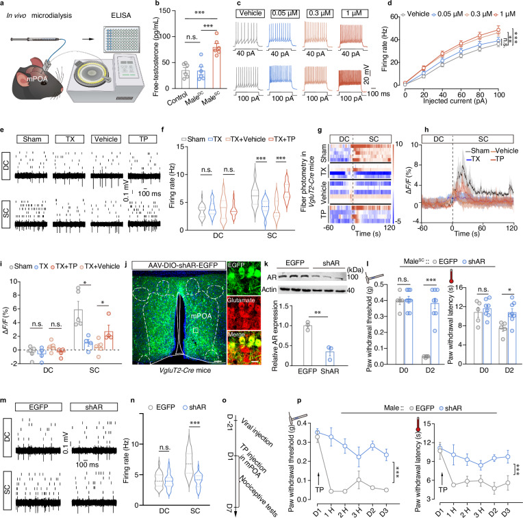
Free testosterone increases in male mPOA under the contextual condition. In slices, some tested analogue concentrations make excitatory cells fire more readily; spontaneous inhibitory-current frequency decreases while spontaneous excitatory-current frequency and amplitude show no significant changes.
Reducing androgen-receptor expression in these excitatory cells alleviates contextual mechanical and thermal sensitivity and decreases firing in the same setting. The hormonal relationship gains a cellular basis involving receptor expression and local neural activity as well as circulating signals.
Source: Zhang M et al. · Nature communications · CC BY-NC-ND
Original figure and English caption ↗
Excitatory input reaches inhibitory midbrain cells
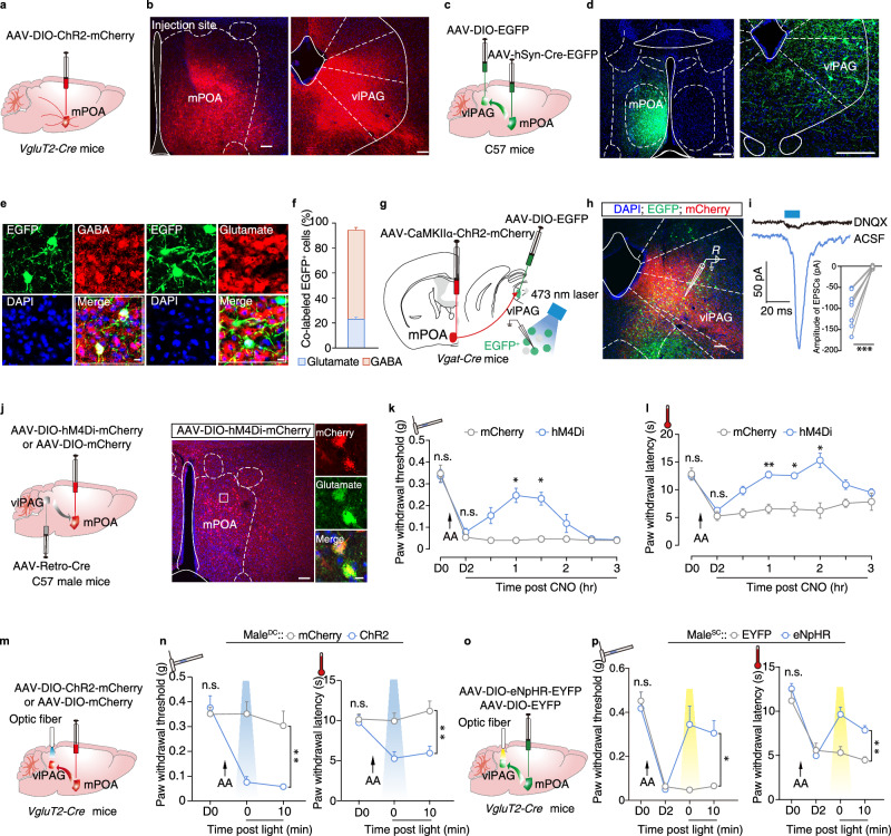
mPOA projects to several regions, with prominent labeling in ventrolateral periaqueductal gray (vlPAG). Input recipients are predominantly GABAergic and also include glutamatergic cells. Slice recordings confirm excitatory responses in vlPAG GABA cells during mPOA-terminal activation.
Inhibiting vlPAG-projecting cells or mPOA terminals there reduces male contextual hypersensitivity. Terminal activation increases sensitivity in both sexes, and anatomical labeling is similar. The difference involves recruitment rather than one sex simply lacking the connection.
Source: Zhang M et al. · Nature communications · CC BY-NC-ND
Original figure and English caption ↗
The same stimulus enters different brain states
This adult male-and-female mouse study shows how environmental cues, hormonal state and defined neural connections jointly influence responses after a sensory experience. Similar initial discomfort gives way to different sensitivity on returning to the same place, linking current responses with the cellular state recruited by that setting.