During pain sensitization, two cortical populations do not rise together
Nerve injury raises average anterior cingulate activity, but cells differ. Pyramidal firing increases while the interneuron population average does not change significantly, with richer variation at single-cell resolution.
When pain sensitivity increases, heightened cortical activity offers an intuitive explanation. But which cells are more active, and do excitatory and inhibitory populations increase together? A study published in Molecular Brain on January 29, 2022 examined the anterior cingulate cortex, ACC, in male mice. Increased average firing after nerve injury was concentrated in the putative pyramidal population, without a significant interneuron-average increase. Input-stimulation comparisons also revealed increased, decreased and stable cells within each population.
Activity labeling and in vivo firing both point to enhanced ACC pyramidal activity after injury without a matching increase in the interneuron population average. Strengthening relevant thalamic input increased mechanical sensitivity and further raised mean pyramidal firing.
Contents · 6 sections
A higher average can contain different changes
ACC participates in pain-related processing. Excitatory pyramidal cells and inhibitory interneurons interact locally to influence output. Injured mice showed lower mechanical withdrawal thresholds, shorter heat-withdrawal latency and higher brush-evoked response scores, indicating sensitization across several tested inputs.
Previous work had identified cortical excitatory changes during chronic pain, while observations of inhibitory adjustment varied. Comparing the populations in vivo helps reveal the cellular composition behind increased cortical activity.
Increased FOS appears in pyramidal-associated labeling
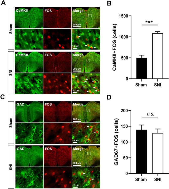
After injury, more ACC cells were colabeled for FOS and the pyramidal-associated marker CaMKII. FOS/GAD67 colabeling did not change significantly. Increased regional activity thus showed different cellular contributions.
FOS reflects activity-related changes over a time window, while cellular markers provide identity clues. Colabeling identifies activated groups without directly specifying moment-to-moment firing. In vivo recordings therefore tested whether electrical activity showed a corresponding difference. The approaches provide complementary views across different timescales.
Source: Zhu DY et al. · Molecular brain · CC BY
Original figure and English caption ↗
Pyramidal activity rises without a significant interneuron-average change
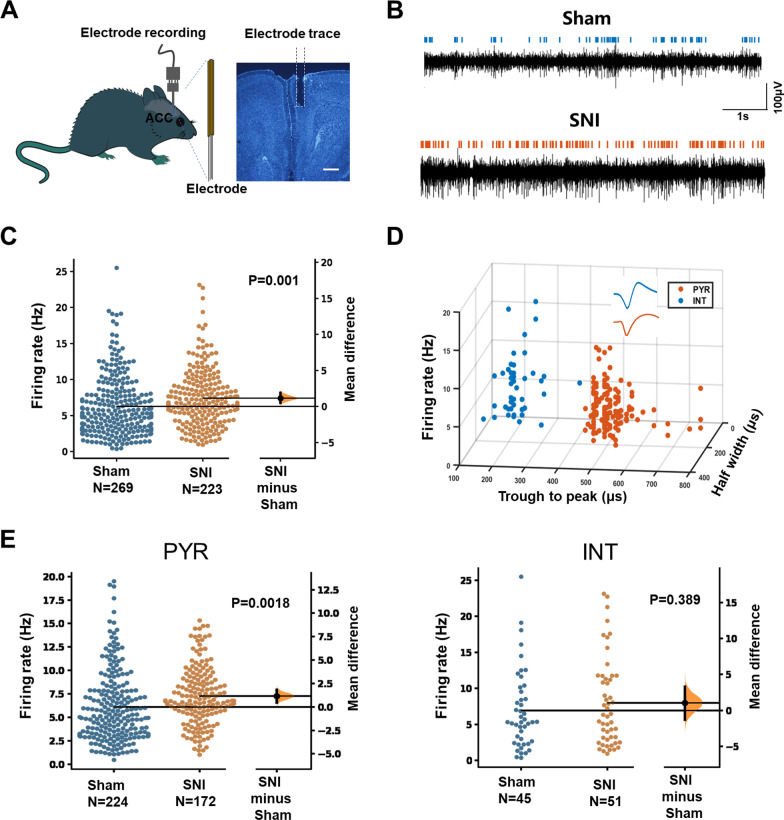
In awake mice, overall mean firing of recorded ACC units was higher after injury. Classification by firing properties localized the increase mainly to putative pyramidal cells, while the putative interneuron mean did not change significantly. Labeling and electrical evidence therefore converged.
A population mean and electrophysiological classification each have limits. The recording comparison involved three mice per group; multiple cells do not replace independent animals. The findings support a cellular difference under these conditions while motivating finer subtype identification and broader replication.
Source: Zhu DY et al. · Molecular brain · CC BY
Original figure and English caption ↗
Thalamic input changes increase sensitivity and pyramidal firing
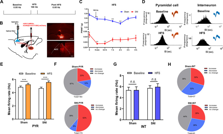
Enhancing relevant MD thalamic input to ACC lowered mechanical thresholds and raised mean pyramidal firing in both sham and injured groups. Neither group showed a significant interneuron-average change. Input changes thus influenced behavior while reproducing the population distinction.
The authors linked this enhancement with plasticity-like changes. Direct observations concerned post-stimulation firing and behavior, without identifying each altered synapse or the pyramidal subtype most important for sensitization. Input, activity and behavior were connected while leaving finer mechanisms unresolved.
Source: Zhu DY et al. · Molecular brain · CC BY
Original figure and English caption ↗
An unchanged mean can contain changing cells
After stronger input, about 30% of sham interneurons increased firing and 32% decreased it; corresponding injured-group fractions were about 28% in each direction. Opposing changes can coexist with a similar mean. Pyramidal populations also included decreasing and stable cells.
This adds detail to the absence of a matching interneuron-average increase. It preserves local cellular diversity and suggests that pain-related network adaptation may involve rate, temporal organization and coordination across subtypes.
Understanding excitation and inhibition requires cellular detail
These male-mouse findings identify a cellular bias in heightened ACC activity, without a matching increase in both population averages. They measure activity rather than the full ratio of excitatory and inhibitory synaptic currents and do not establish that interneurons are irrelevant to nociception.
Which inhibitory subtypes participate across disease stages, how brief responses differ from sustained activity and whether selective modulation benefits sensitization remain open questions. The study provides a cellular picture of cortical sensitization without establishing human treatment efficacy.