Another role for red nucleus: spinal projections contribute to post-injury hypersensitivity
Magnocellular red nucleus contributes to post-injury heat and mechanical hypersensitivity in male mice; spinal-projection interventions reveal the descending route’s function.
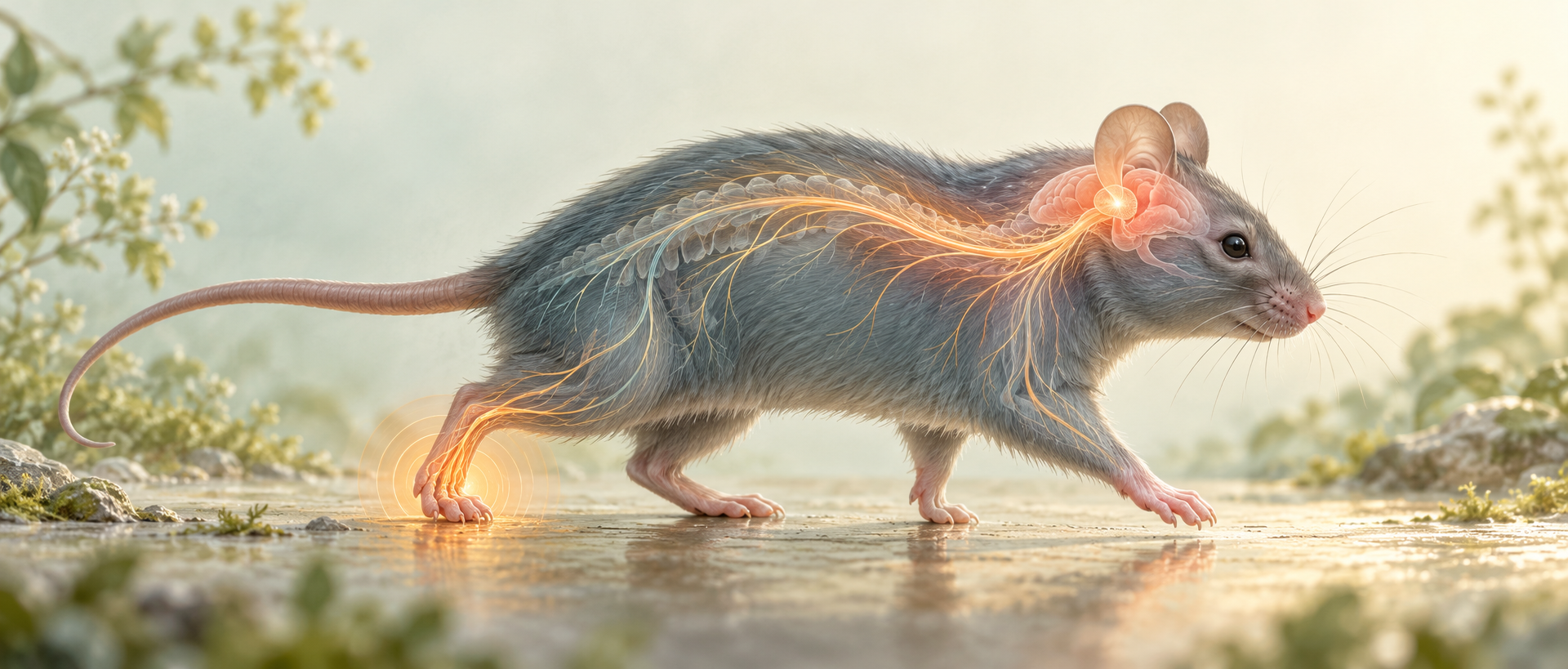
Descending signals regulate both movement and sensation. A Neuroscience Bulletin study examines magnocellular red nucleus, or RMC, in adult male mice after common peroneal nerve ligation. Activity associated with CaMKIIα-labeled cells increases, and reducing RMC activity improves heat and mechanical responses. Spinal-projection tests reveal additional differences among interventions.
RMC inhibition improves model hypersensitivity and healthy photostimulation induces heat/mechanical sensitivity. Projection-cell inhibition improves heat responses with nonsignificant mechanical improvement; spinal-end inhibition improves both.
Contents · 6 sections
RMC responses increase after nerve ligation
Red nucleus contains distinct subdivisions. Its magnocellular part is associated with the rubrospinal system, while the parvicellular part more strongly relates to inferior-olivary circuitry. Focusing on the magnocellular component allows examination of spinal connections in hypersensitivity.
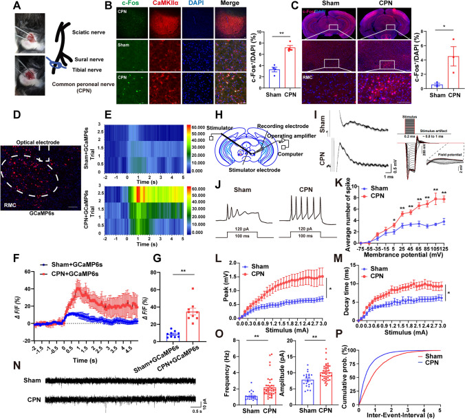
One week after left common-peroneal ligation, RMC c-Fos increases and largely overlaps CaMKIIα. Temporally controlled tagging also identifies more recruited cells. A region known for motor function changes after peripheral nerve injury.
Pinch evokes stronger RMC calcium. Slice evoked firing and miniature excitatory-current frequency/amplitude increase. Altered inputs and stronger output motivate behavioral tests.
Source: Shi J et al. · Neuroscience bulletin · CC BY
Original figure and English caption ↗
Suppressing recruited cells improves responses
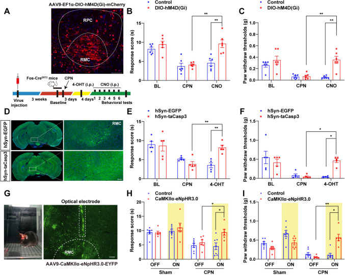
Activity tagging selects recruited RMC cells whose inhibition raises heat latencies and mechanical thresholds. Removing tagged cells through apoptosis also improves both measures, providing complementary evidence. Inhibition begins one week after ligation and is assessed over subsequent days, revealing a contribution during persistent post-injury hypersensitivity.
Photoinhibition of CaMKIIα-labeled cells also improves model mechanical and heat responses. Different interventions converge on a functional RMC contribution.
Source: Shi J et al. · Neuroscience bulletin · CC BY
Original figure and English caption ↗
Two activation methods differ
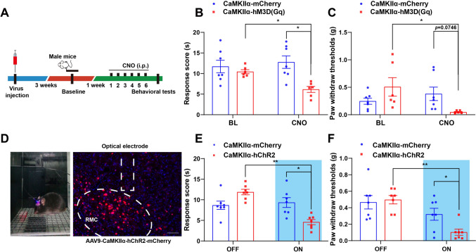
Chemogenetic activation increases heat sensitivity in healthy males, while mechanical decreases are nonsignificant. Photostimulation lowers both thresholds and heat latencies. The population can promote sensitivity with method-dependent outcomes.
Photostimulation follows light timing while chemogenetics changes activity over longer periods. Heat sensitivity increases with both; mechanical changes are clearer with optical activation. Different activity manipulations reveal different behavioral magnitudes.
Source: Shi J et al. · Neuroscience bulletin · CC BY
Original figure and English caption ↗
Spinal-end inhibition reduces hypersensitivity
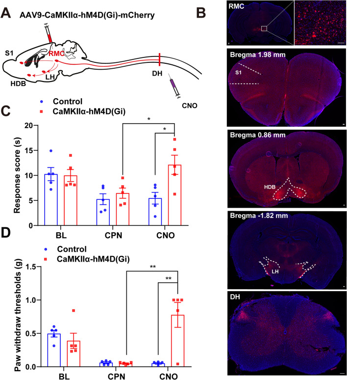
RMC axons reach several regions including spinal dorsal horn, or DH, an early sensory-processing site receiving descending modulation. Intrathecal CNO targeting receptor-expressing RMC spinal axons improves heat and mechanical hypersensitivity.
The spinal intervention connects broader RMC effects with a descending output. Midbrain signals contribute to spinally related sensory responses after peripheral injury.
Source: Shi J et al. · Neuroscience bulletin · CC BY
Original figure and English caption ↗
Heat effects stand out in DH-projecting cells
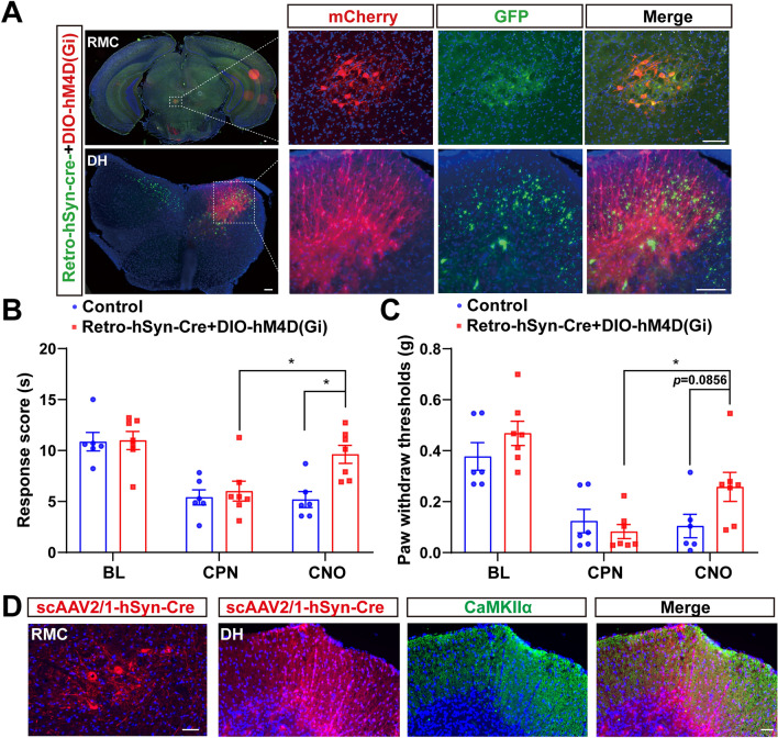
Retrogradely selecting DH-projecting RMC cells and inhibiting them improves heat responses, with nonsignificant mechanical improvement. Forward labeling associates RMC input with CaMKIIα-labeled DH cells, adding cellular detail.
Broad RMC, spinal-terminal and projection-body manipulations have different scopes. Heat effects align across these inhibitory interventions, while the projection-cell mechanical effect is weaker. Heat and mechanical changes do not fully track each other within this descending route.
Source: Shi J et al. · Neuroscience bulletin · CC BY
Original figure and English caption ↗
Sensory modulation extends to a motor-related midbrain node
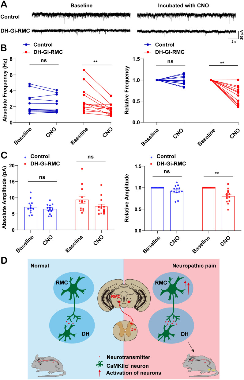
Projection-associated RMC recordings show reduced miniature-current frequency after inhibition. Relative amplitude falls, while absolute-amplitude comparisons are nonsignificant. These synaptic events complement activity and behavioral findings.
In adult male nerve-ligation mice, red-nucleus function extends from motor coordination to hypersensitivity modulation. RMC activity and spinal projections implicate descending signals, with intervention differences revealing functional organization.
Source: Shi J et al. · Neuroscience bulletin · CC BY
Original figure and English caption ↗