An asymmetric jaw, an altered brain: ACC links pain with anxiety-like behavior
A mouse study finds that persistent orofacial pain accompanies anterior-cingulate synaptic remodeling and heightened excitation. Suppressing its CaMKII-positive cells improves mechanical sensitivity and avoidance behavior.
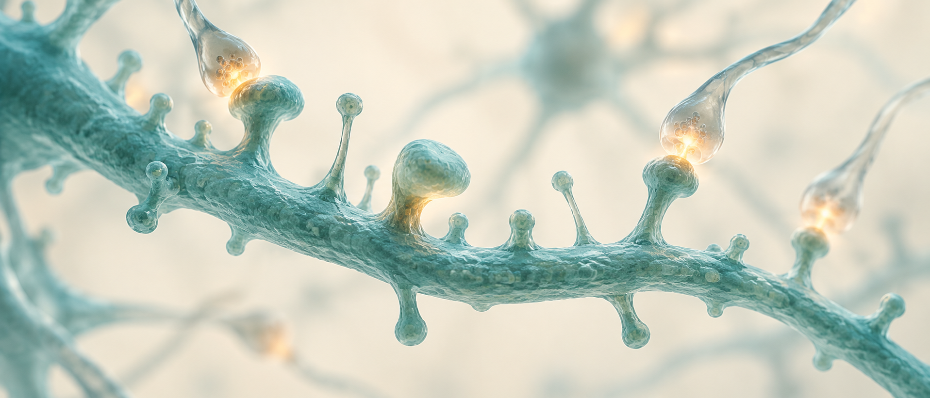
Why would a change in jaw structure leave a mark in a distant brain region? Persistent orofacial pain extends beyond local tissue and often accompanies affective changes. A study published on September 24, 2025 in Advanced Science connects bone and cartilage changes, spontaneous behavior, brain activity and synaptic structure in a mandibular-asymmetry mouse model. Anterior cingulate cortex (ACC) emerges as a key node, with heightened excitatory responses and remodeled synapses. Reducing activity in these cells improves both mechanical sensitivity and anxiety-like behavior.
Orofacial structural changes accompany excitatory synaptic remodeling in ACC. Cellular suppression further shows that ACC activity contributes to pain sensitivity and anxiety-like behavior in model mice.
Contents · 7 sections
Structural changes accompany persistent mechanical sensitivity
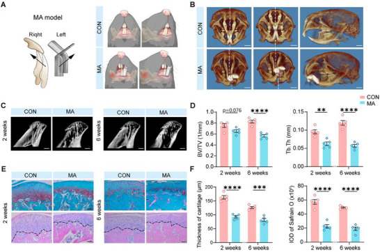
Altering dental occlusion establishes a mandibular-asymmetry model. Three-dimensional imaging shows clear structural differences after two weeks. Subsequent trabecular-bone and cartilage measurements reveal reduced bone-volume fraction and trabecular thickness, with thinner cartilage, providing a persistent peripheral-change context for neural investigation.
From the second week, model mice become more mechanically sensitive, with changes persisting to the fourth-week test. They spend less time in the open-field center and elevated-plus-maze open arms, with corresponding phenotypes in females. Mechanical sensitivity and avoidance of exposed spaces emerge together in this model.
Source: Zhang Z et al. · Advanced science (Weinheim, Baden-Wurttemberg, Germany) · CC BY
Original figure and English caption ↗
Less exploration, more curling: changes within action sequences
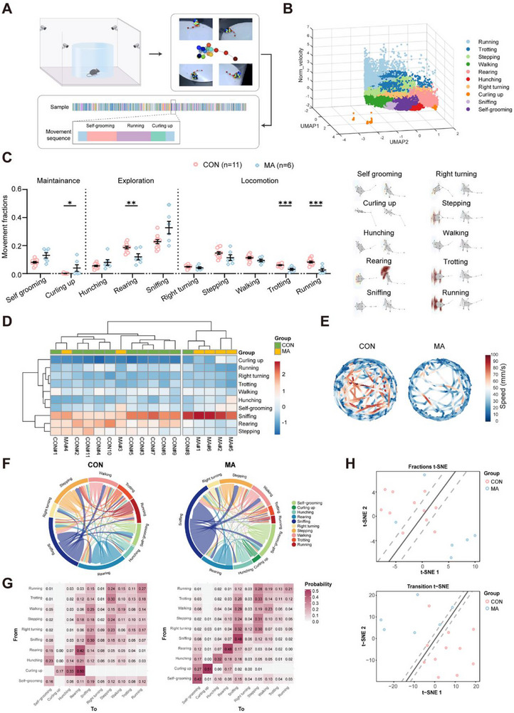
Multiview tracking adds detail beyond conventional tests. Model mice rear less, run and trot less, and curl up more. Changes concern not only distance traveled but transitions between actions: model mice more often maintain their current action, whereas controls more often move from curling to rearing.
This action pattern links persistent discomfort with reduced exploration, although activity level can influence anxiety-related measures. In the elevated plus maze, comparable total distance accompanies reduced open-arm exploration, separating changes in spatial choice from overall reduced movement. The study then turns to associated changes inside the brain.
Source: Zhang Z et al. · Advanced science (Weinheim, Baden-Wurttemberg, Germany) · CC BY
Original figure and English caption ↗
The same stimulus elicits a stronger cellular response
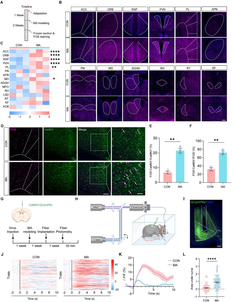
Brain-wide maps reveal changes across multiple regions, with ACC among the strongly responding areas. Further staining finds more activity-marker recruitment among its CaMKII-positive cells. As part of networks connecting sensation with behavioral context, altered ACC activity motivates the question of how a local orofacial problem enters broader neural processing.
In vivo recording shows stronger calcium responses in model ACC CaMKII-positive cells to mechanical stimuli of equal intensity. Tissue activity markers and stimulus-evoked dynamics converge, indicating both ongoing recruitment and enhanced responses to relevant sensory input.
Source: Zhang Z et al. · Advanced science (Weinheim, Baden-Wurttemberg, Germany) · CC BY
Original figure and English caption ↗
Expression maps point toward synaptic remodeling
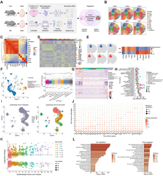
Spatial expression maps and single-nucleus sequencing resolve changes across cell types. Gene-expression changes differ among ACC populations. In glutamatergic cells, protein-synthesis-related pathways increase while gene sets associated with synaptic organization, structure and postsynaptic specialization decrease. Greater activity does not mean every intracellular process rises together.
Expression-based analyses also suggest rearranged signaling patterns, including among glutamatergic cells and between them and astrocytes. These inferences motivate a structural question: do the tiny contact sites receiving information change shape alongside altered synaptic gene expression?
Source: Zhang Z et al. · Advanced science (Weinheim, Baden-Wurttemberg, Germany) · CC BY
Original figure and English caption ↗
More spines, but molecular changes in different directions
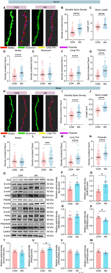
Microscopy finds denser, longer spines on both apical and basal dendrites of ACC pyramidal cells, with changes in several morphological categories. As important sites receiving excitatory input, spines indicate reorganization of neuronal contact structures rather than merely a transient activity increase.
Synaptic proteins change in different directions: Syn, GluR1 and PKMζ rise, PSD95 falls, and others remain comparable. Analgesic treatment improves mechanical sensitivity, anxiety-like behavior and ACC activity while attenuating some synaptic-protein changes. This links persistent pain with neural remodeling, but structural counts alone do not reveal transmission strength.
Source: Zhang Z et al. · Advanced science (Weinheim, Baden-Wurttemberg, Germany) · CC BY
Original figure and English caption ↗
Functional recordings connect structure to signaling
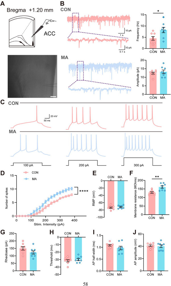
Electrophysiology supplies the functional link. Excitatory synaptic-current events occur more frequently in model ACC, with comparable peak event amplitude. Under the same injected current, pyramidal cells fire more action potentials and show higher membrane resistance. More frequent inputs and greater cellular responsiveness jointly characterize heightened excitation.
The study also finds increased basolateral-amygdala (BLA) activity, stronger BLA-to-ACC evoked excitatory currents and lower paired-pulse ratio, suggesting increased release probability along this input. ACC changes are thus connected to a broader input network, extending persistent orofacial pain beyond one cortical region.
Source: Zhang Z et al. · Advanced science (Weinheim, Baden-Wurttemberg, Germany) · CC BY
Original figure and English caption ↗
Suppressing ACC improves both sensitivity and avoidance
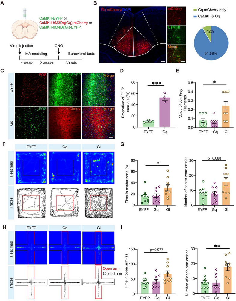
Finally, suppressing ACC CaMKII-positive cells raises mechanical-response thresholds and increases center activity and open-arm exploration, with corresponding improvements in female models. In the elevated plus maze, total distance remains comparable while the fraction of open-arm activity rises, again reflecting altered action choices.
From jaw structure to brain synapses, the study describes linked changes in adult mice. ACC modulation affects both sensory sensitivity and anxiety-like behavior, while the specific downstream outputs remain unresolved. Local tissue changes are processed by neural networks, making these distributed changes part of understanding pain.
Source: Zhang Z et al. · Advanced science (Weinheim, Baden-Wurttemberg, Germany) · CC BY
Original figure and English caption ↗