How can an RNA mark influence pain? Molecular cooperation in sensory cortex
A mouse study finds that nerve injury enhances SERBP1–PCIF1 binding and changes Maf1 RNA modification and protein expression, linking a molecular chain with sensory-cortical excitation, hypersensitivity and anxiety-like behavior.
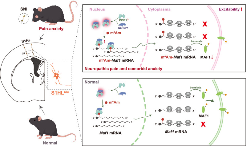
How cells use genetic information depends on more than the RNA sequence. Chemical marks on RNA can also influence protein expression. A study published on August 5, 2025 in Nature Communications connects such a mark with hypersensitivity after nerve injury. In the hindlimb region of mouse primary somatosensory cortex (S1HL), PCIF1 and its partner SERBP1 regulate m6Am modification and affect MAF1 protein. The molecular changes extend to cellular excitation and behavior, outlining one route by which injury changes cortical handling of sensory input.
After nerve injury, PCIF1 increases in layer-five glutamatergic cells of S1HL. Reducing PCIF1 or restoring downstream MAF1 attenuates excessive excitation, hypersensitivity and anxiety-like behavior.
Contents · 7 sections
Changes concentrate in a region and cell layer
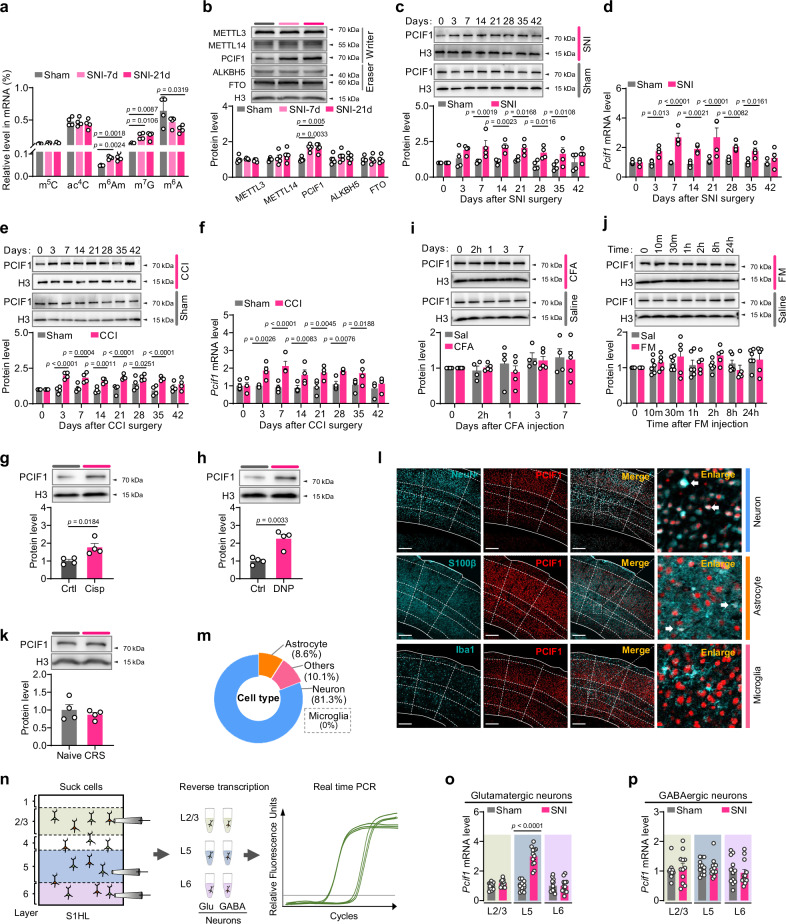
After peripheral nerve injury, mice become more sensitive to mechanical and thermal stimuli and later show anxiety-like behavior. The corresponding contralateral S1HL has increased m6Am and PCIF1. PCIF1 writes this modification near the RNA cap, with changes extending from early to later post-injury stages.
The increase is not found across all tested sites. Adjacent motor cortex, ipsilateral S1HL and other cortical layers lack equivalent changes, with PCIF1 elevation concentrated in layer-five glutamatergic cells. Related changes occur in several neuropathic-pain models, whereas PCIF1 remains broadly unchanged in tested inflammatory-pain and stress-only models, linking the effect to injury type and cellular location.
Source: Huang Y et al. · Nature communications · CC BY-NC-ND
Original figure and English caption ↗
Lowering PCIF1 attenuates sensitivity and excitation
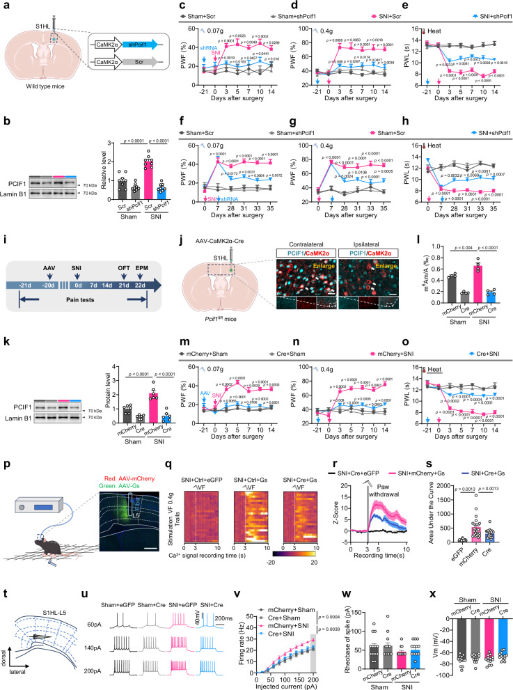
To test PCIF1 participation, the study reduces it in S1HL glutamatergic cells. Mechanical and thermal hypersensitivity attenuate when targeting either development or maintenance after symptoms emerge. Conditional deletion likewise reduces m6Am elevation and improves related behavior.
In vivo and slice recordings find that heightened stimulus-evoked calcium signals and firing are attenuated by reducing PCIF1. Resting membrane potential and current needed to initiate firing remain comparable, and basal sensory responses are not broadly suppressed. PCIF1 affects heightened responsiveness in the injury context rather than changing every neuronal property together.
Source: Huang Y et al. · Nature communications · CC BY-NC-ND
Original figure and English caption ↗
Hypersensitivity-like changes can arise without peripheral injury
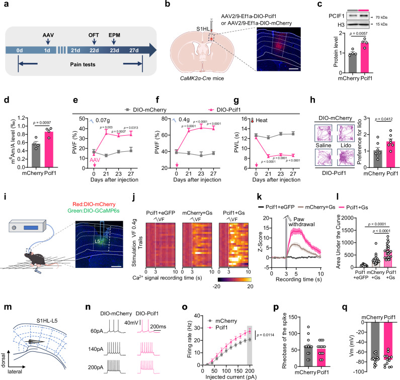
The reverse experiment extends the account. Increasing PCIF1 in S1HL glutamatergic cells of uninjured mice raises m6Am, enhances contralateral mechanical and thermal responses, and produces anxiety-like behavior. Preference for a space paired with local analgesia supplies behavioral evidence related to ongoing discomfort.
Locomotor function remains broadly normal, whereas stimulus-evoked cortical calcium responses and firing strengthen. Equivalent molecular elevation in neighboring motor cortex does not produce corresponding sensory hypersensitivity. A molecule’s effect depends on its cellular and regional setting, making sensory-cortical location particularly important here.
Source: Huang Y et al. · Nature communications · CC BY-NC-ND
Original figure and English caption ↗
Binding changes even without more partner protein
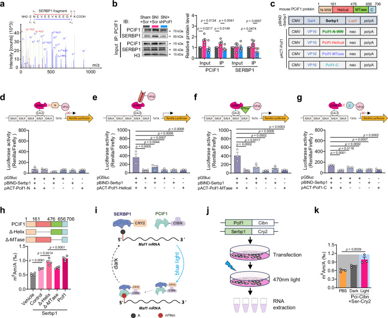
How does PCIF1 write the mark more effectively? The study identifies the RNA-binding protein SERBP1. After injury, total SERBP1 remains comparable while its binding to PCIF1 increases. Protein-function experiments show that SERBP1 improves PCIF1 modification efficiency, and enhancing their association increases m6Am.
Reducing SERBP1 attenuates m6Am elevation and hypersensitivity even when PCIF1 is increased. Molecular-network changes therefore concern not only protein abundance but how proteins cooperate. SERBP1 enhances efficiency rather than independently writing the modification.
Source: Huang Y et al. · Nature communications · CC BY-NC-ND
Original figure and English caption ↗
Maf1 links the modification with protein expression
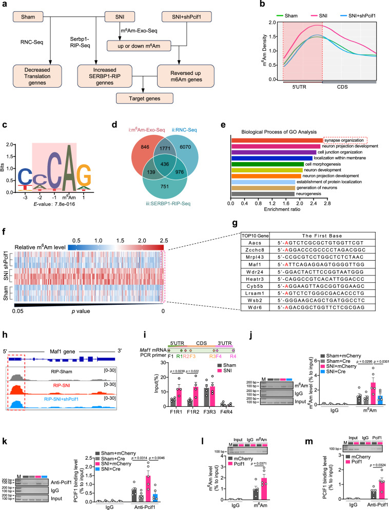
Integrating RNA modification, SERBP1 binding and translation-related datasets identifies targets changing across all three dimensions. Maf1 emerges as one lead: nerve injury increases its m6Am and reduces MAF1 protein. Lowering PCIF1 or SERBP1 prevents the associated rise in modification.
Because MAF1 is associated with regulation of neuronal excitability, its decrease provides a directional link: altered RNA modification may lower a regulatory protein and change cellular responses to sensory input. The study then narrows the question to Maf1 itself, asking whether this relationship can be reproduced directly.
Source: Huang Y et al. · Nature communications · CC BY-NC-ND
Original figure and English caption ↗
Targeted Maf1 modification lowers the protein
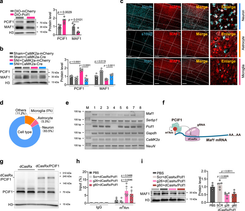
Targetedly increasing m6Am near the start of Maf1 RNA lowers MAF1 protein and enhances mechanical and thermal responses. Two other candidate RNAs lack equivalent modification changes, focusing the result on Maf1 rather than a simultaneous shift across all RNAs.
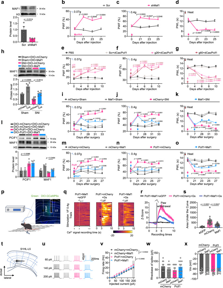
Directly lowering MAF1 likewise produces hypersensitivity and anxiety-like behavior and increases firing. The screening association thus gains functional support in a consistent direction. RNA modification affects multiple targets, with Maf1 representing a specifically tested link rather than the sole endpoint of all downstream changes.
Source: Huang Y et al. · Nature communications · CC BY-NC-ND
Original figure and English caption ↗
Restoring MAF1 attenuates excitation along the molecular chain
Increasing MAF1 attenuates hypersensitivity and anxiety-like behavior in injured mice. Moreover, restoring MAF1 while PCIF1 remains elevated improves behavior and reduces heightened calcium responses and firing. Restoring a downstream component can therefore attenuate effects associated with the upstream molecular change.
This account connects an RNA chemical mark with protein expression, cortical excitation and sensory behavior. Mechanistic experiments primarily use male mice, while injury-associated PCIF1 elevation is also observed in females. It reveals a fine regulatory layer: following peripheral injury, the brain changes not only activity but how cells use RNA information.
Source: Huang Y et al. · Nature communications · CC BY-NC-ND
Original figure and English caption ↗