After withdrawal, will the mouse avoid? Sensory inputs diverge in the spinal cord
Two sensory populations can evoke withdrawal without the same avoidance. After nerve injury, an input previously dominated by reflexes recruits licking, aversion and superficial spinal activity.
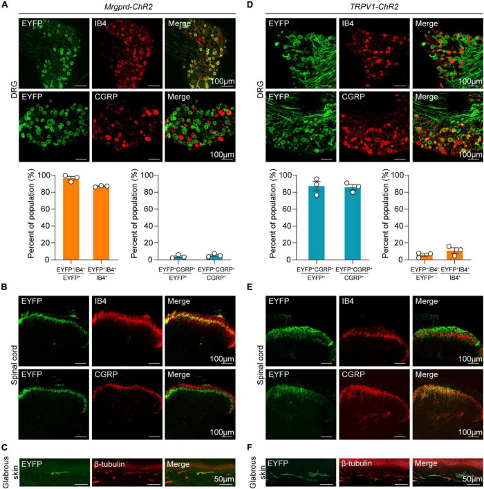
Does lifting a paw imply the same experience as licking it or avoiding a place? Animals cannot report their feelings directly, so distinct behaviors provide complementary evidence. A study published in Frontiers in Cellular Neuroscience on May 24, 2022 compares Mrgprd- and TRPV1-marked sensory populations and separates reflexive from aversion-related behavior. After nerve injury, the same input accompanies a different behavioral and spinal activity pattern. Rapid withdrawal, sustained attention to the body and avoidance of an associated environment occur in different behavioral and temporal contexts. Examining them separately reveals defensive movements without the same place-choice response.
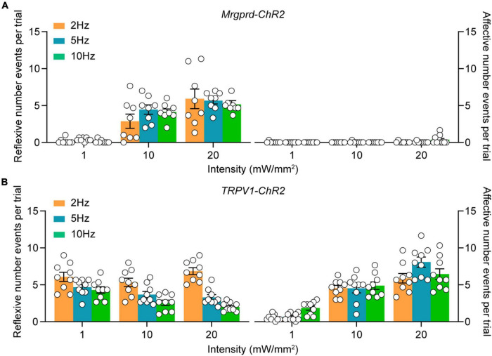
Under normal conditions, Mrgprd-associated input mainly evokes reflexes, whereas TRPV1-associated input also recruits licking and avoidance. After nerve injury, Mrgprd input recruits those behaviors and superficial spinal–parabrachial activity as well.
Contents · 7 sections
Similar withdrawal, different combinations of behavior
Mrgprd and TRPV1 identify two sensory populations, predominantly associated with nonpeptidergic and peptidergic markers, respectively. Their skin endings convey peripheral information to the spinal cord, providing two entry points for comparing defensive responses.
Under normal conditions, enhancing Mrgprd-associated skin input evokes lifting, holding or fluttering the paw, with few licking-like responses. TRPV1-associated input evokes reflexes alongside more licking and guarding. Similar withdrawal can therefore accompany different subsequent responses.
Source: Wang LB et al. · Frontiers in cellular neuroscience · CC BY
Original figure and English caption ↗
Place choice adds another perspective to the movements
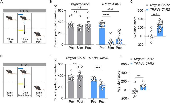
Will an animal return to a place associated with an unpleasant event? Place-choice comparisons add this perspective. Normal Mrgprd mice show reflexive movements without significant avoidance, while TRPV1 mice spend markedly less time in the associated location.
TRPV1 avoidance appears both during the event and in subsequent context-associated testing. Neither comparison is significant in the normal Mrgprd group. Combining movement and place choice reveals dimensions that a single behavior misses.
Source: Wang LB et al. · Frontiers in cellular neuroscience · CC BY
Original figure and English caption ↗
After nerve injury, the same input recruits a new combination
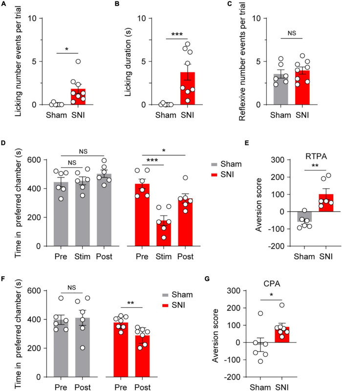
In a nerve-injury model, Mrgprd-associated input has a different outcome. Affected-paw licking becomes more frequent and prolonged, while reflexive-event counts do not increase significantly. The change is therefore selective rather than a rise in every movement.
Place choice changes too: injured mice show avoidance in both real-time and subsequent association tests. The same class of peripheral input can accompany licking and environmental aversion that are not prominent under normal conditions.
Source: Wang LB et al. · Frontiers in cellular neuroscience · CC BY
Original figure and English caption ↗
Different spinal depths show different activity patterns
Activity labeling tracks where the signals engage the spinal cord. Normal Mrgprd mice show labeling predominantly in deeper dorsal horn regions, without a significant superficial increase. Normal TRPV1 mice also show prominent superficial activity.
After nerve injury, Mrgprd mice show increased superficial labeling while deeper activity remains visible. This redistribution parallels the behavioral change and provides a spatial clue to how the same input can have different outcomes.
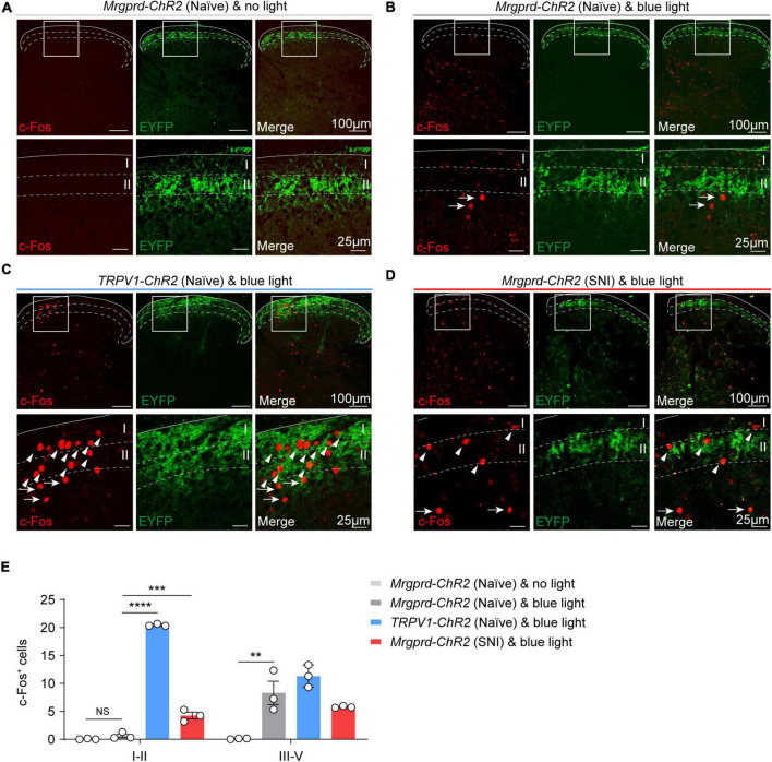
Source: Wang LB et al. · Frontiers in cellular neuroscience · CC BY
Original figure and English caption ↗
Superficial cells projecting to parabrachial nucleus join the response
Some superficial spinal cells project to lateral parabrachial nucleus, or lPBN. Normal Mrgprd input recruits few of these cells, whereas TRPV1 input and Mrgprd input after injury accompany activity labeling in more projection cells.
lPBN itself shows a corresponding activity pattern. This identifies a candidate ascending connection between spinal cord and brainstem: the superficial activity accompanying licking and avoidance extends beyond a local spinal change.
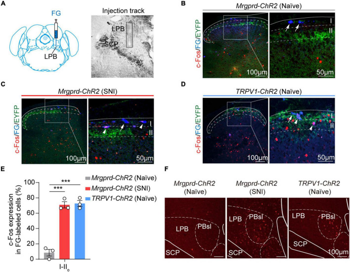
Source: Wang LB et al. · Frontiers in cellular neuroscience · CC BY
Original figure and English caption ↗
Excitation can arrive without producing firing
Cellular recordings in normal Mrgprd mice show that many spinal cells receive excitatory input without firing. Some also receive inhibitory input, while other cells convert input into spikes, revealing different response patterns.
In more superficial layers, excitatory responses are detected in more cells than actual firing. Signal arrival does not ensure onward transmission, supporting a gating account in which local networks limit conversion of input into output.
In inner lamina II, seventeen of twenty recorded cells receive detectable excitation, but only seven of those fire. The cells do not form one uniform transmission unit. Concurrent inhibition in some cells provides another route to different outcomes from the same peripheral input.
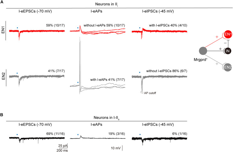
Source: Wang LB et al. · Frontiers in cellular neuroscience · CC BY
Original figure and English caption ↗
A gating model links behavioral differences to spinal networks
The authors propose that a normally restricted superficial pathway becomes more engaged after injury. Weaker inhibition and altered cellular excitability are candidate explanations; their specific contributions remain unresolved.
The study does not observe abnormal extension of the relevant afferents into superficial layers, or significant changes in labeled-cell density and Mrgprd identity proportions. Its interpretation therefore emphasizes processing of existing input rather than simply more afferent fibers.
The clearest finding is that reflexes, licking and avoidance do not always move together. The mouse study links input type, spinal depth and injury state while broadening behavioral interpretation: withdrawal answers one question, and licking and avoidance answer additional ones.
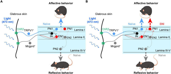
Source: Wang LB et al. · Frontiers in cellular neuroscience · CC BY
Original figure and English caption ↗