Weaker withdrawal responses point to anterior cingulate dendritic spines
With age, 5×FAD mice show weaker inflammatory mechanical responses, delayed anterior cingulate activity and fewer dendritic spines. Changing neuronal activity alters withdrawal behavior.
When disease-model mice withdraw less often than controls, which part of sensory processing has changed? A study published in Pain on April 16, 2022 links mechanical responses in 5×FAD Alzheimer-model mice to anterior cingulate cortex, or ACC, activity and dendritic structure. At six months, inflammatory withdrawal responses weaken, some cells respond later with less enhancement, and dendritic spines become sparser. The study follows behavior into its cellular correlates.
Mechanical-response changes in six-month-old 5×FAD mice accompany altered ACC pyramidal responses and fewer spines. ACC excitatory activation increases withdrawal, while activity suppression or spine-related pharmacological changes reproduce aspects of the phenotype.
Contents · 7 sections
Age and inflammation determine when differences appear
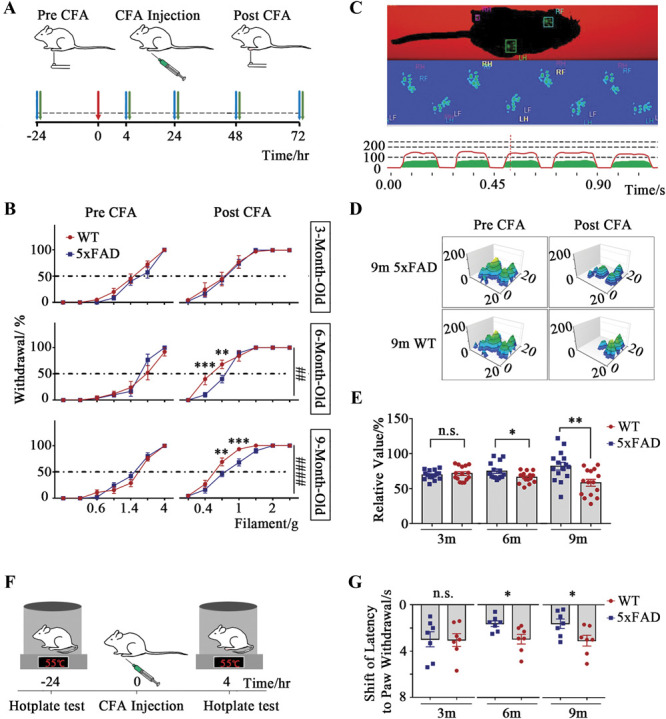
At three months, model and control mice have similar mechanical withdrawal responses. At six and nine months, model mice withdraw less after inflammation. Both genotypes nevertheless become more sensitive after inflammation, and model mice still respond to mechanical stimulation.
The difference has a temporal scope and does not persist uniformly at later observations. Paw swelling does not differ significantly, and later gait-contact comparisons show no significant group differences. The study focuses on early mechanical responses and their relationship to brain activity.
Source: Cui Z et al. · Pain · CC BY-NC-ND
Original figure and English caption ↗
At six months, anterior cingulate responses begin later
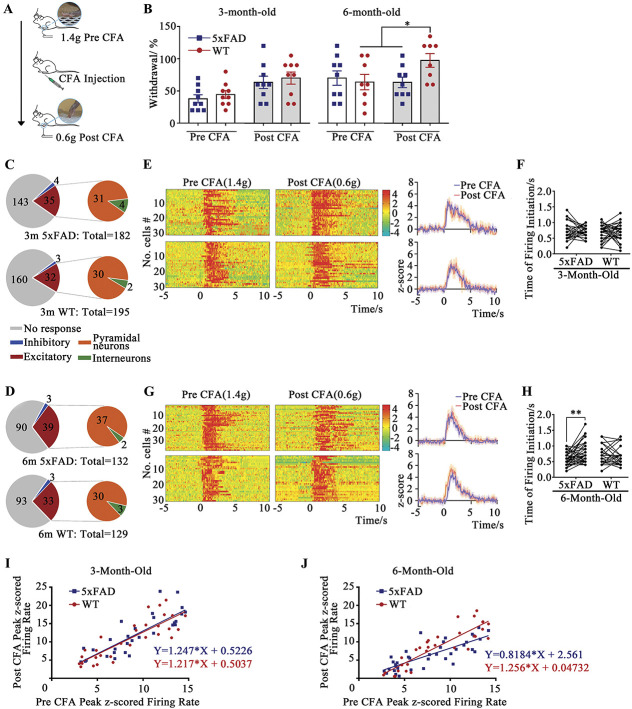
ACC recordings reveal increased, decreased and absent responses across cells. Among units classified as excitatory-responsive, response onset does not differ significantly at three months but is delayed in six-month-old model mice.
At six months, the relationship between post- and pre-inflammatory peak firing is flatter, indicating weaker response enhancement. Activity labeling provides a related clue: ACC labeling decreases in model mice at six and nine months, but not significantly at three months.
Multiple cells are recorded within each animal, describing a distribution of responses. The model group does not lose all activity; timing and response enhancement change.
Source: Cui Z et al. · Pain · CC BY-NC-ND
Original figure and English caption ↗
Similar cell proportions leave connectivity as a candidate
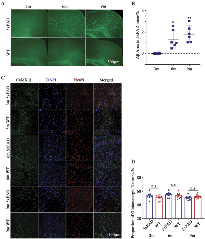
Amyloid-associated deposition increases with age, while the proportion of CaMKII-associated cells among ACC neurons does not differ significantly from controls. Reduced activity is therefore not simply matched by a lower proportion of the marked population.
Neurons receive inputs through dendritic branches and their spines. The study turns to these small structures to ask whether input-associated morphology differs despite similar population proportions.
Source: Cui Z et al. · Pain · CC BY-NC-ND
Original figure and English caption ↗
Dendrites become simpler and their spines sparser
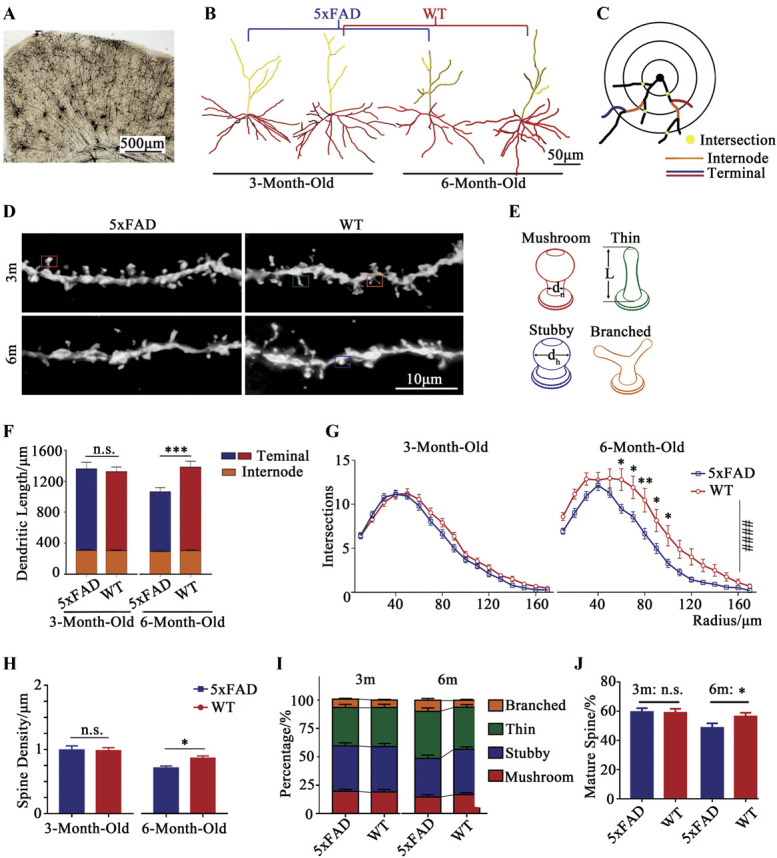
At six months, model ACC pyramidal cells have shorter dendrites, lower branching complexity and fewer spines per dendritic length. The proportion classified as morphologically mature is lower. These differences are not evident at three months.
Structural changes align with age-related differences in behavior and cellular responses. Spines receive many excitatory inputs, making their density a candidate contributor to weaker responsiveness. This correspondence motivates functional comparisons.
Source: Cui Z et al. · Pain · CC BY-NC-ND
Original figure and English caption ↗
Increasing anterior cingulate activity increases withdrawal
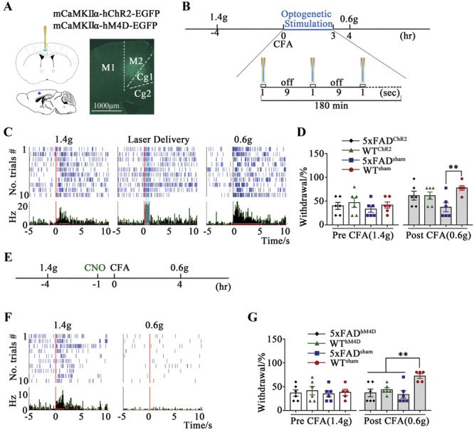
Activating ACC excitatory cells raises inflammatory withdrawal in model mice toward control levels. The same activation does not significantly alter withdrawal in normal controls, establishing a modifiable link between activity and behavior.
Conversely, reducing ACC-associated activity in normal mice lowers inflammatory withdrawal. These effects support ACC involvement in mechanical responding. The restoration concerns the tested behavior; dendritic regrowth and cognitive recovery are not measured here.
Source: Cui Z et al. · Pain · CC BY-NC-ND
Original figure and English caption ↗
Spine-related perturbation reproduces only part of the structural change
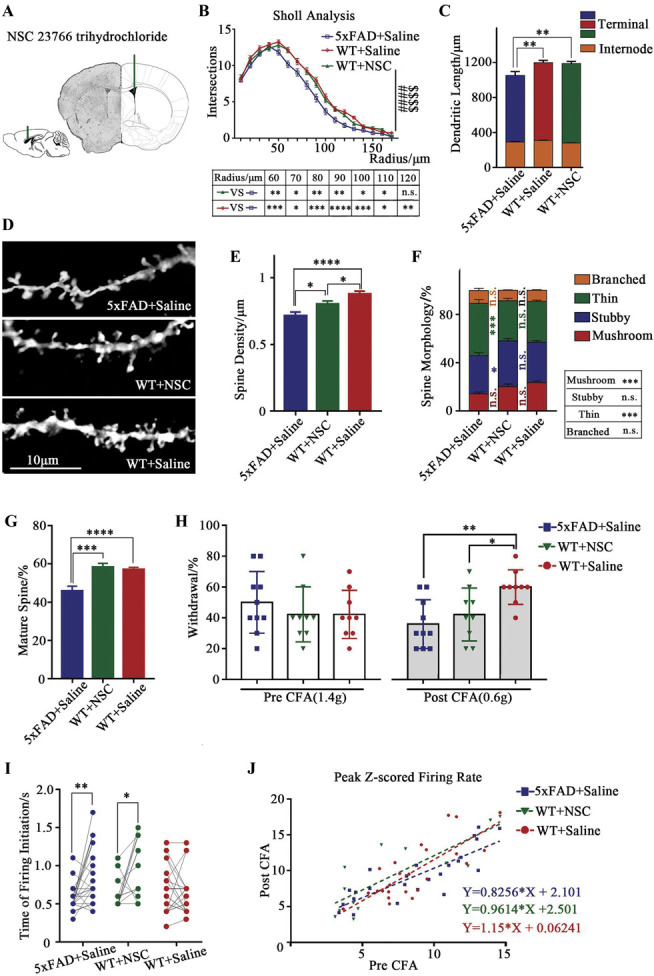
In normal mice, suppressing a Rac1-related regulatory process reduces spine density and mechanical withdrawal, delays responses and weakens peak enhancement. These changes resemble aspects of the model phenotype.
Dendritic length, branching complexity and mature-form proportion do not change significantly relative to normal controls. Structural features therefore do not all move together, and the pharmacological condition reproduces only part of the model phenotype.
Source: Cui Z et al. · Pain · CC BY-NC-ND
Original figure and English caption ↗
Structure, activity and behavior provide converging evidence
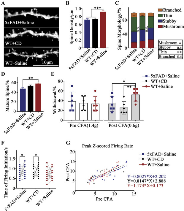
A pharmacological condition affecting actin polymerization likewise accompanies lower spine density, less inflammatory withdrawal, delayed ACC responses and weaker peak enhancement, providing another line of evidence linking structure-related processes and behavior.
Both pharmacological conditions can affect cellular functions beyond spines, leaving the precise path from structure to behavior unresolved. The study separates model-mouse responses into bodily movements, neural responsiveness and small input-receiving structures, revealing relationships beyond withdrawal counts alone.
Source: Cui Z et al. · Pain · CC BY-NC-ND
Original figure and English caption ↗