What septal cells reveal about memory difficulties after epilepsy
Clinical imaging and male-mouse experiments implicate medial-septal cholinergic cells. Epilepsy models show reduced cell labeling and hippocampal acetylcholine; activating remaining cells improves object-location and recognition tasks.
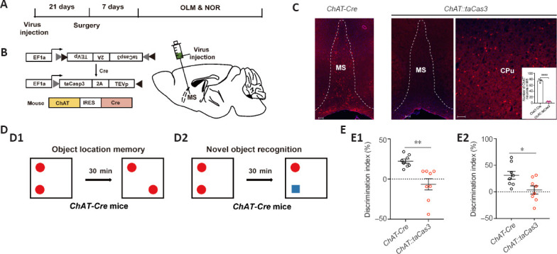
Will a mouse spend more time examining an object that has changed location? This simple question helps examine memory-related changes after epilepsy. A study published online in Neural Regeneration Research on March 15, 2023 begins with imaging and memory assessments in temporal-lobe epilepsy patients with hippocampal sclerosis, then turns to two male-mouse models. It focuses on medial-septal cholinergic cells connected with hippocampus. Their labeled counts decline in the model, while activating the remaining cells improves object-location and recognition performance.
Ablating medial-septal cholinergic cells is sufficient to reduce discrimination in two mouse object-memory tasks. Activating the cells improves performance in two epilepsy models. Separate recordings show increased hippocampal acetylcholine-sensor signals during activation.
Contents · 7 sections
Patient imaging points beyond hippocampus
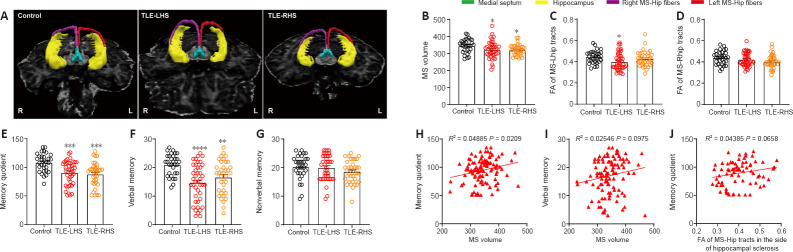
The study includes 78 temporal-lobe epilepsy patients with left- or right-sided hippocampal sclerosis and 31 healthy controls. Patients have smaller medial-septal volume and lower memory quotient and verbal-memory scores, without a significant nonverbal-memory difference. Different memory components are affected differently.
The left-sclerosis group has a lower diffusion metric in the septum–left-hippocampus pathway, unlike the corresponding right-sclerosis comparisons. Septal volume correlates positively with overall memory scores, while its correlation with verbal memory and the pathway metric’s correlation with overall memory are not significant. Imaging motivates subsequent cellular experiments.
Source: Chen J et al. · Neural regeneration research · CC BY-NC-SA
Original figure and English caption ↗
Less transmitter in hippocampus and fewer labeled septal cells
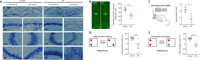
The kainate-induced chronic epilepsy model shows altered hippocampal cellular architecture and fewer ChAT-positive medial-septal cells. ChAT marks acetylcholine synthesis, and extracellular hippocampal acetylcholine is also lower, indicating changes in this long-range modulatory system.
Discrimination falls in object-location memory and novel-object recognition tasks. One compares exploration of a moved object, the other exploration of a new object, allowing spatial and object changes to be examined in separate tasks. Location memory depends more closely on hippocampus, while novel-object recognition also involves other regions. Their shared decline motivates studying a modulatory system that reaches across regions.
Source: Chen J et al. · Neural regeneration research · CC BY-NC-SA
Original figure and English caption ↗
Removing the cells reduces task performance
Cholinergic, glutamatergic and GABAergic cells intermingle in medial septum and all project to hippocampus. Different cell types can influence different network activities, motivating a focus on the cholinergic population. Association alone does not establish a contribution to task performance. In another group of mice, selective ablation greatly reduces medial-septal cholinergic-cell labeling while neighboring striatal cholinergic labeling remains similar.
Without the preceding epilepsy induction, these mice still show lower discrimination in both object tasks. Losing the population is sufficient to produce similar task difficulties, directly testing a cellular contribution beyond co-occurring pathology.
Source: Chen J et al. · Neural regeneration research · CC BY-NC-SA
Original figure and English caption ↗
Remaining cells can support improved performance
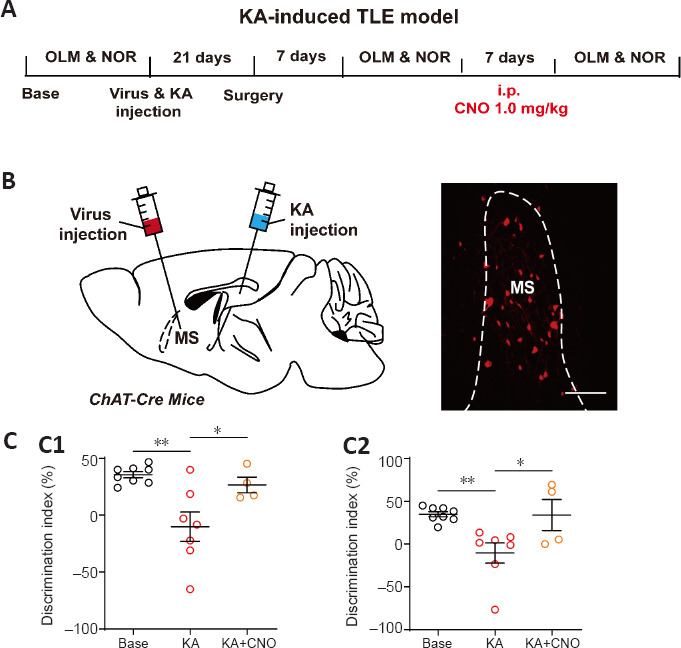
Can remaining cells still contribute after labeled counts fall? In the chronic kainate model, increasing septal cholinergic activity improves both object-task discrimination measures relative to the model condition, without significant differences from baseline.
The improvement reflects changed performance after enhancing the activity of an existing population, rather than regrowth of missing cells. The study next examines whether the effect appears in another epilepsy model.
Source: Chen J et al. · Neural regeneration research · CC BY-NC-SA
Original figure and English caption ↗
A second model reveals task-dependent improvement
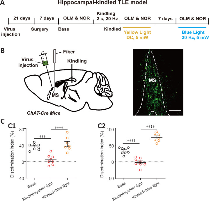
In a hippocampal-kindling model, rapid activation enhances the same cell type during behavioral tests. Object-location performance improves and is similar to baseline. Novel-object recognition also increases and exceeds baseline. The two tasks do not change identically.
Activation on different timescales in two models shifts performance toward higher discrimination. This shared direction supports a contribution from septal cholinergic cells, while differing magnitudes across tasks suggest that recognition preference involves multiple processes.
Source: Chen J et al. · Neural regeneration research · CC BY-NC-SA
Original figure and English caption ↗
Hippocampal acetylcholine adds a functional link
Separate sensor recordings show increased acetylcholine-related fluorescence in dorsal hippocampus during medial-septal cholinergic activation. This complements lower acetylcholine in the model, connecting population activity with remote transmitter changes.
The connection supplies a candidate mechanism: acetylcholine may contribute to improved task performance. The study does not test this step by blocking a hippocampal receptor class or directly follow synaptic-plasticity or oscillation changes during improvement.

Source: Chen J et al. · Neural regeneration research · CC BY-NC-SA
Original figure and English caption ↗
From clinical observations to a testable cellular contribution
Patient imaging raises the question, while ablation and activation experiments in male mice supply functional evidence. These observations concern clinical memory scores and mouse object-exploration tasks, with related but distinct measurement scopes.
The study follows memory-related difficulties after epilepsy to a modulatory population beyond hippocampus. Reduced cell labeling does not mean the remaining network has lost all regulatory capacity: strengthening this input can still alter behavioral performance in the tested models.