Activating zona incerta inhibitory cells improves movement in model mice
In a 6-OHDA lesion model, activating zona incerta GABAergic cells improves movement, balance and selected gait measures. Repeated activation also raises striatal tissue dopamine, while the downstream mechanism remains unresolved.
When movement slows and balance worsens, which cells can still help the motor network adjust? A study published in iScience on June 15, 2023 focuses on the zona incerta (ZI), a subthalamic region. Activating its GABAergic cells improves movement in male mice modeling parkinsonian motor deficits. Repeated activation also increases striatal tissue dopamine, linking local cellular activity to broader motor-system changes.
Inhibiting ZI GABAergic cells impairs movement and balance in unlesioned mice, while activation improves motor performance in the 6-OHDA model. Repeated activation also increases striatal tissue dopamine and HVA.
Contents · 7 sections
Looking beyond the injured nigrostriatal system
Parkinsonian movement deficits are commonly linked to damage in the nigral dopamine system. ZI sits beyond that classic focus but connects widely with cortex, thalamus, basal ganglia and brainstem, contributing to posture and locomotion. It may therefore influence how motor networks respond to damage.
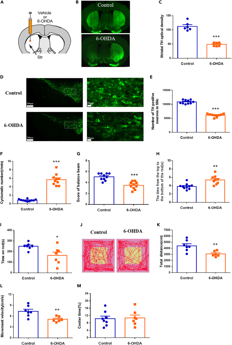
The 6-OHDA model shows reduced striatal TH fiber labeling and fewer TH-positive nigral cells. Movement distance and speed decline, alongside poorer balance and coordination, while open-field center time does not significantly change. These measures provide the baseline for testing interventions.
Source: Chen F et al. · iScience · CC BY-NC-ND
Original figure and English caption ↗
Inhibitory markers in ZI also change
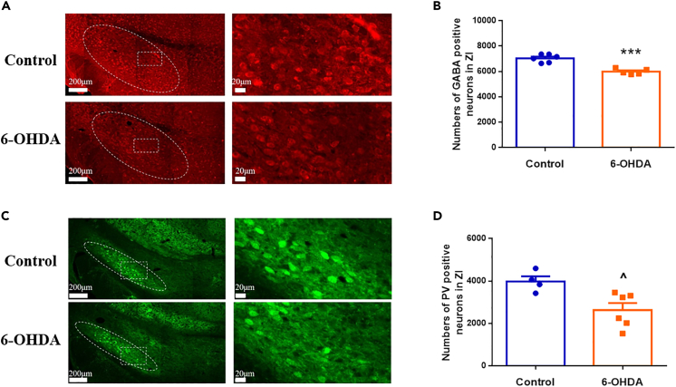
In ZI, GABA-positive cell counts fall by about 13%, while counts positive for parvalbumin (PV), a related population marker, fall by about 34%. The model thus involves changes in local inhibitory-cell labeling beyond the dopamine pathway.
The labeling results lead to a functional question: can changing the activity of ZI GABAergic cells alter movement? The study then examines inhibition in unlesioned animals and activation in lesioned animals, connecting cellular observations with behavioral consequences.
Source: Chen F et al. · iScience · CC BY-NC-ND
Original figure and English caption ↗
Balance and coordination are affected in unlesioned mice
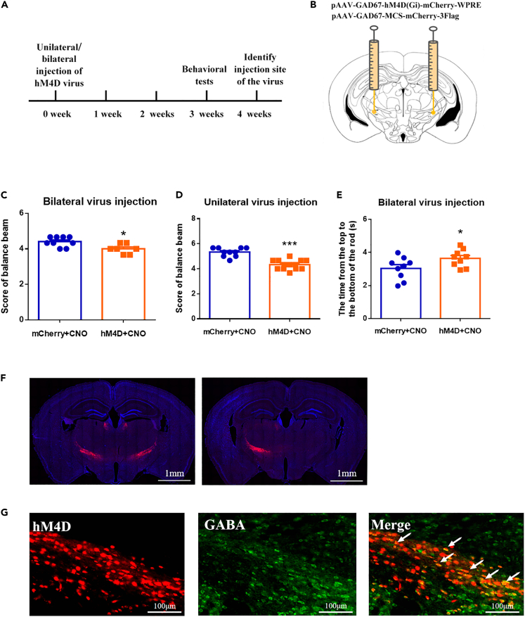
In unlesioned males, either unilateral or bilateral inhibition of ZI GABAergic cells lowers balance-beam scores. Bilateral inhibition also increases pole-descent time. The population therefore contributes to ordinary motor performance, rather than acting only after damage.
Brief optogenetic inhibition produces similar directional findings: movement distance, speed and balance performance decrease during illumination. Observing reduced cellular activity alongside behavioral changes within the same time window adds evidence for a functional contribution.
Source: Chen F et al. · iScience · CC BY-NC-ND
Original figure and English caption ↗
Distance and speed increase in the lesion model
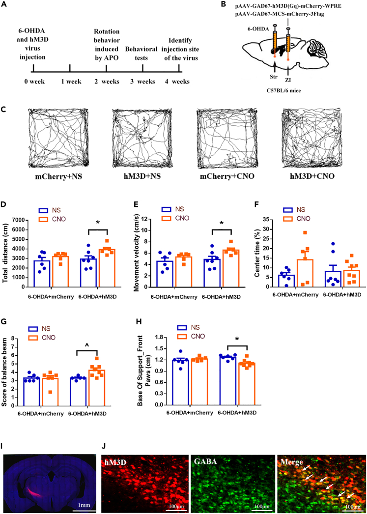
Activating ZI GABAergic cells in the 6-OHDA model increases movement distance and speed and improves balance-beam scores. Gait analysis also finds a narrower forepaw base of support, showing effects on how the body is supported as well as how much the animal moves.
Center exploration time does not change significantly. Distance, speed and balance measures jointly reveal the motor effect, which is not similarly enhanced in control-vector animals.
Source: Chen F et al. · iScience · CC BY-NC-ND
Original figure and English caption ↗
Behavioral improvement accompanies higher tissue dopamine
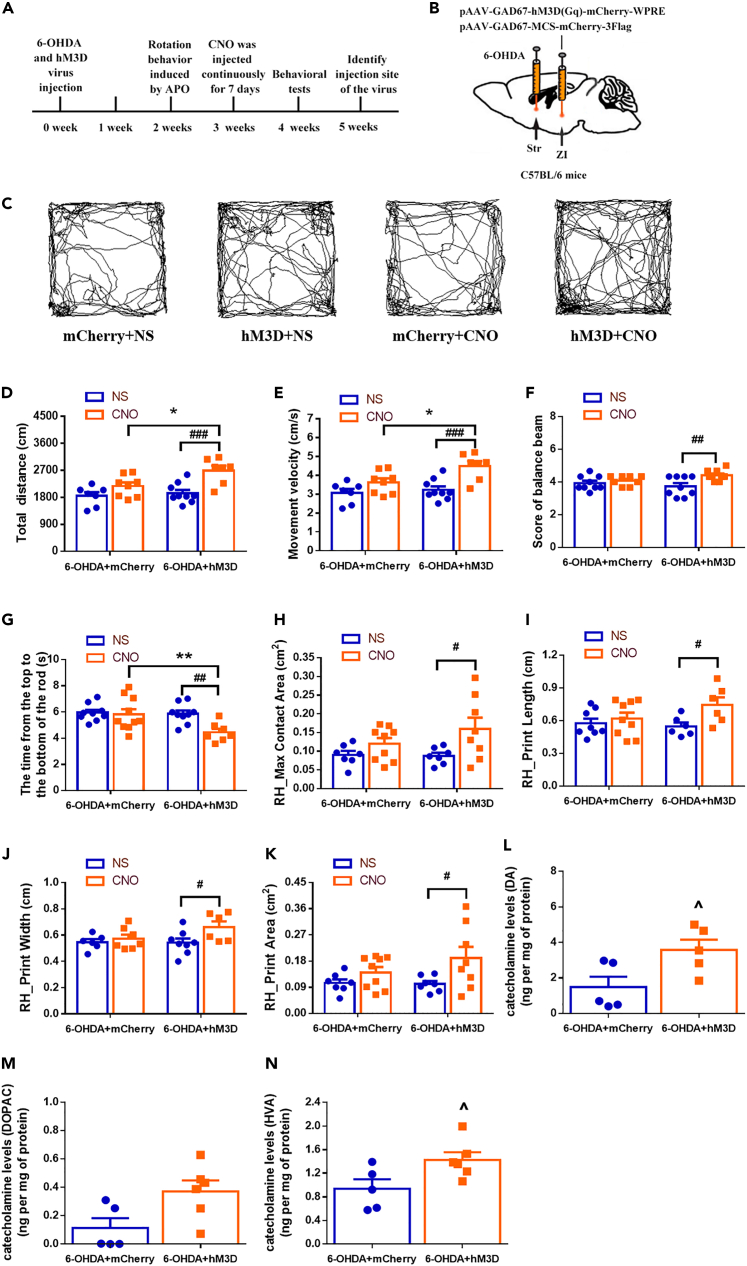
After one week of repeated activation, model mice continue to show improvements in movement, balance and coordination measures. Maximum contact area and print length, width and area of the right hind paw also increase. Footprint data add detail to the motor change: animals move more and place their paws differently.
Tissue measurements show increased striatal dopamine and its metabolite homovanillic acid (HVA). These measurements concern accumulated tissue content in the repeated-activation group. They link ZI activity with the chemical state of the nigrostriatal system without identifying the downstream connection responsible.
Source: Chen F et al. · iScience · CC BY-NC-ND
Original figure and English caption ↗
Movement also increases during optical activation
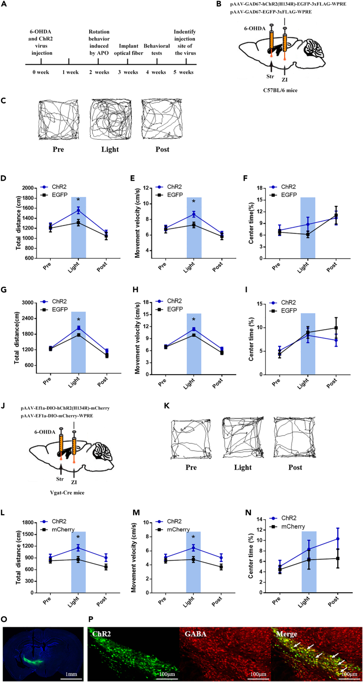
Optogenetic activation supports the same direction of effect. In unilateral and bilateral lesion models, ZI GABAergic activation increases distance and speed during illumination without significantly changing center time. A cell-type-related validation in Vgat-Cre mice also shows enhanced movement.
The change accompanies the activation period, showing that modulating the population can rapidly influence motor output. Together with chemogenetic and repeated-activation findings, the study links ZI GABAergic cells to model motor performance across different timescales.
Source: Chen F et al. · iScience · CC BY-NC-ND
Original figure and English caption ↗
Motor modulation extends beyond the classic pathway
ZI is widely connected. Local inhibition and outputs to basal ganglia or brainstem could influence movement, and the authors propose these possibilities without identifying a specific projection responsible for improvement. The direct finding is that changing this population’s activity can alter motor output after damage.
The evidence comes from male mice in a 6-OHDA model, and increased tissue transmitter content does not demonstrate reconstruction of damaged neurons. The study broadens the question from how much dopamine is missing to how other networks can modulate movement, adding cellular evidence for ZI function.