How do striatal D1 populations change during dyskinetic movements?
In a rat dyskinesia model, D1-associated population calcium accompanies abnormal movements. Inhibition reduces some movements; longer-lasting modulation delays onset and shortens duration without significantly changing peak scores.
Which cells change activity at the moment an abnormal movement occurs? A study published in Frontiers in Aging Neuroscience on February 3, 2022 follows striatal cells expressing D1 dopamine receptors in a rat model of levodopa-induced dyskinesia. It moves from activity labeling to real-time calcium and then behavioral effects of inhibition. The outcome is not disappearance of every movement: movement types, onset, duration and peak severity change differently.
Striatal D1-associated population activity accompanies dyskinetic movements. Inhibition reduces some movements, and longer-lasting modulation delays onset and reduces duration and total scores without significantly changing peak scores.
Contents · 6 sections
Severity accompanies the number of active cells
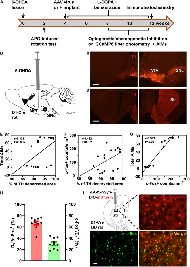
In rats with marked dopaminergic denervation and dyskinesia, levodopa is followed by increased striatal c-Fos labeling. Active-cell counts correlate with total abnormal involuntary movement scores, while denervation extent does not correlate significantly with scores within this cohort. Denervation is already severe in this cohort, so the comparison concerns behavioral variation within that restricted background. Activity labeling adds information about current cellular state beyond lesion extent.
About two-thirds of activity-labeled cells carry a D1-associated marker, while roughly three in ten D1-marked cells carry the activity marker. These complementary denominators describe active-population composition and recruitment within the D1 population.
The change is not uniform across the striatum. Active cells without colabeling and D1-associated cells without activity labeling remain, indicating a broader, heterogeneous network.
Source: Gao S et al. · Frontiers in aging neuroscience · CC BY
Original figure and English caption ↗
Real-time signals align population activity with individual movements
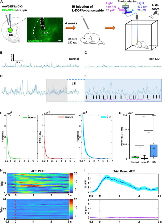
After levodopa, the dyskinetic group shows greater power of D1-associated population fluorescence in the analyzed band than normal and nondyskinetic controls. The signal reflects stronger time-varying population calcium activity.
Aligning traces to each dyskinetic movement reveals corresponding population calcium peaks. A calcium-independent reference lacks the same movement-related change, clarifying the temporal association beyond a single activity-label snapshot.
A population peak sums many cells and does not reveal each cell’s contribution. An increase near movement onset also leaves open its source and whether it predicts the next movement.
Source: Gao S et al. · Frontiers in aging neuroscience · CC BY
Original figure and English caption ↗
Inhibition reduces some movements without a significant orolingual effect
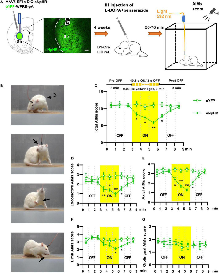
When marked dyskinesia is already present, transient inhibition of striatal D1-associated cells reduces abnormal locomotion, axial movements and total scores. Limb-score reductions are more restricted to particular observations, while orolingual signs do not change significantly.
The movement types do not decline uniformly, and scores move back toward prior levels after inhibition ends. This links D1 population activity to some dyskinetic features while suggesting different movement types may depend on different conditions.
Source: Gao S et al. · Frontiers in aging neuroscience · CC BY
Original figure and English caption ↗
Direct-pathway cells remain embedded in a broader network
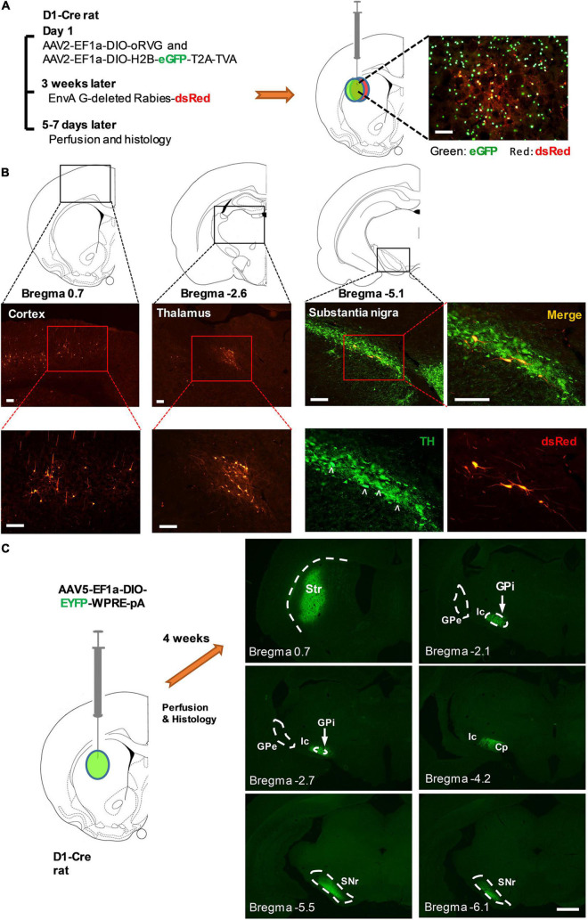
Tracing identifies cortical, thalamic and substantia nigra pars compacta inputs, with fibers reaching basal-ganglia output regions including internal globus pallidus and substantia nigra pars reticulata. Sparse external pallidal fibers fit direct-pathway anatomy.
D1 receptor identity describes a way cells receive dopamine-related signals, while the input distribution shows additional sources of information. Stronger dyskinetic activity may involve several connections, whose individual contributions are not established here.
Source: Gao S et al. · Frontiers in aging neuroscience · CC BY
Original figure and English caption ↗
Longer-lasting modulation changes onset and duration
Reducing D1-associated activity before levodopa delays dyskinesia, shortens its duration and lowers total scores. Peak scores do not change significantly, so the effect is not uniform suppression throughout the entire episode.
This complements transient inhibition of ongoing movements: one comparison tests immediate changes, the other the subsequent episode. Separating onset, duration and peak identifies where changes occur and preserves a peak that a total score could obscure.
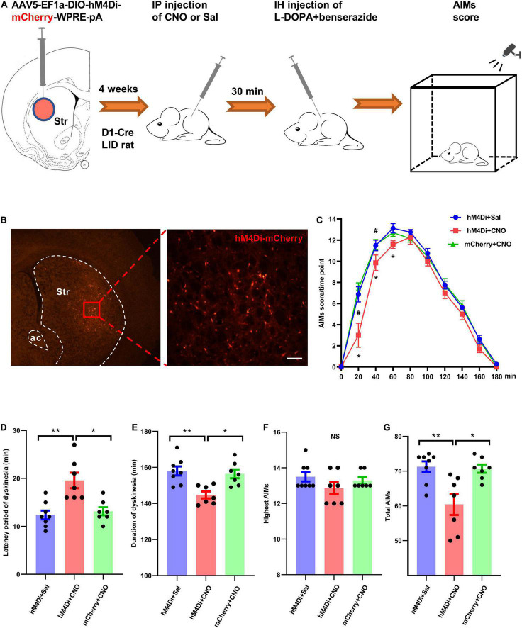
Source: Gao S et al. · Frontiers in aging neuroscience · CC BY
Original figure and English caption ↗
The timing of abnormal movements refines the circuit question
The rat-model study establishes a functional connection between heightened D1-associated activity and some dyskinetic features. Population calcium reveals movement-associated timing, while inhibition exposes a contribution to behavior.
The inputs generating abnormal activity, differing orolingual responses and effects on normal movement remain unresolved. The findings refine a broad overactivity account by cell identity, movement type and temporal course.