Reducing a dorsal raphe population slows hippocampal seizure progression
A mouse study finds increased dorsal raphe serotonergic activity during hippocampal seizures. Suppressing the population slows seizure progression, while activation does not produce a mirror-image effect.
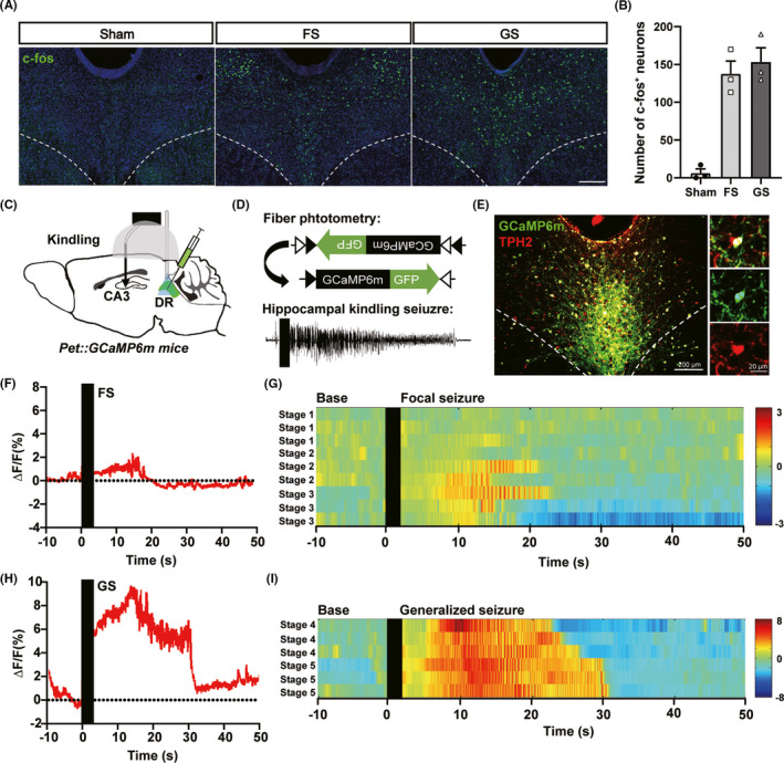
Changes during a hippocampal seizure extend beyond hippocampus. Brainstem systems involved in emotion, sleep and other functions can also be recruited. A mouse study published in CNS Neuroscience & Therapeutics on May 6, 2021 finds marked activity in dorsal raphe (DR) serotonergic cells during hippocampal seizures. Suppressing these cells slows the worsening of seizures during repeated induction, revealing a contribution that depends on the population and seizure stage.
DR serotonergic activity increases with seizure severity. Suppression slows progression mainly at later stages. The tested low-frequency electrical stimulation produces a similar benefit, which is lost when the population is concurrently activated.
Contents · 6 sections
The seizure is accompanied by brainstem activity
DR is a major source of serotonin-related signals to forebrain. After hippocampal seizures, its activity-associated c-Fos labeling increases. Separate calcium recordings from the serotonergic population also show a rise around seizures. These observations suggest recruitment of this brainstem network.
Calcium signals grow as seizure stages progress from focal to generalized events. This measures population activity rather than serotonin release itself. Whether the recruited cells oppose or facilitate the seizure requires testing what happens when their activity changes.
Source: Cheng H et al. · CNS neuroscience & therapeutics · CC BY
Original figure and English caption ↗
Suppressing activity slows seizure progression
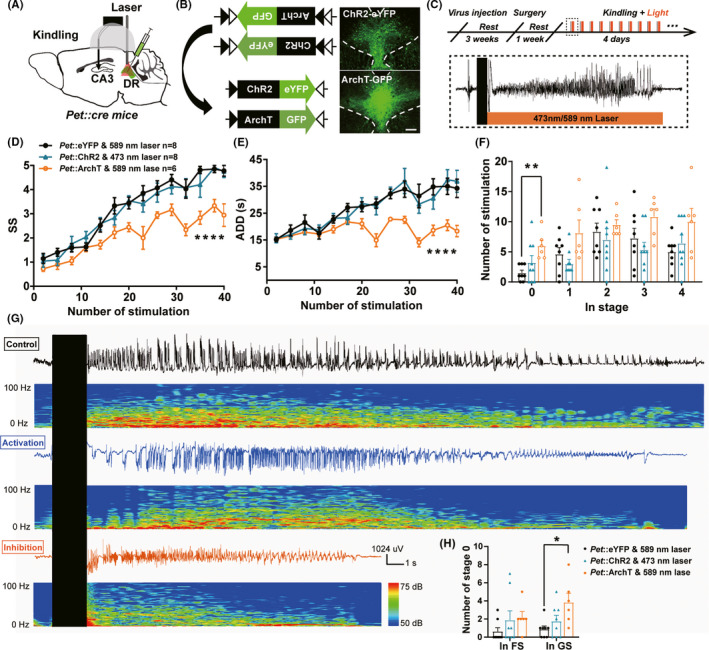
In hippocampal kindling, seizures worsen across repeated induction. Suppressing DR serotonergic cells slows the rise in seizure stage and shortens electrographic afterdischarge. Afterdischarge reflects how long abnormal electrical activity persists after the triggering event, complementing the behavioral stage measure.
The effect is clearest later in progression, during the generalized-seizure period, rather than in earlier focal stages. Activating the population does not significantly alter progression. The asymmetric effects of suppression and activation connect the population’s contribution with the network state during seizures.
Source: Cheng H et al. · CNS neuroscience & therapeutics · CC BY
Original figure and English caption ↗
A tested low-frequency stimulation condition produces a similar pattern
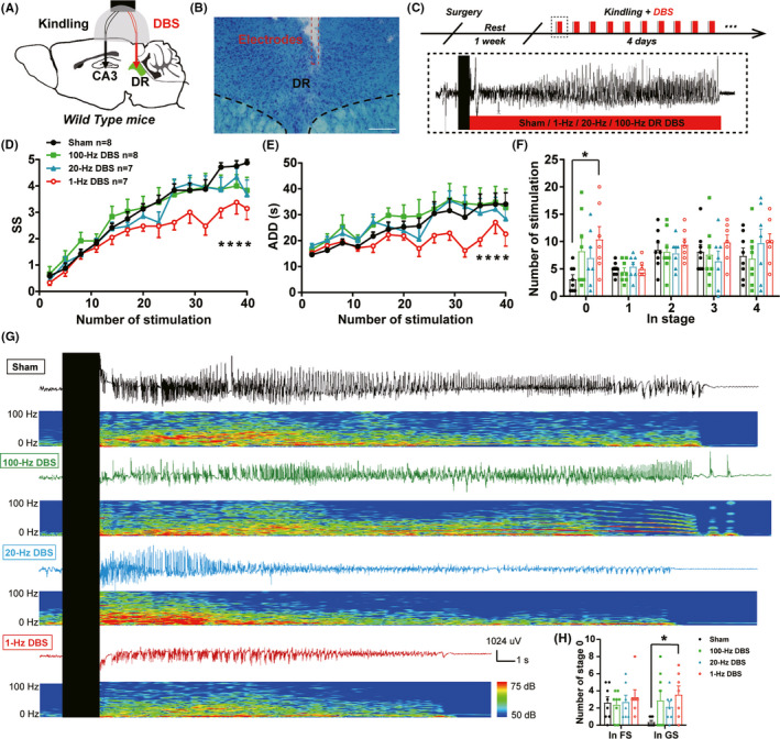
The team next examines regional DR electrical stimulation. One tested low-frequency condition likewise slows stage progression, shortens afterdischarge and increases seizure-free events in the late period. Two other tested frequency conditions do not significantly change progression, making the effect condition-dependent.
The direction and timing resemble serotonergic-cell inhibition. Regional electrical stimulation, however, can influence a broader set of neurons and passing fibers. Similar seizure outcomes suggest a possible shared mechanism and motivate a closer link between regional stimulation and the defined population.
Source: Cheng H et al. · CNS neuroscience & therapeutics · CC BY
Original figure and English caption ↗
Concurrent activation removes the stimulation benefit
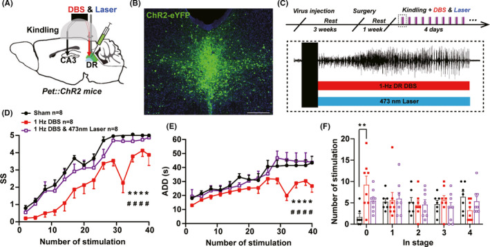
When DR electrical stimulation is combined with serotonergic-cell activation, its benefits for stage, afterdischarge and seizure-free events are lost, with outcomes approaching controls. The population’s state can therefore alter the effect of regional stimulation.
The authors propose that reducing excessive serotonergic activity during seizures may contribute to the stimulation effect. How those cells change during stimulation, and whether other populations or connections participate, remains unresolved. Synaptic mechanisms such as long-term depression remain hypotheses in this setting.
Source: Cheng H et al. · CNS neuroscience & therapeutics · CC BY
Original figure and English caption ↗
Seizure changes do not coincide with mood-related behavioral changes
DR is also associated with emotion regulation, making mood-related behavior relevant to epilepsy comorbidity. After kindling acquisition, the study compares sucrose preference, grooming-related and tail-suspension behavior. Regional stimulation does not significantly change these measures, separating the seizure effect from the tested behavioral outcomes.
Kindling itself also fails to produce significant differences from controls on these measures, so an established depression-like phenotype was not demonstrated and then rescued. Seizure-aligned regional modulation and subsequent behavioral assessment address a different setting from interventions directed at the behavioral test itself.
A transmitter system does not have a single uniform role
Previous serotonin-related studies report differing effects across seizure models. This work focuses on a DR population during hippocampal kindling: recruitment and inhibition effects both depend on stage. Transmitter identity, cellular origin and disease-model phase together define the circuit contribution.
The mouse findings identify a brainstem node for further study, within a region containing diverse subpopulations and broad projections. Which connections carry the seizure-related influence, and whether other seizure types follow the same pattern, remain open. The current evidence concerns experimentally induced progression rather than long-term control of spontaneous seizures.