A prefrontal inhibitory population responds during social approach
Reduced social preference in male Magel2-deficient mice accompanies lower somatostatin-cell excitability and calcium responses. Activating these cells or their lateral-septal output improves tested behavior.
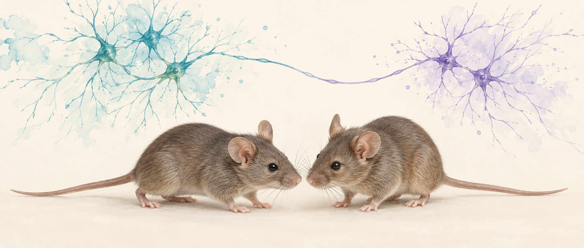
An empty cage on one side, an unfamiliar mouse on the other—which does a test mouse approach more? This choice makes social-approach preference observable. A study published in Molecular Autism in 2025 finds reduced preference for an unfamiliar conspecific in male Magel2-deficient mice. It follows somatostatin-marked, SOM, neurons in medial prefrontal cortex: although inhibitory, increasing their activity improves social-approach behavior in this model.
Prefrontal SOM-cell responses during social interaction are blunted in Magel2-deficient mice. Suppressing the cells reduces preference in controls, whereas activating them or their lateral-septal terminals increases preference in the model, supporting regulation by this inhibitory output.
Contents · 6 sections
Preference changes while overall movement remains similar
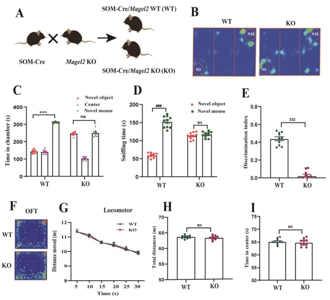
Controls spend more time in the chamber containing an unfamiliar mouse and sniff it longer than the empty cage. In the Magel2-deficient group, chamber and sniffing times do not differ significantly between targets, and the social-preference index is lower. The difference involves both approach and subsequent investigation.
Open-field distance, activity over time and center time remain similar, distinguishing the preference change from the measured overall movement. With whole-animal Magel2 deficiency in this model, the next question is which cellular responses connect with altered preference.
Source: Wang X et al. · Molecular autism · CC BY-NC-ND
Original figure and English caption ↗
The population becomes harder to recruit
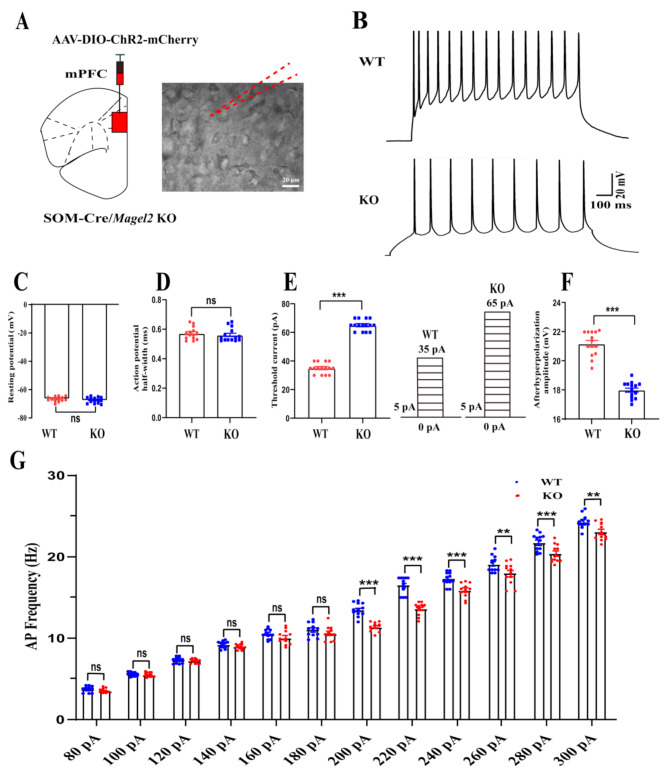
Slice recordings identify differences in prefrontal SOM-cell firing. Deficient cells require more current to initiate spikes and produce fewer action potentials with stronger input, indicating lower intrinsic excitability. The inhibitory population itself has an altered response threshold.
The change is selective: firing at smaller injected currents, resting voltage and action-potential half-width do not differ significantly. Afterhyperpolarization amplitude is lower. Together, these measurements describe an altered firing pattern rather than a total loss of activity.
Recruitment during stronger input may matter beyond the resting state. The study therefore returns to the moments when mice actually approach a conspecific.
Source: Wang X et al. · Molecular autism · CC BY-NC-ND
Original figure and English caption ↗
Conspecific-related calcium responses are blunted
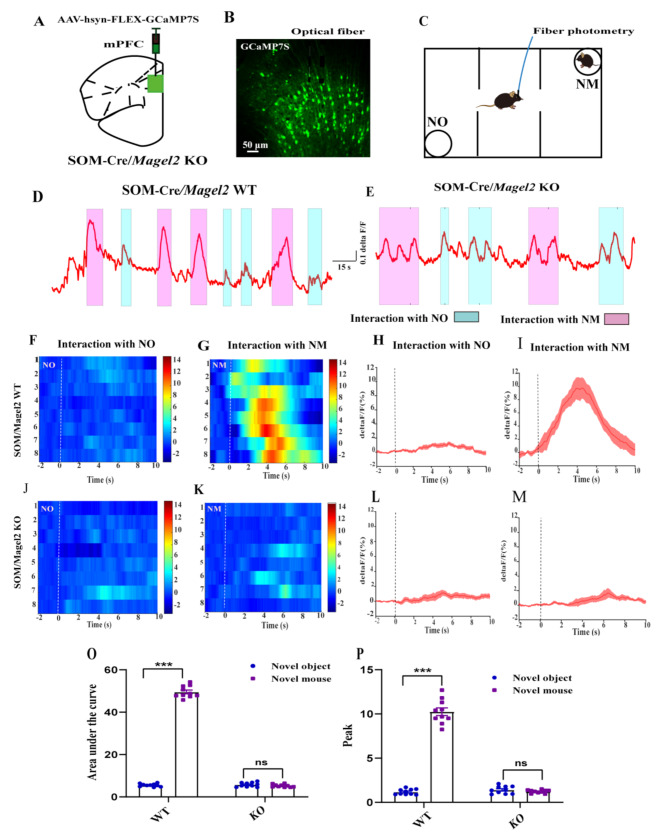
In controls, interaction with an unfamiliar mouse produces a strong prefrontal SOM-population calcium response, while empty-cage investigation produces a smaller change. Deficient mice show weak responses to both targets, without significant differences in integrated response or peak. The target distinction seen in controls is blunted in the model.
The recordings align cellular responses with social behavior. Combined with lower slice excitability, they motivate a specific test: does reducing or increasing this population’s activity change preference for the conspecific?
Source: Wang X et al. · Molecular autism · CC BY-NC-ND
Original figure and English caption ↗
Suppressing control cells reduces social preference
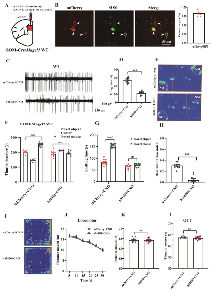
Chemogenetic suppression lowers prefrontal SOM-marked activity in controls with intact Magel2. Once reduced firing is confirmed, the usual chamber and sniffing preference for an unfamiliar mouse is no longer significant, and the preference index falls. The intervention connects cellular activity with behavior.
Open-field movement and center time do not clearly change. Reducing this inhibition does not promote social approach. The result illustrates how inhibitory-cell effects depend on circuit position and connectivity rather than the name of their transmitter alone.
Source: Wang X et al. · Molecular autism · CC BY-NC-ND
Original figure and English caption ↗
Activation reinstates conspecific preference in the model
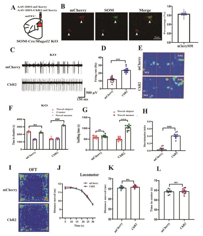
Increasing the same population’s activity in Magel2-deficient mice shifts behavior in the opposite direction. Mice spend longer in the conspecific chamber and sniff the mouse more than the empty cage, with an increased preference index. Increased firing accompanies these changes in the intervention condition.
Total distance and center time remain similar, concentrating the improvement in the tested social choice. Suppression in controls and activation in deficient mice provide complementary evidence across different backgrounds, leading the study to the population’s output destinations.
Source: Wang X et al. · Molecular autism · CC BY-NC-ND
Original figure and English caption ↗
Following the output to lateral septum
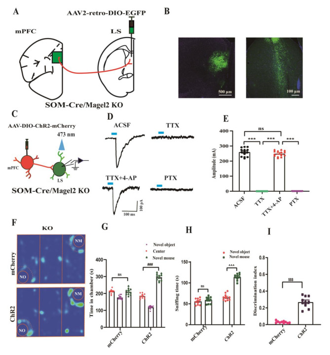
Tracing identifies a long-range projection from some prefrontal SOM-marked neurons to lateral septum. Terminal activation evokes inhibitory currents there, and blockade-and-rescue experiments support direct GABAergic connectivity. The population’s influence thus extends beyond its local network.
Activating prefrontal-derived terminals locally in the lateral septum of deficient mice also increases chamber and sniffing preference and the preference index. This output’s regulatory capacity connects cellular excitability and interregional communication with the same behavior.
Using male mice aged eight to ten weeks, the study measures conspecific-approach choices. It shows that an inhibitory output can support social preference, and recruiting that output in one genetic model can change the behavior measured at the time.
Source: Wang X et al. · Molecular autism · CC BY-NC-ND
Original figure and English caption ↗