Why do hyperactivity, impulsivity, and inattention appear together? Two outputs from substantia nigra PV neurons
A mouse study traces histamine H2 receptor loss in substantia nigra pars reticulata PV neurons, showing how two downstream pathways separately shape attention and locomotion.
Attention-deficit/hyperactivity disorder (ADHD) is characterized by core symptoms of locomotor hyperactivity, impulsivity, and impaired sustained attention. While central histaminergic signaling regulates arousal and cognition, the specific postsynaptic histamine receptor subtype, neuronal population, and downstream circuit architecture that coordinate these distinct behavioral dimensions have remained unclear. Published in Neuron on January 8, 2025, a study led by Prof. Weiwei Hu and Prof. Zhong Chen at Zhejiang University and Zhejiang Chinese Medical University ("Deficiency of histamine H2 receptors in parvalbumin-positive neurons leads to hyperactivity, impulsivity, and impaired attention") combined conditional genetics (PV-Cre;Hrh2fl/fl), DAT-HET mouse and human postmortem brain analyses, the 5-choice serial reaction time task (5-CSRTT), in vivo multichannel optrode recordings, GRAB_DA2h dopamine fiber photometry, and pathway-specific chemogenetics to show that histamine H2 receptors (H2Rs) in substantia nigra pars reticulata parvalbumin-positive (SNr PV+) neurons prevent ADHD-like phenotypes through segregated downstream projections to the SNc and mSC.
Loss of histamine H2 receptors in SNr PV+ neurons reduces their basal and task-locked firing activity, causing sustained attention deficits, premature impulsive responses, and impaired bright-light risk assessment via disinhibition of SNc dopamine neurons (SNr→SNc PV), while driving open-field hyperactivity and impaired bright-light risk assessment via disinhibition of the medial superior colliculus (SNr→mSC PV).
Contents · 6 sections
Conditional Hrh2 Deletion in PV+ Neurons Induces Methylphenidate-Reversible Hyperactivity, Impulsivity, and Attention Deficits
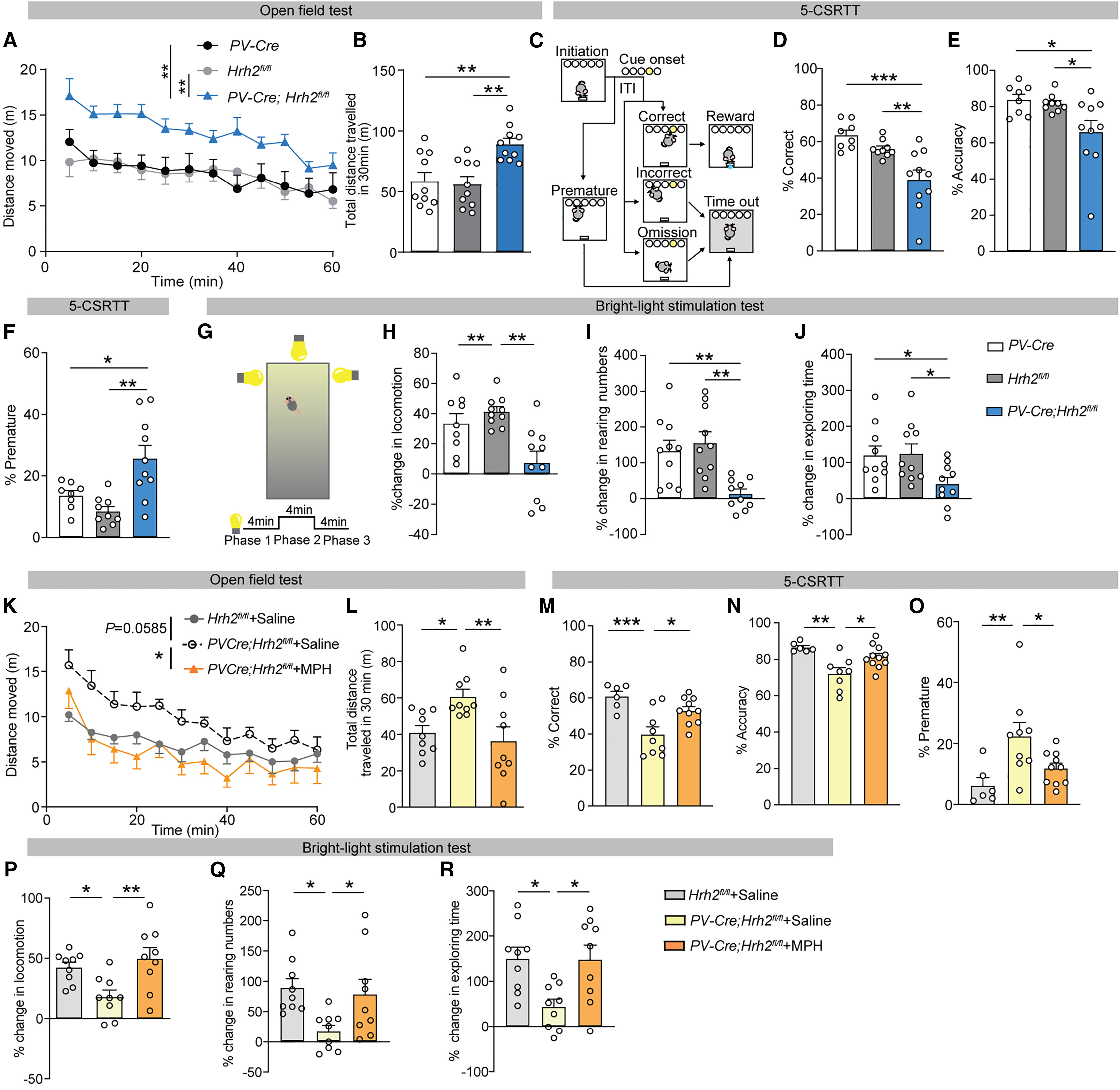
The authors generated conditional knockout mice lacking Hrh2 in parvalbumin-positive neurons (PV-Cre;Hrh2fl/fl) and compared them against PV-Cre and Hrh2fl/fl controls. In the open field test, PV-Cre;Hrh2fl/fl mice exhibited robust locomotor hyperactivity across 5-minute bins and 60-minute total distance. In the bright-light stimulation test—which assesses pre-encounter defensive risk assessment and attention—control mice significantly increased locomotion, rearing frequency, and exploring time during bright-light illumination (Phase 2), whereas these adaptive Phase-2 increases were blunted in PV-Cre;Hrh2fl/fl mice.
In the 5-choice serial reaction time task (5-CSRTT, featuring a pseudorandom 4, 5, or 6 s inter-trial waiting period before cue onset), PV-Cre;Hrh2fl/fl mice displayed significantly reduced % Correct and % Accuracy alongside elevated premature nose-poke responses (% Premature), reflecting impaired sustained attention and heightened motor impulsivity. Systemic administration of the frontline ADHD medication methylphenidate (MPH, 0.5 mg/kg, i.p.) rescued open-field hyperactivity, 5-CSRTT inattention and impulsivity, and Phase-2 behavioral responses in the bright-light test.
Source: Dadao An et al. · Neuron · © Elsevier Inc.
Original figure and English caption ↗
SNr PV+ Neurons Exhibit Selective H2R Downregulation in DAT-HET Mice and Postmortem Brains With ADHD Symptoms
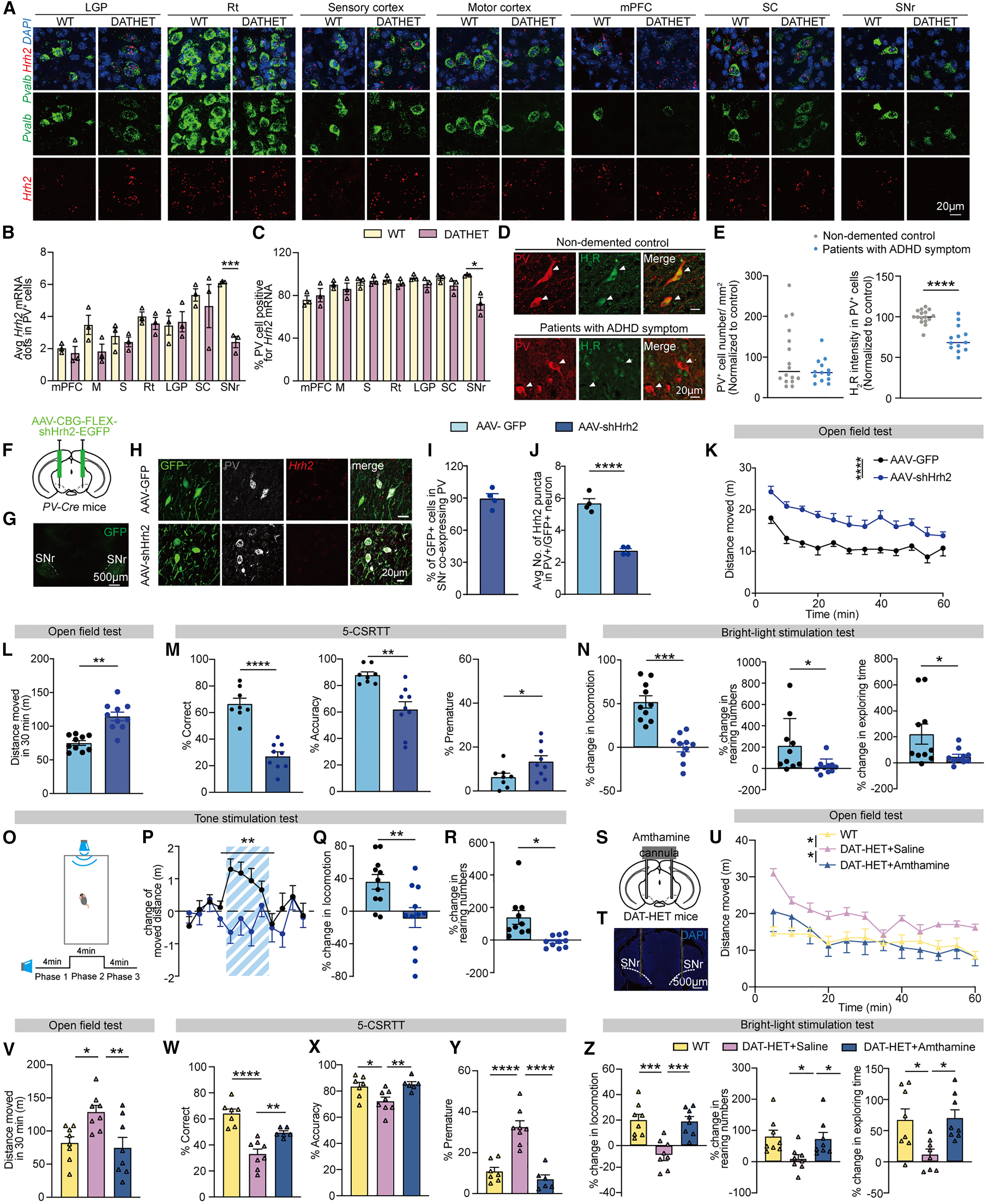
To pinpoint the anatomical locus of H2R dysfunction, the authors performed RNAscope fluorescence in situ hybridization across seven brain regions (LGP, Rt, sensory cortex, motor cortex, mPFC, SC, and SNr) in dopamine transporter heterozygous (DAT-HET) mice, a classic genetic model of ADHD. Hrh2 mRNA expression was selectively reduced in PV+ neurons of the substantia nigra pars reticulata (SNr). Consistent with this finding, postmortem brain tissue from individuals with ADHD symptoms exhibited a significantly lower percentage of H2R-expressing PV+ neurons in the SNr compared with matched controls.
Selective knockdown of Hrh2 in SNr PV+ neurons of PV-Cre mice via AAV-FLEX-shHrh2-EGFP (89.590% ± 4.508% knockdown efficiency) recapitulated open-field hyperactivity, decreased % Correct and % Accuracy, increased % Premature responses in the 5-CSRTT, and blunted behavioral responses in the bright-light and tone stimulation tests. Conversely, intra-SNr microinjection of the selective H2R agonist amthamine in DAT-HET mice alleviated hyperactivity, inattention, impulsivity, and bright-light response deficits.
Source: Dadao An et al. · Neuron · © Elsevier Inc.
Original figure and English caption ↗
H2R Deficiency Blunts Basal and Task-Locked Firing of SNr PV+ Neurons, and Chemogenetic Activation Rescues ADHD-like Behaviors
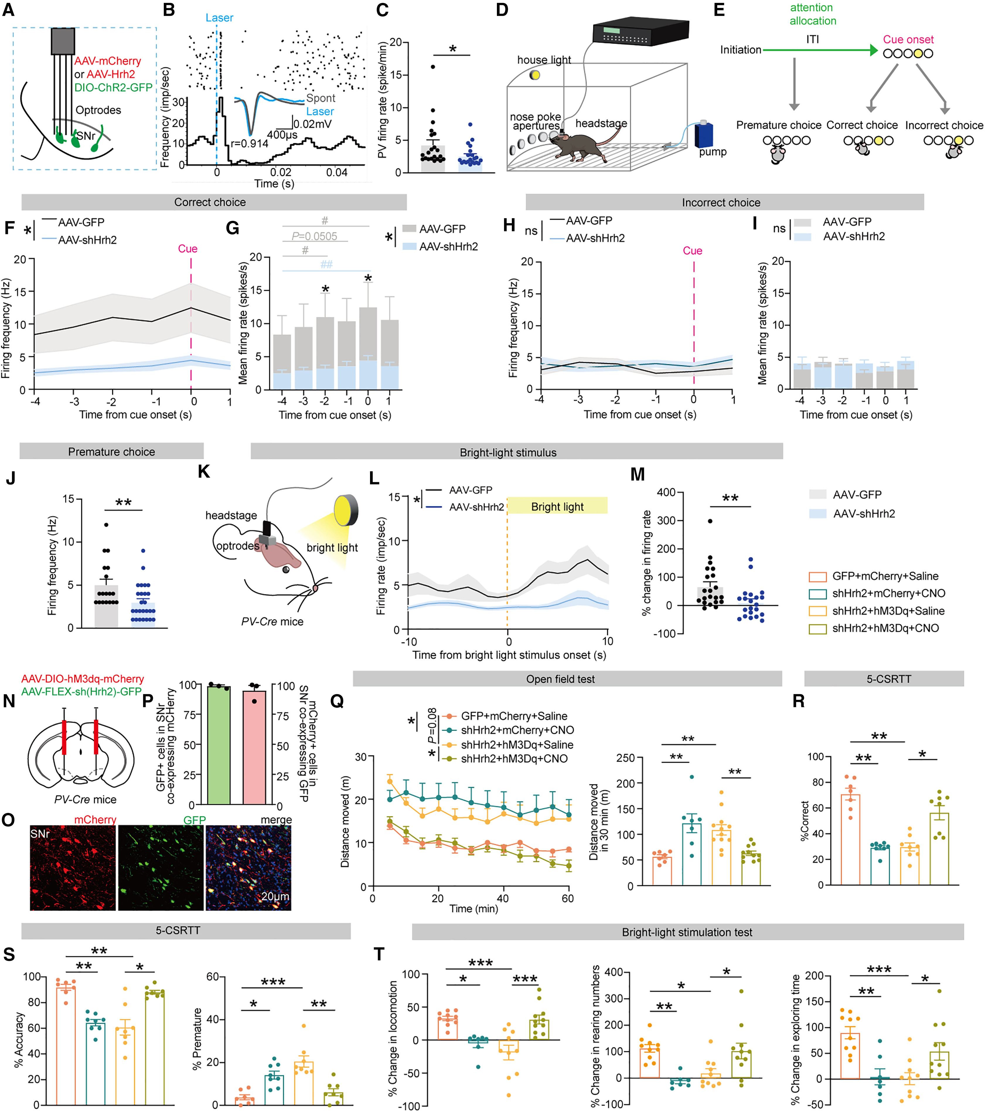
Using in vivo multichannel optrode recordings of opto-tagged SNr PV+ neurons (waveform correlation r = 0.914) in freely moving PV-Cre mice, the authors found that Hrh2 knockdown significantly reduced basal firing rates. During the 5-CSRTT, control SNr PV+ neurons exhibited robust firing increases during the -4 s to 0 s attention-allocation window preceding cue onset in correct trials; this pre-cue recruitment was markedly blunted in AAV-shHrh2 mice (whereas firing was unchanged before incorrect choices), and firing rates 1 s prior to premature responses were significantly lower. Firing increases during Phase 2 of the bright-light test were similarly blunted.
To test whether restoring SNr PV+ neuronal activity rescues these behavioral deficits, the authors co-expressed AAV-DIO-hM3Dq-mCherry and AAV-FLEX-shHrh2-EGFP in the SNr of PV-Cre mice. Chemogenetic excitation with CNO (5 mg/kg, i.p.) normalized open-field locomotion, rescued % Correct, % Accuracy, and % Premature metrics in the 5-CSRTT, and restored Phase-2 behavioral responses in the bright-light test.
Source: Dadao An et al. · Neuron · © Elsevier Inc.
Original figure and English caption ↗
H2R Loss in SNr PV+ Neurons Disinhibits SNc Dopamine Neurons and Elevates Dorsal Striatal Dopamine Release
Anterograde transsynaptic tracing (scAAV2/1-Syn-FLEX-GFP) and monosynaptic rabies virus retrograde tracing from SNc dopamine starter cells in DAT-Cre mice demonstrated that 76.750% ± 5.490% of presynaptic SNr neurons innervating SNc dopamine neurons are PV+. Fiber photometry with the dopamine sensor Syn-GRAB_DA2h in the dorsal striatum (dStr) showed that optogenetic inhibition (DIO-Arch-YFP) of SNr PV+ neurons elevated striatal dopamine levels and delayed post-illumination recovery.
In vivo multichannel recordings of putative SNc dopamine neurons (pDAs) revealed that SNr Hrh2 knockdown elevated basal pDA firing rates. During correct 5-CSRTT trials, pre-cue suppression of SNc pDA firing was blunted (with higher firing rates 2 s before cue onset), and pDA firing 1 s prior to premature responses was elevated. In the bright-light test, SNc pDAs in SNr Hrh2-knockdown mice exhibited elevated Phase-1 firing rates, spikes within bursts, and burst duration, preventing further recruitment during Phase 2.
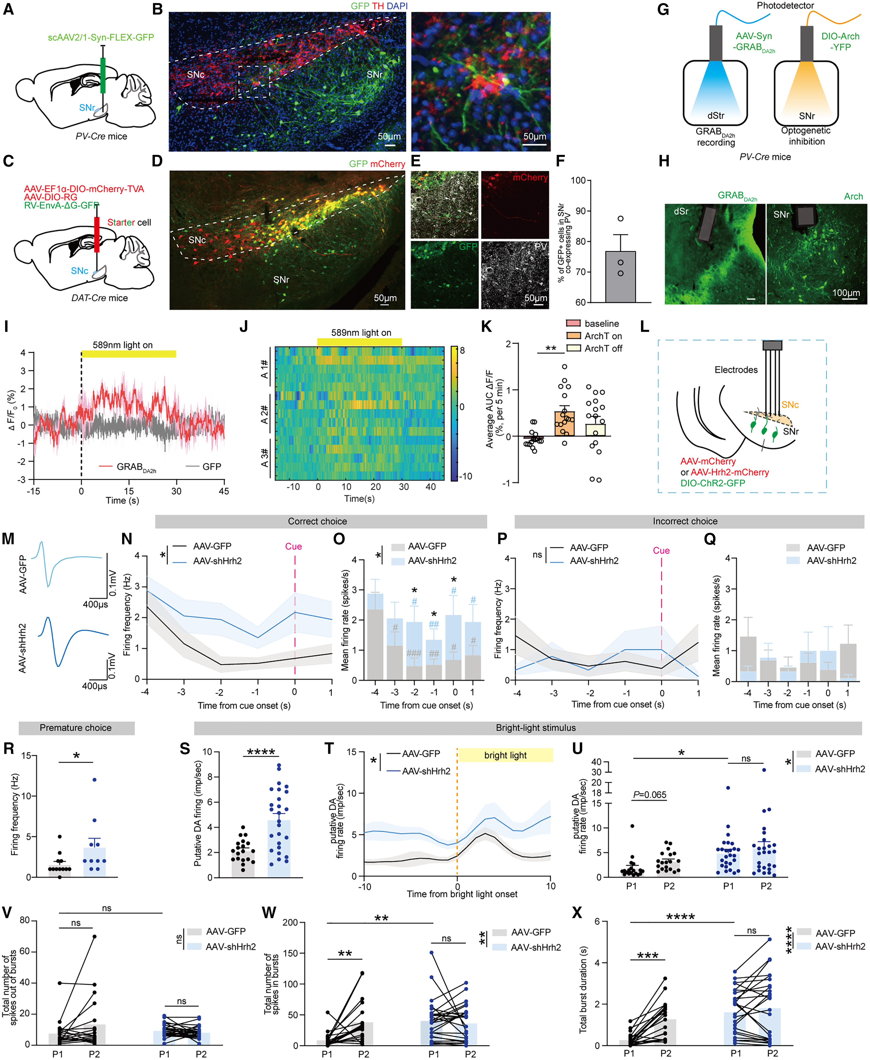
Source: Dadao An et al. · Neuron · © Elsevier Inc.
Original figure and English caption ↗
Activating SNr→SNc PV Terminals or Inhibiting Nigrostriatal DA Neurons Rescues Attention, Impulsivity, and Bright-Light Deficits Without Affecting Hyperactivity
To define the behavioral role of the SNr→SNc PV pathway, the authors co-injected DIO-hM3Dq-mCherry and AAV-shHrh2 into the SNr of PV-Cre mice and locally infused CNO (5 μM, 100 nL) into the SNc to selectively excite SNr→SNc PV terminals. Terminal activation rescued % Correct, % Accuracy, and % Premature responses in the 5-CSRTT and restored Phase-2 behavioral responses in the bright-light test, but had no effect on open-field hyperactivity.
Complementarily, intersectional chemogenetic inhibition of nigrostriatal dopamine neurons—achieved by injecting retrograde rAAV2retro-hSyn-FLP into the dStr and AAV-fDIO-hM4Di-mCherry into the SNc (94.570% ± 1.319% TH+ specificity)—similarly rescued bright-light stimulation deficits in SNr Hrh2-knockdown mice without reducing open-field hyperactivity.
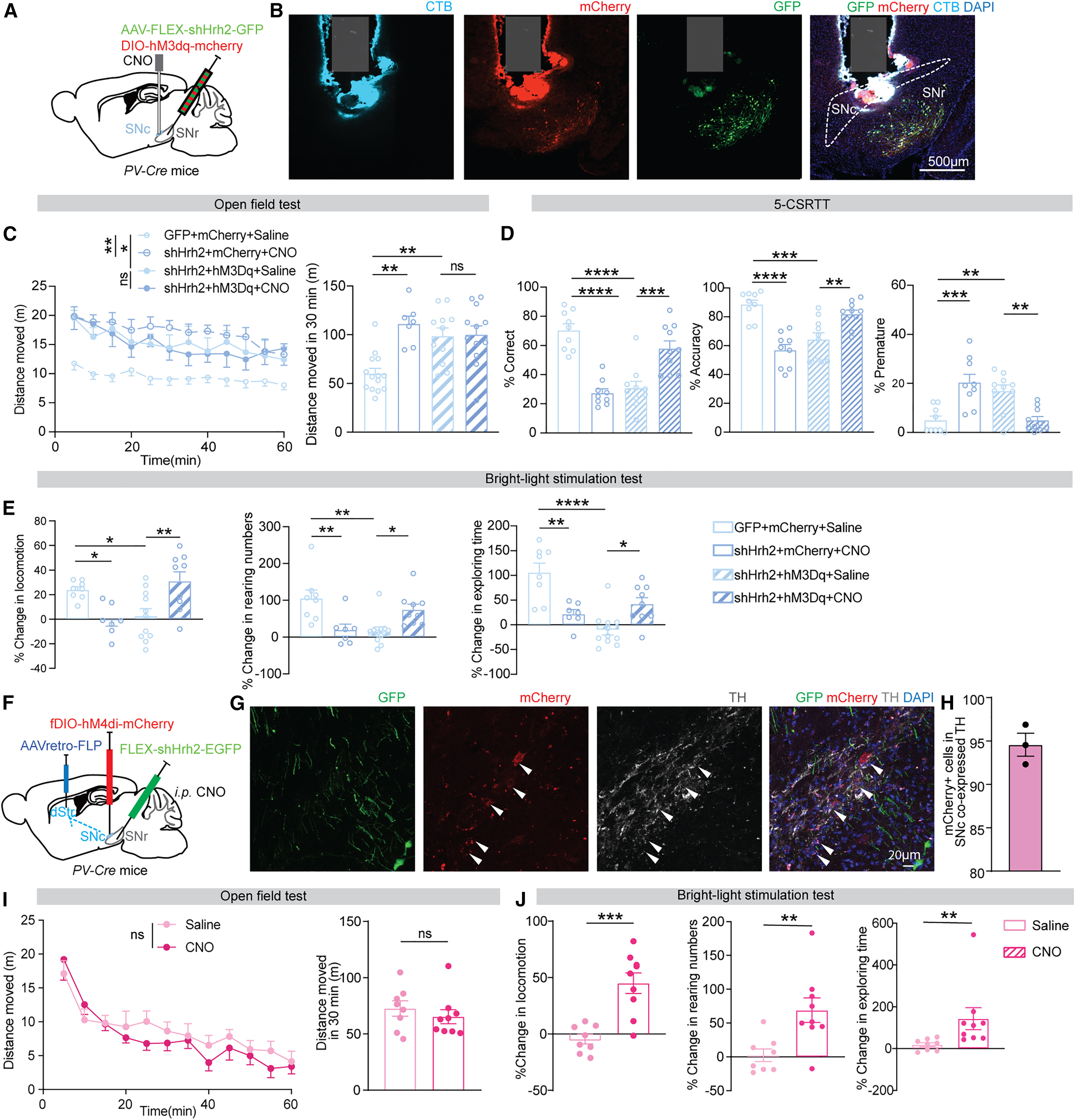
Source: Dadao An et al. · Neuron · © Elsevier Inc.
Original figure and English caption ↗
The SNr→mSC PV Pathway Regulates Locomotor Hyperactivity and Bright-Light Risk Assessment, Completing a Dual-Pathway Model
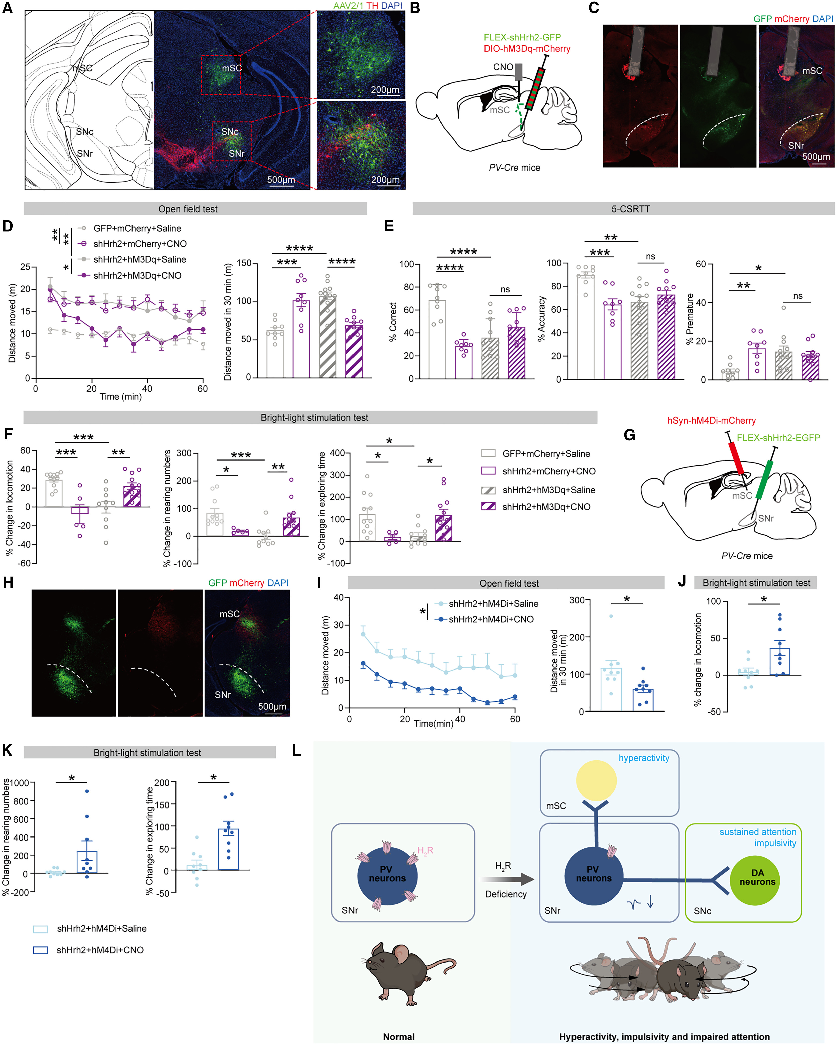
To identify the circuit mediating locomotor hyperactivity, the authors examined SNr PV+ projections to the medial superior colliculus (SNr→mSC PV). In PV-Cre mice co-expressing DIO-hM3Dq-mCherry and AAV-shHrh2 in the SNr, local CNO microinfusion into the mSC selectively reversed open-field hyperactivity and restored Phase-2 behavioral responses in the bright-light test, without improving % Correct, % Accuracy, or % Premature metrics in the 5-CSRTT.
Consistently, direct chemogenetic inhibition of mSC neurons (AAV-hSyn-hM4Di-mCherry) in SNr Hrh2-knockdown mice reversed open-field hyperactivity and bright-light response deficits. Together, these findings establish a dual-pathway model (Figure 6L) in which H2R deficiency in SNr PV+ neurons drives inattention and impulsivity via disinhibition of the SNr→SNc PV pathway, drives locomotor hyperactivity via disinhibition of the SNr→mSC PV pathway, and impairs bright-light defensive risk assessment through both pathways.
Source: Dadao An et al. · Neuron · © Elsevier Inc.
Original figure and English caption ↗