A node in the seizure network: how posterior amygdala engages wider circuits
A male mouse study links posterior amygdala excitatory cells with insula and inhibitory feedback. Reducing this population attenuates acute and chronic seizures in a hippocampal-onset model.
The amygdala contains multiple nuclei with distinct functions. Identifying its critical contribution to seizure networks requires resolving particular cells and connections. A 2024 Advanced Science study focuses on posterior basolateral amygdala (pBLA). Its excitatory neurons connect both to seizure-engaged insula and to regions providing inhibitory feedback. Facilitation and restraint coexist in one network, highlighting a node distant from the hippocampal origin.
pBLA excitatory cells contribute to seizure generation and propagation, with insula as an important downstream component and BNST/CeA providing inhibitory feedback. Reducing pBLA cells attenuates seizures in the tested hippocampal-onset model.
Contents · 7 sections
When posterior cells are activated
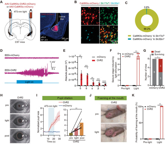
Activating pBLA glutamatergic neurons evokes prominent epileptiform discharges and severe seizures, with some deaths. Activating corresponding anterior basolateral-amygdala cells does not produce the same outcome. Moving from anterior to posterior changes the functional consequence substantially.
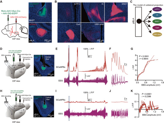
During hippocampally induced seizures, calcium activity in BNST-projecting pBLA cells strongly tracks EEG discharges, unlike the corresponding anterior population. Activation and recordings during model seizures independently connect posterior cells to the seizure network.
Source: Sun YH et al. · Advanced science (Weinheim, Baden-Wurttemberg, Germany) · CC BY
Original figure and English caption ↗
One population, several destinations
pBLA cells send collaterals to insula, BNST, central amygdala (CeA) and other areas. Retrograde labeling shows substantial overlap among target-defined populations. Stimulating terminals at one destination can therefore recruit more than a single local connection, including other branches of the same cells.
Terminal stimulation in insula, BNST or CeA induces seizures, whereas stimulation at the tested prefrontal or hypothalamic targets does not. Comparing terminal activation with activation of input-recipient cells then reveals different results.
Source: Sun YH et al. · Advanced science (Weinheim, Baden-Wurttemberg, Germany) · CC BY
Original figure and English caption ↗
Insula reveals a different role
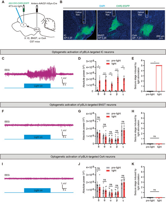
Activating insular cells receiving pBLA input evokes discharges and seizures, unlike activating the corresponding BNST or CeA recipient cells. Reducing insular cells attenuates pBLA-driven seizures, whereas reducing BNST or CeA cells does not provide the same attenuation.
Why does BNST-terminal stimulation still induce seizures? Simultaneous recordings show increased pBLA somatic calcium during activation of those terminals, suggesting upstream recruitment and effects through collaterals. Insular facilitation and broader effects of terminal stimulation thus receive distinct explanations.
Source: Sun YH et al. · Advanced science (Weinheim, Baden-Wurttemberg, Germany) · CC BY
Original figure and English caption ↗
Inhibition returning upstream
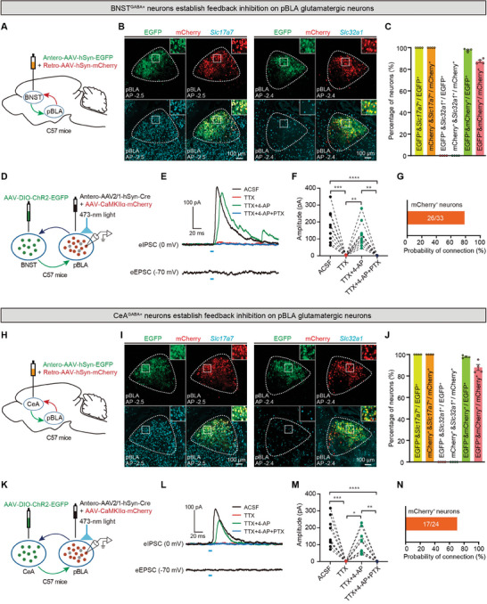
BNST and CeA do more than participate in outward propagation. Tracing shows input-recipient cells projecting back to pBLA, and many excitatory cells receiving those connections also send output to the two regions. The network contains reciprocally connected routes.
Activating the returning terminals in slices evokes direct GABAergic inhibitory currents in pBLA excitatory cells. BNST and CeA thus provide feedback brakes: excitation travels outward and inhibition returns to restrain the population.
Source: Sun YH et al. · Advanced science (Weinheim, Baden-Wurttemberg, Germany) · CC BY
Original figure and English caption ↗
The network after losing its brakes
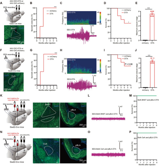
Reducing BNST or CeA GABAergic cells is followed by recurrent sporadic discharges and seizures, with some later deaths. Seizures also occur readily in new environments. Losing these populations makes abnormal network activity more likely.
When pBLA excitatory cells are reduced concurrently, the corresponding seizures and deaths are not observed during monitoring. The combined intervention links loss of feedback inhibition with pBLA engagement, distinguishing this node from a view in which all downstream regions equally facilitate seizures.
Source: Sun YH et al. · Advanced science (Weinheim, Baden-Wurttemberg, Germany) · CC BY
Original figure and English caption ↗
Hippocampal input reaches this node
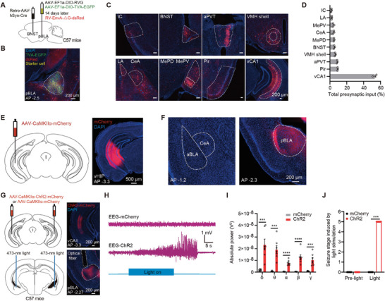
Upstream tracing identifies ventral hippocampal CA1 as a major excitatory source to pBLA. Activating CA1 terminals there induces epileptiform discharges and seizures, unlike activation of two other tested inputs.
Hippocampus, posterior amygdala and insula form connected components capable of affecting seizures, while BNST and CeA provide restraint. The study then tests whether reducing pBLA engagement changes seizures in a hippocampal-onset epilepsy model.
Source: Sun YH et al. · Advanced science (Weinheim, Baden-Wurttemberg, Germany) · CC BY
Original figure and English caption ↗
Distant from onset, yet influencing seizures
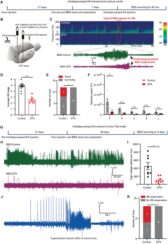
In the acute hippocampal model, reducing pBLA excitatory cells beforehand lowers seizure severity and deaths. In the chronic model, reducing the population after hippocampal induction decreases later spontaneous high-voltage spikes, with no generalized seizures observed during monitoring.
In a supplementary experiment, suppressing pBLA activity without removing cells also reduces acute discharges and seizure severity, although attenuation in some EEG bands is weaker. The extent of network suppression varies with the intervention.
Reducing pBLA does not eliminate all abnormal discharges. It functions as an important network bottleneck influencing progression to severe seizures. These findings in adult male mice and the tested hippocampal-onset model highlight the importance of nodes and feedback connections beyond the initial source.
Source: Sun YH et al. · Advanced science (Weinheim, Baden-Wurttemberg, Germany) · CC BY
Original figure and English caption ↗