One brain region, two circuits: cellular division of labor in the medial mammillary body
A mouse study connects the morphology, electrical activity and wiring of CB and PV cell classes, revealing how neighboring populations can process information through segregated circuits.
As a mouse begins to move, one cell population in the medial mammillary body becomes active, while a neighboring population shows no comparably clear change. This small region, part of networks associated with memory, navigation and emotion, is not internally uniform. A study published on June 23, 2025 in Molecular Psychiatry identifies two major cell classes using the calcium-binding proteins CB and PV as markers. Their shapes, firing properties, inputs and outputs differ, and local connections between them are rarely detected. Neighboring populations can thus form parallel, segregated channels.
The two cell classes form distinct input-output circuits. Activating CB cells induces place avoidance and increases locomotion; activating PV cells does not produce these effects in the same tasks.
Contents · 7 sections
Two cell classes occupy complementary territories
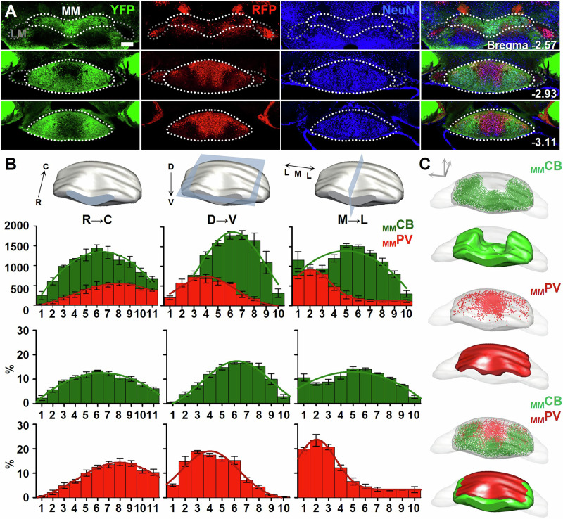
The medial mammillary body belongs to the extended hippocampal system and has often been described as a relatively uniform population of excitatory projection neurons. Labeling CB and PV cells separately reveals that together they account for most neurons here, with little overlap. In this region, PV identifies a cell class rather than the PV inhibitory interneurons familiar from some other brain areas.
Three-dimensional reconstruction shows a ventrolateral bias for CB cells and a dorsomedial bias for PV cells. The CB territory cradles the PV territory from the front, side and below, resembling a calyx around a bud. This spatial pattern becomes the starting point for investigating their division of labor.
Source: Liu H et al. · Molecular psychiatry · CC BY-NC-ND
Original figure and English caption ↗
Different labels identify different cellular properties
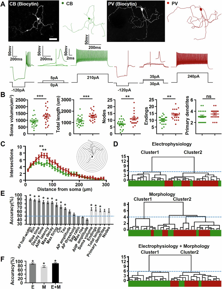
Are the colors merely marking different molecules? Single-cell reconstruction provides a fuller answer. CB cells have smaller somata, shorter dendrites overall and less branching; PV cells have larger somata and more complex dendrites. These differences correspond to distinct electrical properties of their membranes.
CB cells have taller, wider action potentials and a lower maximum firing frequency, whereas PV cells show a different set of properties. Classification using morphology and electrical activity together closely matches the CB and PV labels. The two colors therefore correspond to coordinated differences in cell shape and physiology.
Source: Liu H et al. · Molecular psychiatry · CC BY-NC-ND
Original figure and English caption ↗
Both cell classes can change their activity mode
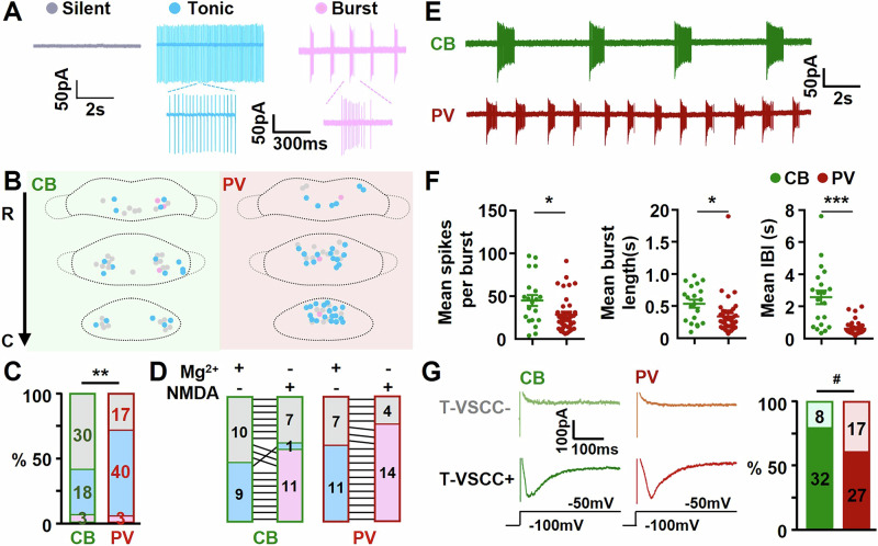
In resting brain slices, some cells remain silent, others fire tonically and others produce bursts. A greater proportion of PV cells is spontaneously active. Activity mode is not an immutable label attached to cell identity: individual cells are also observed switching between tonic and burst firing.
When NMDA-receptor activity is enhanced, both classes more readily fire in bursts, but with different rhythms. CB cells produce more spikes per burst with longer intervals between bursts; PV bursts are comparatively shorter and more frequent. Their division of labor concerns both where signals originate and the temporal patterns used to transmit them.
Source: Liu H et al. · Molecular psychiatry · CC BY-NC-ND
Original figure and English caption ↗
Shared destinations, distinct territories
Following axons outward extends the division of labor. Both classes project to regions including the anterior thalamus, but their terminals occupy largely complementary territories. Recordings from anterior-thalamic cells support functional segregation corresponding to this spatial separation, beyond a boundary between colors in a microscopic image.
Tracing upstream reveals that both classes receive subicular input with different biases: CB cells are more associated with ventral subiculum, and PV cells with dorsal subiculum. Hypothalamic sources also account for a larger share of CB inputs. Neighboring populations therefore receive different combinations of information.
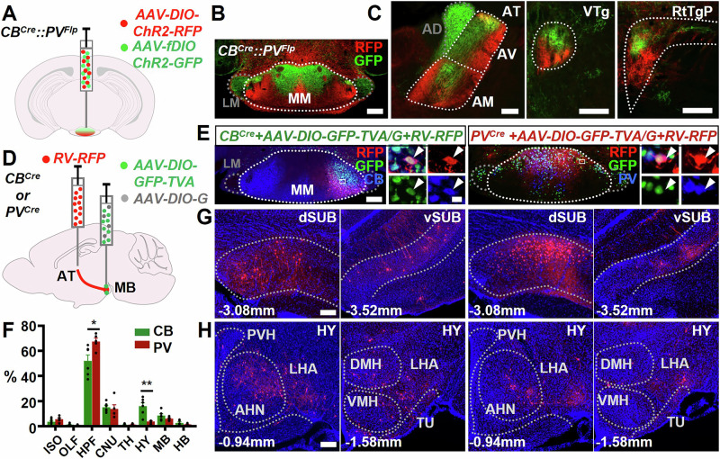
Source: Liu H et al. · Molecular psychiatry · CC BY-NC-ND
Original figure and English caption ↗
Activating CB cells increases movement and avoidance
If the circuits serve different functions, will increasing their activity produce different behavior? The study pairs cell activation with one spatial compartment. Activating CB cells reduces time spent in that compartment while increasing activity within it; activating PV cells does not produce comparable place avoidance.
Open-field observations confirm increased movement time and distance during CB activation compared with the periods before and after it. PV activation and controls show no equivalent change. The coexistence of place avoidance and increased movement shows why time allocation alone can miss changes in how an animal acts.
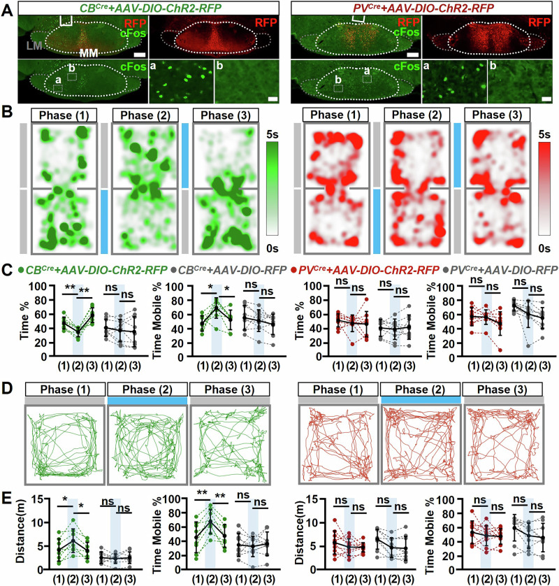
Source: Liu H et al. · Molecular psychiatry · CC BY-NC-ND
Original figure and English caption ↗
CB population signals rise at movement onset
Beyond imposed activation, the researchers record calcium signals as mice move freely. Aligning activity with movement onset reveals a clear increase in CB cells, with no equivalent significant change in PV cells. This agrees with the activation experiments and establishes a movement association during spontaneous behavior.
Together, the results show that CB cells can promote movement and become active when movement starts. The weaker PV response in these tasks leaves room for other roles in behavior or information processing. The mammillary body’s established associations with memory and navigation require more specific task-by-task mapping.
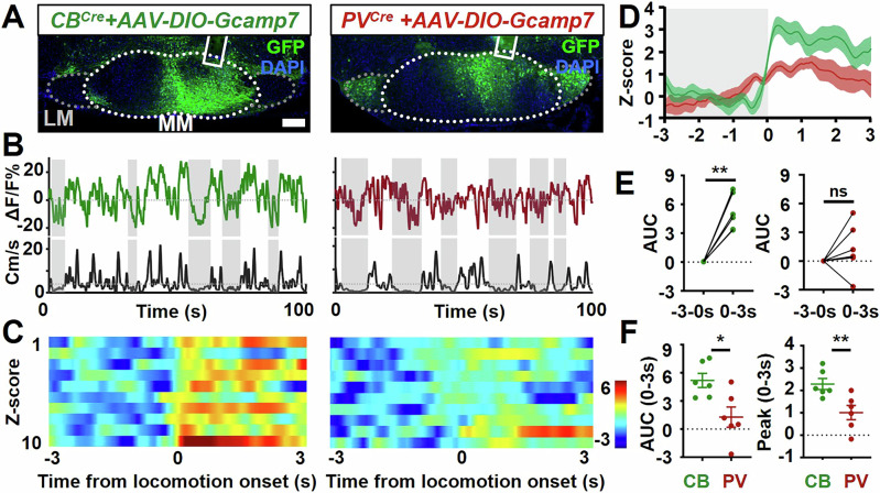
Source: Liu H et al. · Molecular psychiatry · CC BY-NC-ND
Original figure and English caption ↗
Neighbors need not form a serial circuit
The most counterintuitive result concerns local connectivity. Activating one class does not clearly recruit the other. Paired recordings from 123 mammillary-cell pairs detect no local chemical synapses or electrical coupling, consistent with the lack of local axonal branching.
Information processing here therefore resembles adjacent parallel channels more than a local serial relay before output. Behavioral experiments in adult male mice and cellular analyses in both sexes jointly map this organization. Finer divisions within the two major cell classes remain a next layer to uncover.
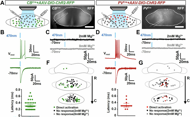
Source: Liu H et al. · Molecular psychiatry · CC BY-NC-ND
Original figure and English caption ↗