How do disrupted cellular powerhouses contribute to morphine withdrawal?
A mouse study connects morphine-responsive neurons, mitochondrial fragmentation and withdrawal, revealing ways to reduce selected withdrawal-related outcomes.
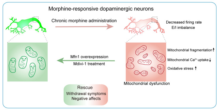
Morphine can relieve pain, yet withdrawal after repeated exposure can bring bodily discomfort, avoidance and anxiety-like behavior. Which brain cells participate in this transition, and what changes inside them? A study published in Journal of Clinical Investigation on January 18, 2024 focuses on a dopaminergic ensemble recruited by initial morphine exposure in the mouse ventral tegmental area (VTA). Its clues lead to mitochondria: cellular powerhouses that continually divide, fuse and remodel. Repeated exposure accompanies more fragmented mitochondria and reduced expression of a fusion-related gene in these responsive cells. Intervening in mitochondrial dynamics reduces selected withdrawal-related behaviors.
Mitochondrial changes in morphine-responsive dopaminergic cells accompany withdrawal development. Increasing their Mfn1 expression or intervening in mitochondrial fission during repeated exposure reduces selected somatic and negative-affect-related outcomes.
Contents · 8 sections
Initially recruited cells later become less active
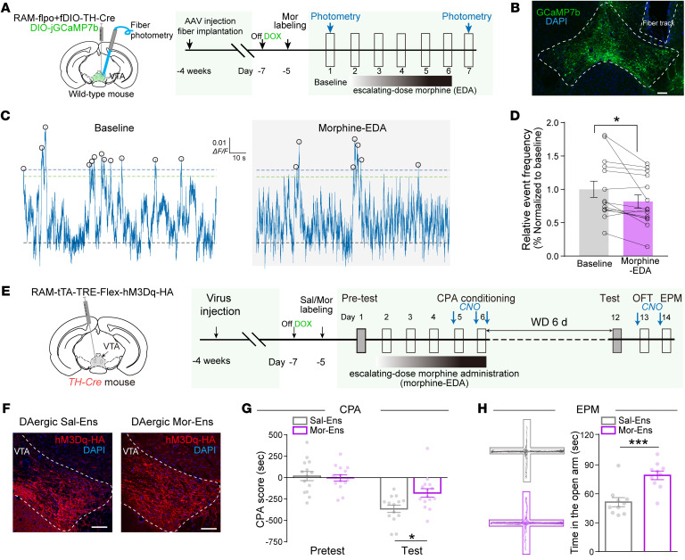
VTA dopaminergic neurons do not respond uniformly to drugs. The Mor-Ens population tracked here was activity-labeled during initial morphine exposure. After repeated administration, its spontaneous calcium-event frequency decreased, indicating an altered population activity state.
Recruiting this population during withdrawal-related tests reduced avoidance of a withdrawal-associated context and increased open-arm time in an elevated plus maze, without significantly changing locomotor distance. These findings connect ensemble activity with negative-affect-related behavior and prompt a deeper question: what changed the cells’ state?
Source: Jiang C et al. · The Journal of clinical investigation · CC BY
Original figure and English caption ↗
The powerhouses change at several levels
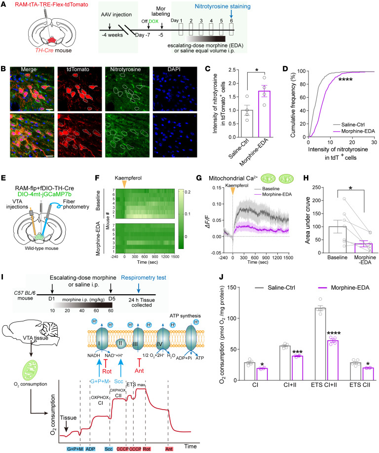
Transcriptomic comparisons highlighted mitochondrial respiration, structure and oxidoreductase-related processes. Mor-Ens showed increased nitrotyrosine signal, consistent with oxidative-stress-related changes, alongside weaker evoked mitochondrial calcium-uptake responses. Mitochondrial alteration thus involved more than appearance.
Several respiratory measures also decreased in VTA tissue. These tissue-level findings complement signals from the defined ensemble: one describes regional respiratory function, while the other localizes changes to morphine-responsive cells. Together, these observations bring mitochondria into focus.
Source: Jiang C et al. · The Journal of clinical investigation · CC BY
Original figure and English caption ↗
Fragmentation persists in dendrites after withdrawal
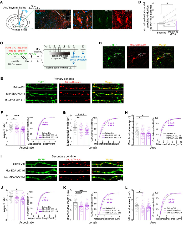
Mitochondria are not static particles. Fission and fusion help them adjust shape and distribute function. Following repeated morphine exposure, mitochondria in Mor-Ens dendrites were shorter and smaller, with reduced aspect ratios. These changes were observed on withdrawal days 1 and 21.
Expression of the fusion-related gene Mfn1 also decreased, with reduced transcript signal persisting for weeks after withdrawal. Mitochondrial autophagy-related signals increased in VTA neurons. Changes in architecture, fusion-related expression and turnover enrich the cellular picture of withdrawal.
Source: Jiang C et al. · The Journal of clinical investigation · CC BY
Original figure and English caption ↗
Increasing Mfn1 reduces selected bodily signs
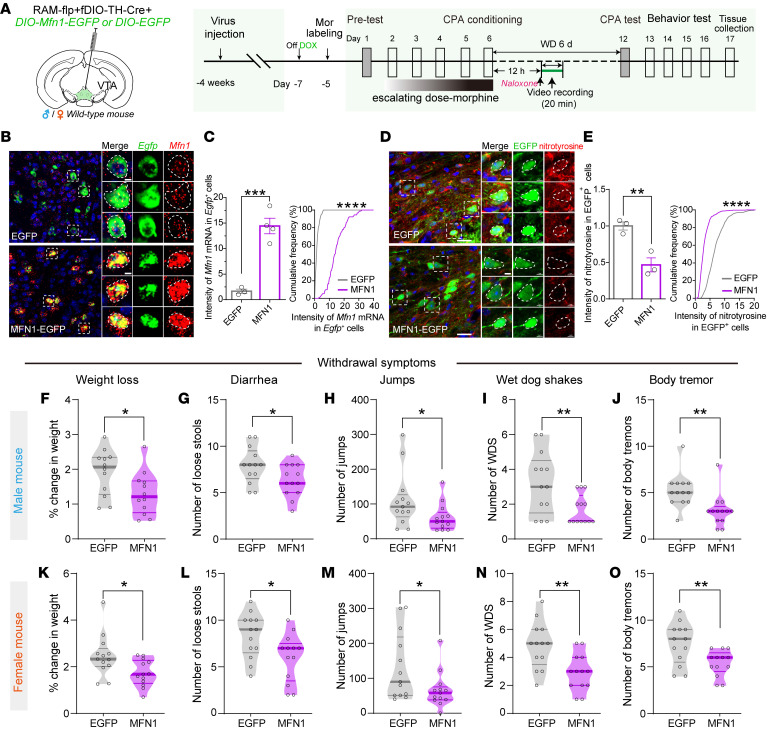
Increasing Mfn1 expression in dopaminergic Mor-Ens reduced nitrotyrosine signal, an oxidative-stress-related marker, and altered somatic withdrawal outcomes. This intervention connects a fusion-related molecule within defined cells to behavior.
In both sexes, naloxone-precipitated withdrawal involved less weight loss, diarrhea, jumping, wet-dog shakes and body tremor, while piloerection did not significantly improve. The molecular intervention affects several aspects of withdrawal, with a specific pattern across outcomes.
Source: Jiang C et al. · The Journal of clinical investigation · CC BY
Original figure and English caption ↗
Less context avoidance and greater social novelty
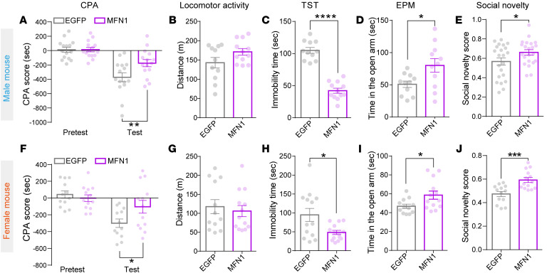
Mfn1 enhancement also affected post-withdrawal behavior. Mice showed less avoidance of withdrawal-associated contexts, less tail-suspension immobility and more elevated-plus-maze open-arm time. These changes occurred in both sexes without a significant change in overall locomotor distance.
Preference for a novel rather than familiar conspecific increased, while basic social preference did not significantly change. Somatic and negative-affect-related outcomes together position Mfn1 in this defined population as a useful entry point into withdrawal development.
Source: Jiang C et al. · The Journal of clinical investigation · CC BY
Original figure and English caption ↗
Another route toward less fragmented mitochondria
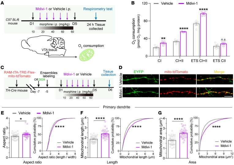
The researchers administered the mitochondrial-fission intervention compound Mdivi-1 during repeated morphine exposure. Several VTA tissue respiratory measures improved, alongside increased mitochondrial length, area and aspect ratio in Mor-Ens dendrites. These structural and functional observations support this intervention route.
Reduced spontaneous and evoked firing and the lower excitatory-to-inhibitory current ratio were also ameliorated. Systemically administered Mdivi-1 can act across multiple cells and targets; changes in the responsive ensemble provide an important observation within that broader picture.
Source: Jiang C et al. · The Journal of clinical investigation · CC BY
Original figure and English caption ↗
Somatic and affect-related effects have distinct profiles
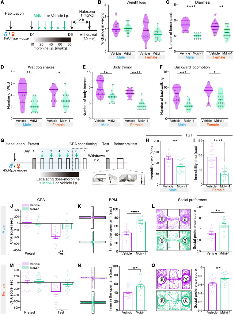
Mdivi-1 reduced diarrhea, shaking, tremor and backward locomotion in both sexes, but did not significantly improve weight loss; reduced jumping was observed mainly in females. Post-withdrawal context avoidance, tail-suspension immobility and open-arm avoidance decreased, social preference increased, and total locomotor distance did not significantly change.
Drug-associated learning and seeking did not change uniformly. Mdivi-1 reduced morphine-conditioned place preference without significantly altering self-administration acquisition or seeking at 24 hours of withdrawal, while reducing active nose pokes at 14 days. Its effects depend on the behavioral measure and time point.
Source: Jiang C et al. · The Journal of clinical investigation · CC BY
Original figure and English caption ↗
An analgesia-related clue and questions ahead
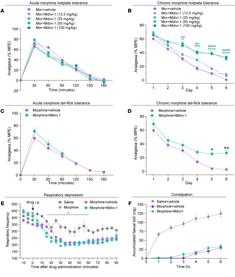
Mdivi-1 did not significantly alter acute morphine analgesia in hot-plate or tail-flick tests and ameliorated analgesic tolerance with repeated exposure. Respiratory depression and constipation did not significantly improve. These results make mitochondrial dynamics a promising research direction while revealing separable opioid-related outcomes.
This mouse study connects morphine-responsive cells, mitochondrial architecture and withdrawal behavior across scales. Its principal interventions during repeated drug exposure provide clues for reducing withdrawal development. Sustained effects after withdrawal is established, greater cellular and molecular specificity, and relevance to human research remain important next questions.
Source: Jiang C et al. · The Journal of clinical investigation · CC BY
Original figure and English caption ↗