Astrocytes help mice recognize a familiar companion
In the adult male mouse ventral hippocampus, NLG3 links astrocyte activity, glutamate handling and adenosine signaling to social recognition.
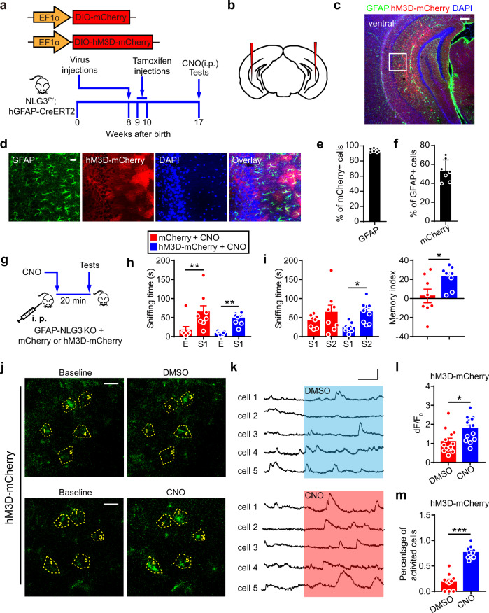
How does the brain distinguish a familiar companion from a new one? Neurons are a natural starting point, but astrocytes, whose fine processes surround synapses, may also shape this memory. A study published in Nature Communications on October 5, 2024 examines neuroligin 3, or NLG3, in the adult male mouse hippocampus. Removing astrocytic NLG3 preserved social approach while disrupting the usual preference for a new companion. The study connects this change with glutamate transport, astrocyte activity and adenosine receptors.
Ventral hippocampal astrocytic NLG3 loss accompanies increased EAAT2 protein, weaker social-evoked astrocyte and adenosine signals, impaired potentiation and social recognition. Several interventions improve these outcomes.
Contents · 8 sections
Social approach persists while recognition changes
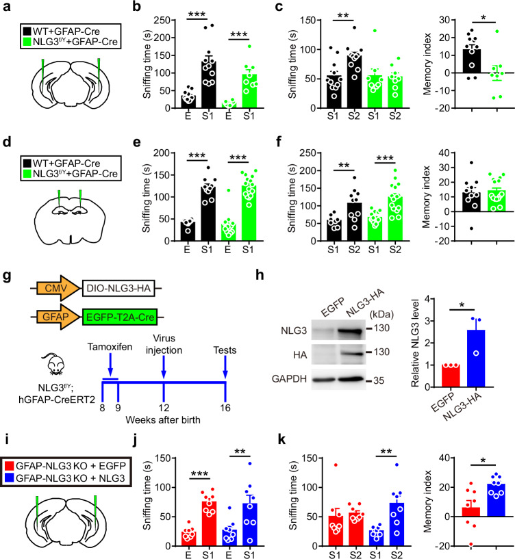
Both groups preferred a companion over an empty cage. When familiar and new companions were presented together, controls spent more time investigating the new animal, whereas the NLG3-deletion group lacked that preference. Measured locomotion, basic olfaction and novel-object recognition did not significantly change, focusing the difference on tested social recognition.
Deleting NLG3 in the ventral hippocampus produced the recognition deficit; the corresponding dorsal manipulation did not. Re-expressing NLG3 in ventral hippocampal astrocytes improved preference for the new companion, connecting a synaptic adhesion protein with glial function in the adult brain.
Source: Dang R et al. · Nature communications · CC BY-NC-ND
Original figure and English caption ↗
Astrocyte responses weaken during social investigation
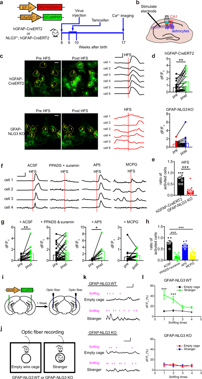
NLG3 loss did not significantly alter measured astrocyte abundance, morphology or basal spontaneous calcium activity. The difference emerged during recruitment: controls responded to high-frequency stimulation, while responses were impaired after deletion. Social investigation in behaving mice revealed a similar separation.
The cells remained present, but their response to surrounding activity changed. Impaired recognition was accompanied by weaker recruitment during social events rather than a general loss of astrocytes.
Source: Dang R et al. · Nature communications · CC BY-NC-ND
Original figure and English caption ↗
Recruiting astrocytes improves recognition
Increasing ventral hippocampal astrocyte activity restored preference for a new companion and improved the recognition index in deletion mice. Complementary slice recordings confirmed stronger calcium signals. Measured locomotion, olfaction and novel-object recognition did not significantly change.
Improved behavior alongside stronger cellular responses made astrocyte activity an intervention point. The next question concerned how NLG3 influences the signals these cells receive, directing attention to glutamate transport around synapses.
Source: Dang R et al. · Nature communications · CC BY-NC-ND
Original figure and English caption ↗
Stronger transport accompanies weaker event signals
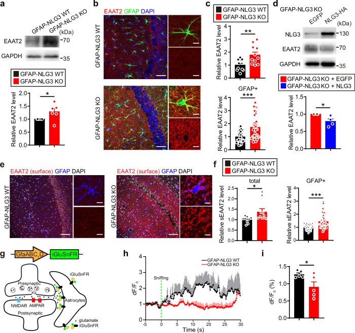
Glutamate must be cleared after signaling. EAAT2 protein and surface labeling increased following NLG3 loss and decreased after NLG3 re-expression. Social-evoked extracellular glutamate signals were also weaker, connecting transporter changes with altered glutamate availability around synapses.
The molecular changes were not uniform: EAAT2 messenger RNA decreased despite higher protein levels. mTOR-related findings implicated translational regulation, adding a link between protein control and extracellular signaling.
Source: Dang R et al. · Nature communications · CC BY-NC-ND
Original figure and English caption ↗
Limiting transport restores cellular recruitment
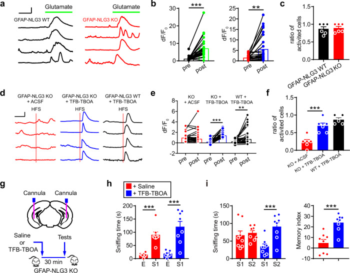
Applied glutamate increased calcium signals in both groups, indicating retained response capacity after deletion. An EAAT1/2 transport inhibitor improved stimulation-evoked calcium responses in deletion slices. Local ventral hippocampal treatment also improved new-companion preference and the recognition index.
These results connect stronger transport, weaker event-related signals and altered behavior. Glutamate clearance supports stability while influencing the signal available during an event; adjusting transport recruited astrocytes again in this model.
Source: Dang R et al. · Nature communications · CC BY-NC-ND
Original figure and English caption ↗
Basal transmission persists while potentiation is impaired
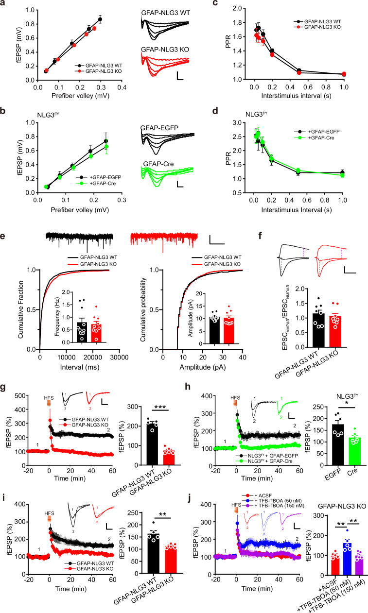
Basal synaptic transmission, paired-pulse responses and miniature excitatory currents did not significantly change, whereas long-term potentiation was impaired. Ordinary transmission persisted while the ability to sustain stronger connections after intense activity was affected.
Transport inhibition also improved potentiation, with a concentration-dependent outcome: 50 nM restored it, whereas 150 nM did not. The balance between clearance and activity-dependent signaling therefore matters.
Source: Dang R et al. · Nature communications · CC BY-NC-ND
Original figure and English caption ↗
A second chemical signal connects to neurons
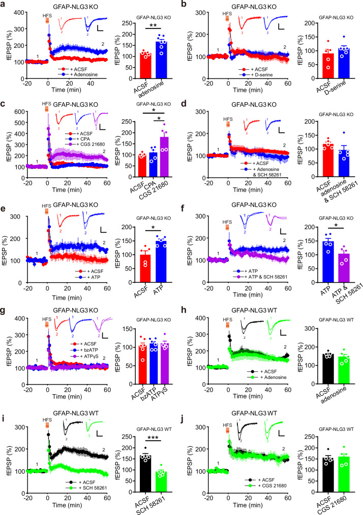
Adenosine added another mechanistic link. It improved potentiation in deletion slices, while the tested D-serine treatment did not. An A2a agonist also improved potentiation, whereas an A1 agonist did not; A2a antagonism blocked the adenosine rescue.
ATP also improved potentiation, but this effect was blocked by A2a antagonism. A nonhydrolyzable ATP analogue did not have the same effect. These findings support regulation through ATP-derived adenosine, connecting astrocyte-associated activity with neuronal receptor responses.
Source: Dang R et al. · Nature communications · CC BY-NC-ND
Original figure and English caption ↗
From chemical responses back to a familiar companion
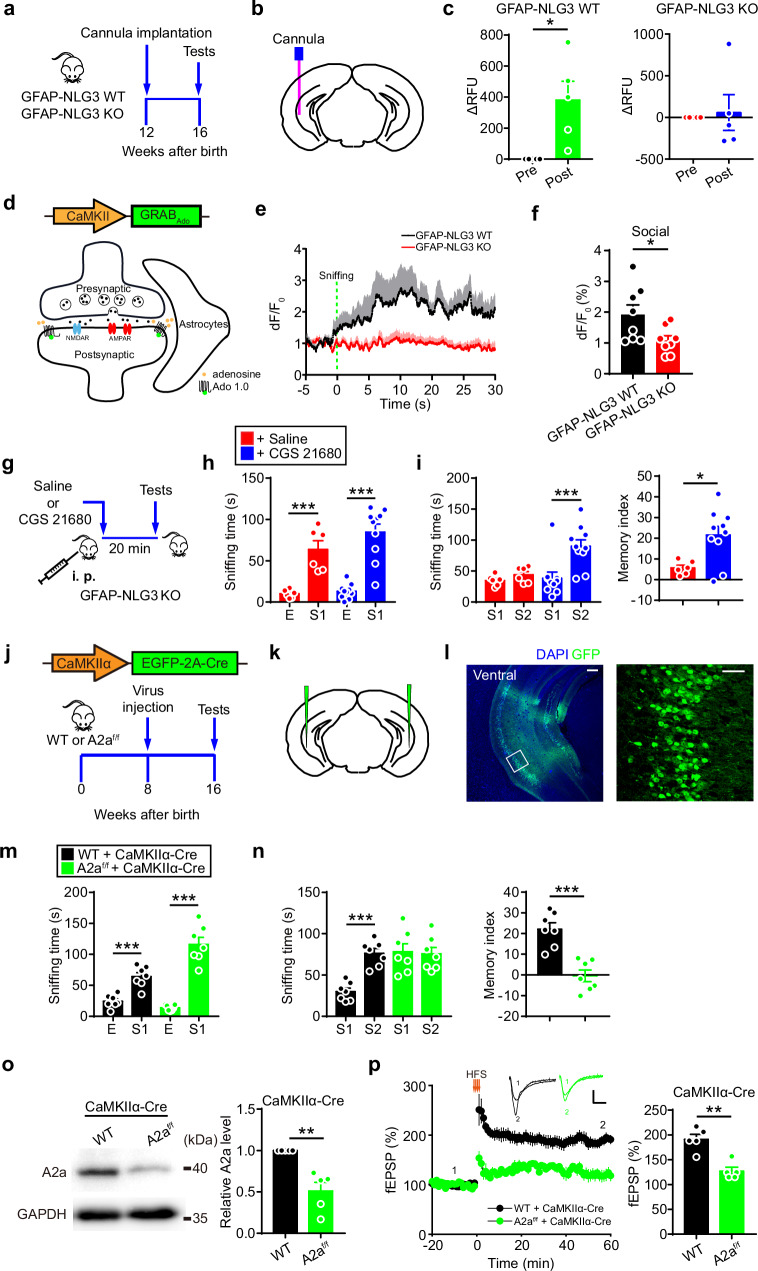
Controls showed stronger adenosine signals during social investigation; deletion mice had weaker event responses despite comparable basal levels. An A2a agonist improved recognition in deletion mice. Removing A2a receptors from ventral hippocampal excitatory neurons impaired recognition and potentiation while preserving social approach.
This adult male mouse study expands the cellular account of remembering a companion, linking NLG3, glutamate transport, astrocyte activity and adenosine receptors. Adenosine may also originate from neurons, and its effects depend on cell type and brain region. Following these links can deepen understanding of how glia participate in retaining experience.
Source: Dang R et al. · Nature communications · CC BY-NC-ND
Original figure and English caption ↗