Cellular roles in withdrawal: YTHDF1 and Sst-marked neurons
A mouse study links RNA regulation to cell type in morphine withdrawal: reducing YTHDF1 in inhibitory or Sst-marked ventrolateral periaqueductal-gray cells weakens somatic scores and place aversion.

Morphine withdrawal can produce somatic responses and lead animals to avoid environments associated with withdrawal. Which cellular processes might regulate both? A 2024 BMC Medicine study begins with YTHDF1 in the ventrolateral periaqueductal gray, vlPAG. This protein recognizes m6A modifications on RNA and participates in regulating how RNA information is used. Comparing a brain region, excitatory and inhibitory populations, then Sst-marked cells reveals that the same molecule has different roles across cell types. RNA modifications act as information-associated marks; proteins that recognize them help cells handle that information differently.
Reducing YTHDF1 in inhibitory vlPAG cells and Sst-marked cells attenuates somatic withdrawal scores and place aversion. Corresponding interventions in excitatory, PV- or Cck-marked populations do not significantly improve these outcomes. NF-κB and local activity changes provide mechanistic clues.
Contents · 7 sections
Somatic responses come with an aversive association
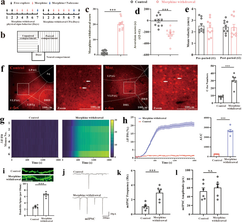
After repeated morphine exposure, naloxone precipitates withdrawal. Mice show somatic signs such as jumping and tremor, with higher composite withdrawal scores. A separate cohort in the place test avoids the withdrawal-paired environment. The measures describe bodily responses and learned environmental association respectively. Place aversion is measured through subsequent free-choice occupancy, following the association left by the experience.
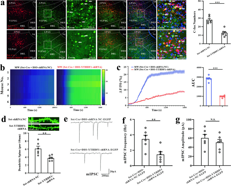
Mean movement speed changes nonsignificantly in the place test. The vlPAG shows more activity-marker-positive cells, stronger population calcium signals and greater dendritic-spine density. Miniature inhibitory currents received by local neurons increase in frequency, while amplitude changes nonsignificantly.
Source: Ou C et al. · BMC medicine · CC BY-NC-ND
Original figure and English caption ↗
YTHDF1 rises, and reducing it weakens responses
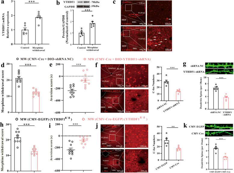
YTHDF1 transcript and protein levels rise in vlPAG in the withdrawal model. Reducing regional YTHDF1 lowers somatic withdrawal scores and weakens place aversion. Knockdown in wild-type mice and conditional genetic intervention yield behavioral changes in similar directions.
Interventions also accompany reductions in activity-marker-positive cell counts and dendritic-spine density. Molecular expression, local cellular changes and behavior become linked, but vlPAG contains several cell types. The next question is which population’s YTHDF1 influences these outcomes.
Source: Ou C et al. · BMC medicine · CC BY-NC-ND
Original figure and English caption ↗
Expression does not make roles identical
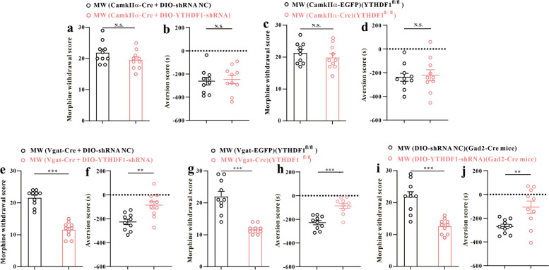
Single-cell data and histological colabeling detect YTHDF1 in excitatory and inhibitory neurons. Reducing it in CaMKIIα-marked excitatory populations changes somatic scores and place aversion nonsignificantly, with the result repeated under two genetic intervention conditions.
In contrast, reducing YTHDF1 in Vgat-marked inhibitory populations weakens both outcomes. Intervention using Gad2 genetic targeting gives similar results. Regional changes thus resolve into a distinction associated with cell type.
Source: Ou C et al. · BMC medicine · CC BY-NC-ND
Original figure and English caption ↗
NF-κB stands out without a uniform decline
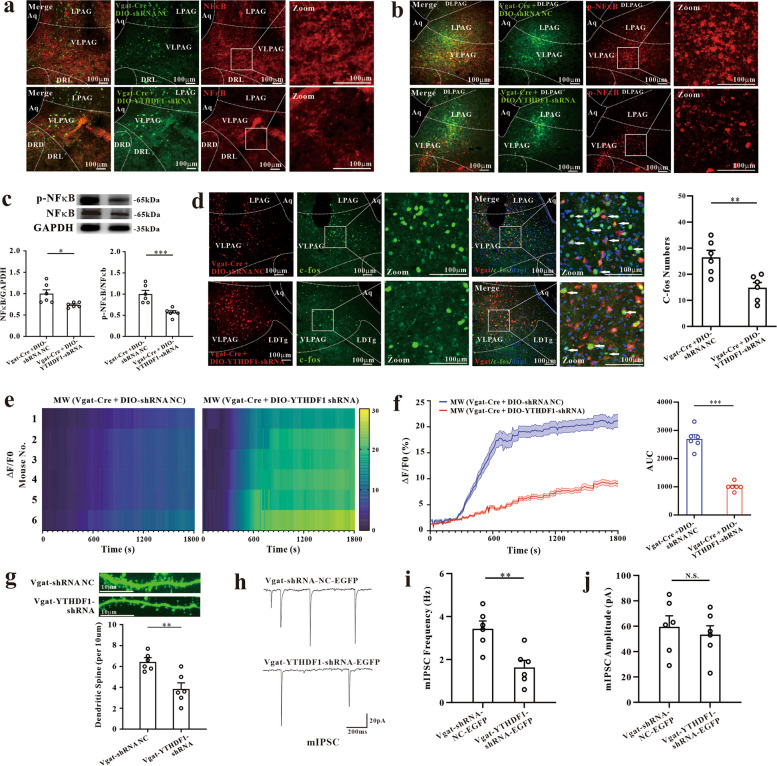
The researchers examine inflammation-related molecules. Single-cell analysis detects NF-κB-related expression in inhibitory populations, while several tested inflammatory cytokines have low expression. Reducing inhibitory-cell YTHDF1 lowers local NF-κB protein and its phosphorylation ratio.
IL-1β, IL-6 and TNF-α levels change nonsignificantly. Mean calcium responses and spine density decline, as does the frequency of received inhibitory currents; amplitude changes nonsignificantly. The findings show selective molecular and activity changes rather than a simultaneous decline in every inflammatory measure.
Source: Ou C et al. · BMC medicine · CC BY-NC-ND
Original figure and English caption ↗
Sst becomes a focus within inhibitory populations
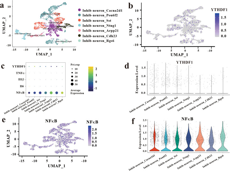
Further clustering of Gad2-positive cells detects YTHDF1 and NF-κB expression in an Sst-associated population. Sst marks somatostatin expression, distinguishing an expression profile within a broader population rather than treating all inhibitory neurons as identical.
YTHDF1 intervention in Sst-marked cells subsequently weakens somatic scores and place aversion. Corresponding interventions in PV- and Cck-marked populations produce no significant improvement. Related cellular populations need not contribute to withdrawal through the same molecule.
Source: Ou C et al. · BMC medicine · CC BY-NC-ND
Original figure and English caption ↗
Changes differ between baseline and withdrawal
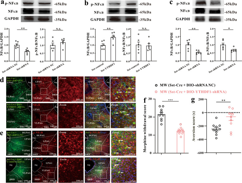
In mice without the withdrawal model, reducing or increasing Sst-marked-cell YTHDF1 changes local NF-κB and phosphorylated protein in the same direction, while the phosphorylation ratio changes nonsignificantly. In the withdrawal model, lowering YTHDF1 reduces both NF-κB and its phosphorylation ratio.
Directly reducing NF-κB in Sst-marked cells also weakens somatic scores and aversion. Interventions in the two molecules support their contribution to the process, while total protein and phosphorylation state show distinct patterns of regulation.
Source: Ou C et al. · BMC medicine · CC BY-NC-ND
Original figure and English caption ↗
Molecular regulation meets cellular activity
After reducing Sst-marked-cell YTHDF1, mean population calcium responses and spine density decline, while received inhibitory currents decrease in frequency without significant amplitude changes. Concurrent changes in incoming currents and cellular activity show that local network regulation is more complex than a simple relation between stronger inhibition and lower activity.
Using male wild-type mice and several genetic models, the study links withdrawal responses to RNA regulation in particular cellular populations. NF-κB is one contributing component, while the full downstream network remains unresolved. Cellular distinctions already help explain why intervention in the same molecule affects one population without producing the same outcome in another.
Source: Ou C et al. · BMC medicine · CC BY-NC-ND
Original figure and English caption ↗