Why does prolonged isolation impair social interaction? Cannabinoid receptor clues at anterior cingulate synapses
A study in male mice shows that chronic social isolation downregulates presynaptic CB1 receptors on anterior cingulate CCK interneurons, increasing GABA release and impairing social behavior.
Published in Neuron on November 21, 2023, this study by Shengxi Wu, Wenting Wang, and Baolin Guo's team at Fourth Military Medical University (with Baolin Guo, Kaiwen Xi, Honghui Mao, and Keke Ren as co-first authors) uncovers how chronic social isolation stress (CSIS) impairs sociability in male mice. Combining Sr2+-evoked quantal asynchronous IPSC (aIPSC) recordings, pre-embedding immunogold electron microscopy, RNAscope, FACS-based single-cell qPCR, in vivo axonal fiber photometry (recorded with an Inper two-color fiber photometry system and 200 μm, 0.37 NA optical fibers, and analyzed via InperDataProcess software), and a chimeric photosensitive opto-CB1R receptor, the authors show that 2-week CSIS selectively downregulates presynaptic cannabinoid type 1 receptors (CB1Rs) at cholecystokinin-positive (CCK+) interneuron synapses in the anterior cingulate cortex (ACC), elevating axonal Ca2+ influx and inhibitory drive onto pyramidal neurons to cause social deficits.
Fourteen-day chronic social isolation stress (CSIS) selectively reduces presynaptic CB1R density at ACC CCK+ interneuron synapses and impairs depolarization-induced suppression of inhibition (DSI), driving elevated axonal Ca2+ influx and quantal GABA release onto L2/3 pyramidal neurons; restoring presynaptic CB1R signaling via opto-CB1R or targeted CB1R agonists/positive allosteric modulators rescues social impairments.
Contents · 7 sections
Fourteen-Day Chronic Social Isolation Stress Selectively Enhances Inhibitory Synaptic Inputs onto ACC L2/3 Pyramidal Neurons
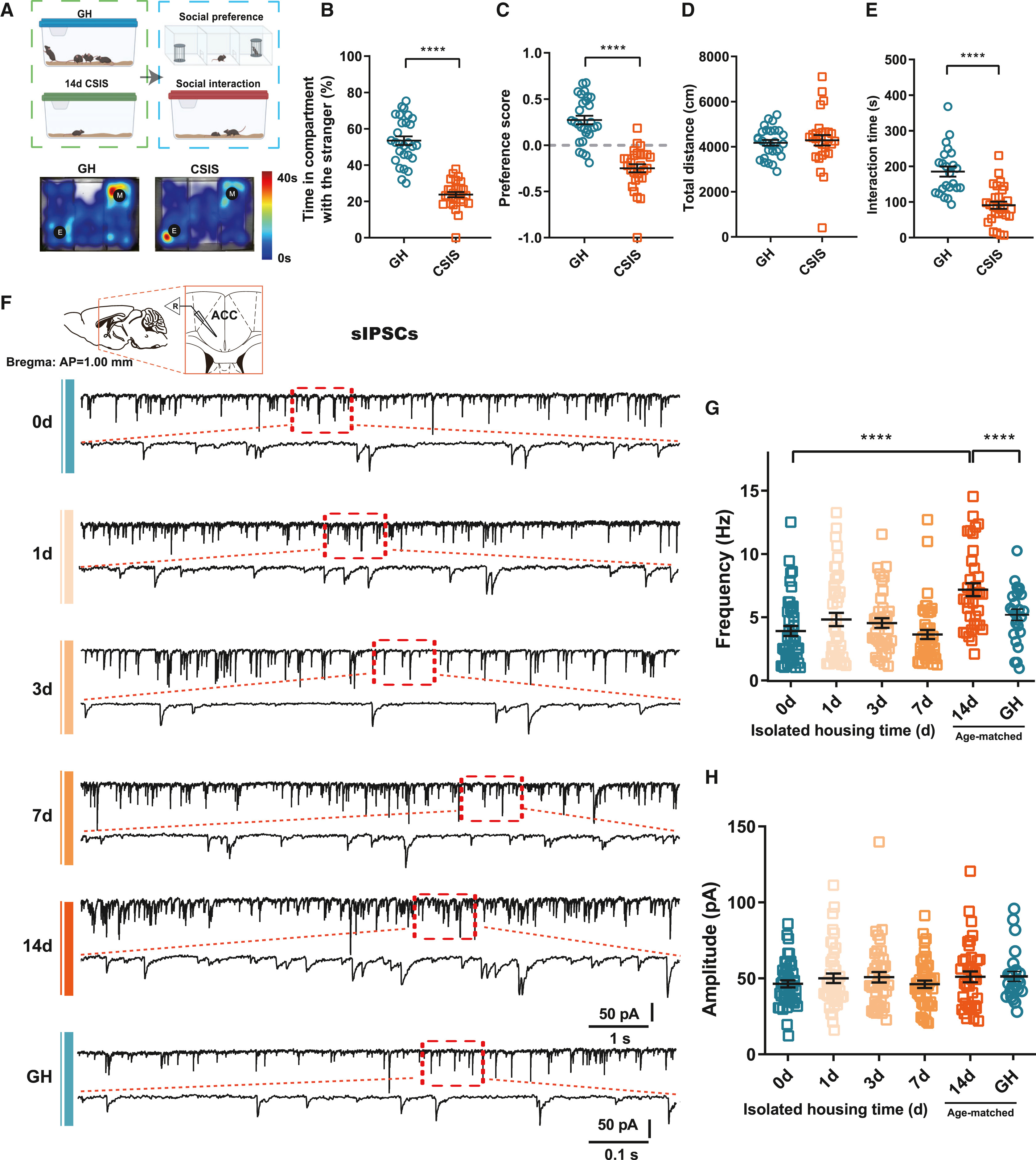
Two-week (14-day, unlike 1-week) chronic social isolation stress (CSIS) in adult male C57BL/6J mice significantly reduced social exploration and preference in the three-chamber assay and active interaction with a juvenile mouse without affecting locomotion, a deficit persisting for at least 28 days after partitioned re-grouping. Screening five isolation-sensitive brain regions (dHPC, dBNST, CeA, DMH, ACC) via social c-Fos mapping and bidirectional somBiPOLES optogenetics identified the anterior cingulate cortex (ACC) as the critical hub where photoactivation rescued CSIS-induced social deficits.
Whole-cell patch-clamp recordings in ACC L2/3 pyramidal neurons revealed that 14-day CSIS selectively increased spontaneous IPSC (sIPSC) frequency without altering sIPSC amplitude, sEPSCs, or intrinsic membrane properties, and selective optogenetic activation of L2/3 pyramidal neurons restored social interaction in CSIS mice.
Source: et al. · Neuron · Original figure reproduced for academic literature review under fair scholarly commentary; copyright remains with the authors/publisher.
Original figure and English caption ↗
Strontium-Evoked Quantal Asynchronous Release Reveals Selectively Elevated GABA Release at ACC CCK+ Interneuron Synapses
Quantification in GAD67-GFP mice showed that CSIS did not alter the total number of GABAergic interneurons in the ACC, and RNAscope for Cck mRNA in PV-tdTomato mice confirmed that PV+ and CCK+ basket interneurons represent non-overlapping populations in the ACC.
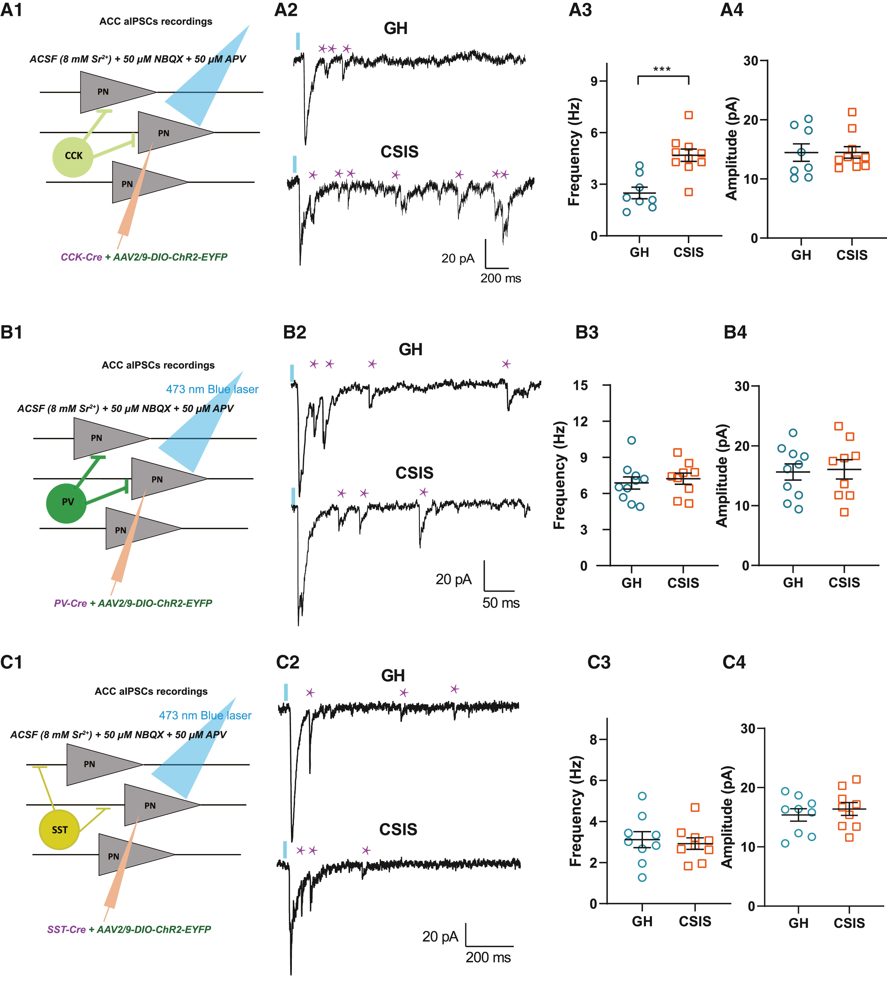
Using cell-type-specific ChR2 expression (AAV-mDlx-DIO-ChR2-mCherry) in CCK-Cre, PV-Cre, and SST-Cre mice combined with 8 mM Sr2+ substitution to record light-evoked quantal asynchronous IPSCs (aIPSCs) in ACC pyramidal neurons, the authors found that CSIS selectively increased aIPSC frequency (without changing amplitude) at CCK+ interneuron synapses, leaving PV+ and SST+ inhibitory inputs unchanged.
Source: et al. · Neuron · Original figure reproduced for academic literature review under fair scholarly commentary; copyright remains with the authors/publisher.
Original figure and English caption ↗
Optogenetic Activation of ACC CCK+ Interneurons in Group-Housed Mice Suffices to Induce CSIS-Like Social Deficits
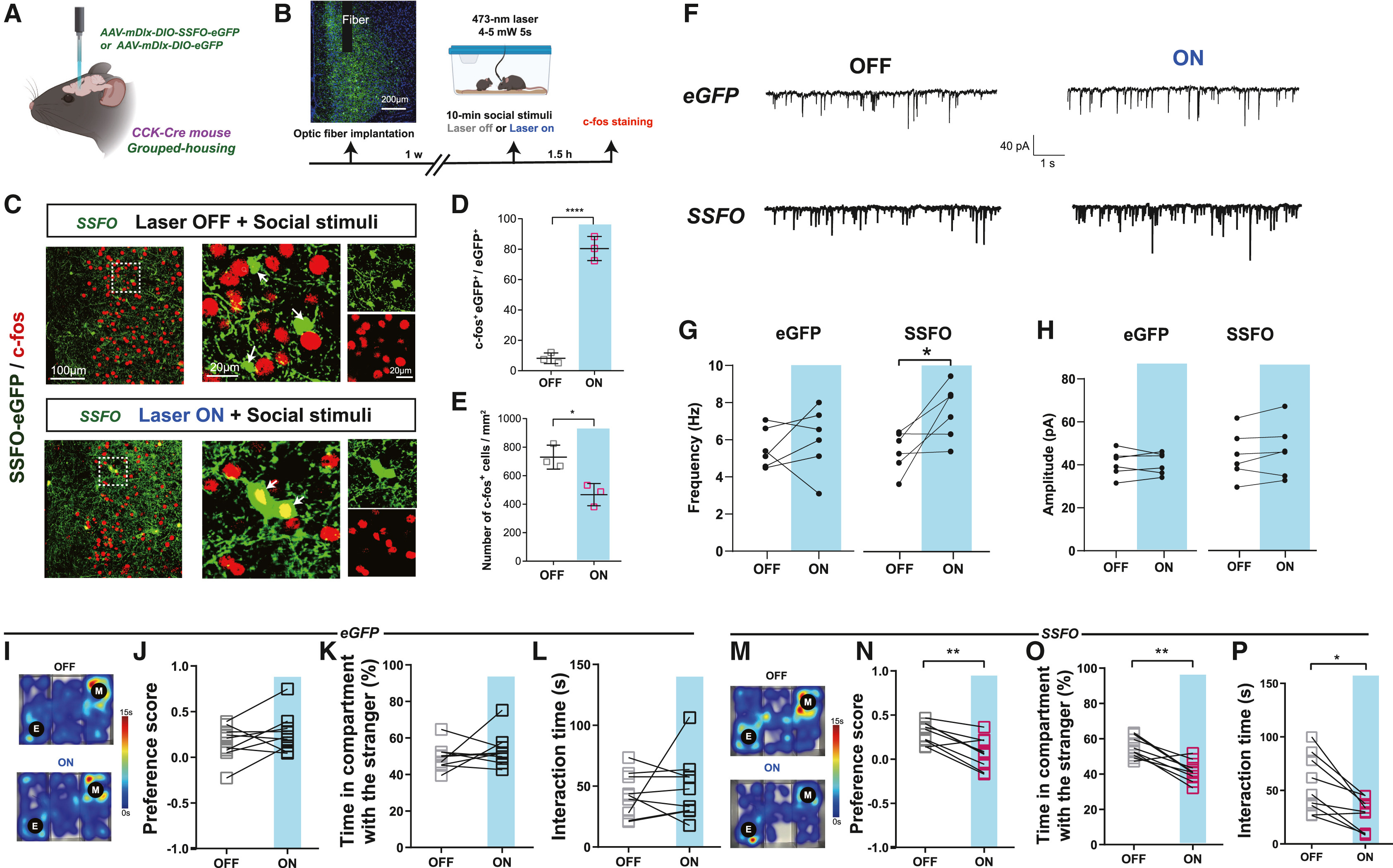
To test whether hyperactivity of ACC CCK+ interneurons suffices to drive social deficits, group-housed (GH) CCK-Cre mice were injected bilaterally in the ACC with an interneuron-specific stabilized step-function opsin (AAV-mDlx-DIO-SSFO-eYFP). Brief blue-light activation of SSFO significantly increased sIPSC frequency in ACC pyramidal neurons and reduced social-evoked c-Fos expression in the ACC.
Behaviorally, SSFO-mediated photoactivation of ACC CCK+ interneurons in group-housed mice reproduced CSIS-like social avoidance in both the three-chamber test and the juvenile social interaction test without affecting locomotion (whereas stGtACR2 photoinhibition of ACC CCK+ interneurons in CSIS mice restored sociability).
Source: et al. · Neuron · Original figure reproduced for academic literature review under fair scholarly commentary; copyright remains with the authors/publisher.
Original figure and English caption ↗
CSIS Downregulates Presynaptic CB1R at Inhibitory Synapses and Impairs DSI, Driving Elevated Axonal Ca2+ Influx and GABA Release
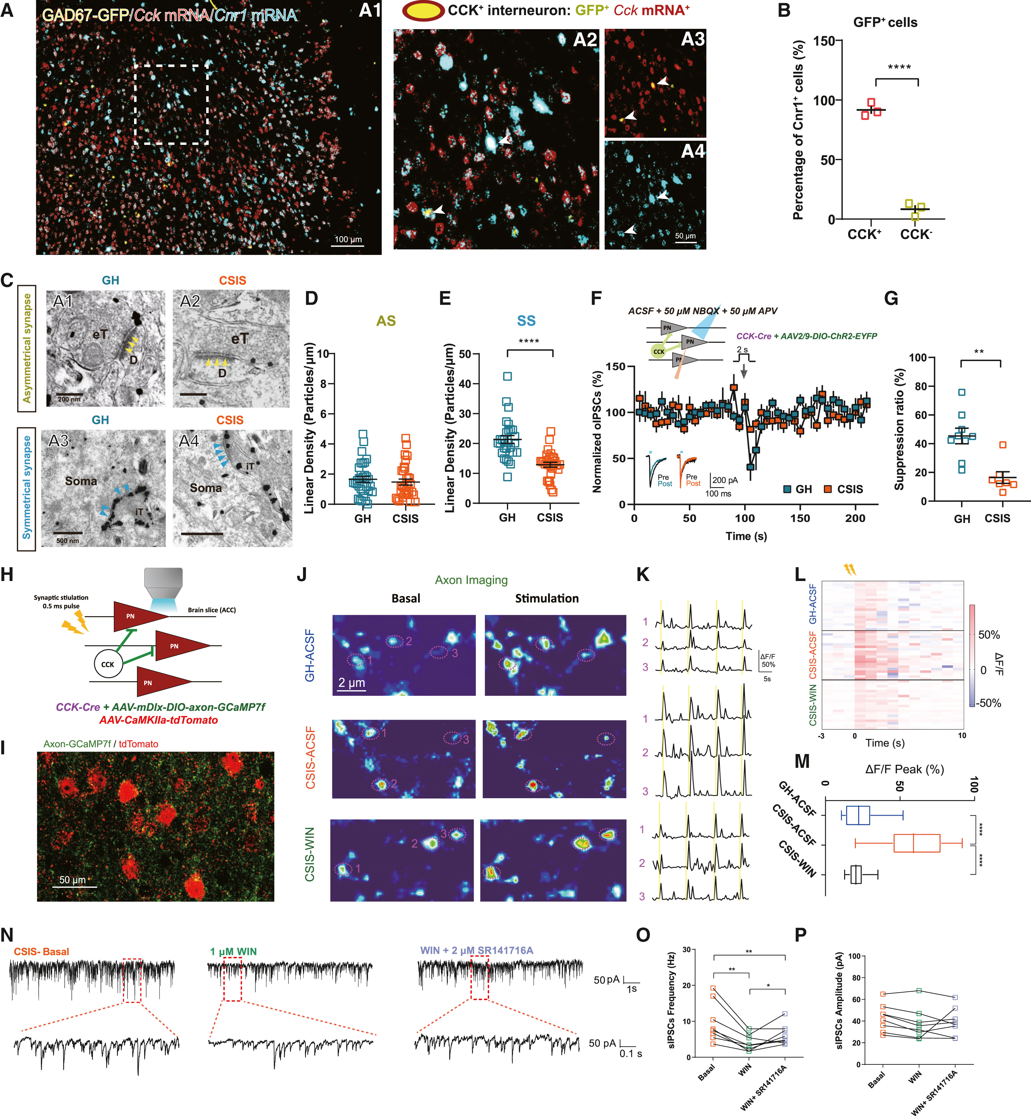
Dual RNAscope for Cck and Cnr1 mRNA in GAD67-GFP mice showed that ~90% of Cnr1-expressing interneurons in the ACC are CCK+. Pre-embedding immunogold electron microscopy revealed that CSIS significantly reduced presynaptic CB1R linear particle density at symmetrical (inhibitory) synapses—but not asymmetrical (excitatory) synapses—in the ACC, accompanied by impaired depolarization-induced suppression of inhibition (DSI, 2 s depolarization at 0 mV, with DSE unchanged).
In vivo axonal fiber photometry (rAAV-mDlx-DIO-axon-jGCaMP7f recorded with an Inper two-color fiber photometry system at 470 nm/410 nm and analyzed with InperDataProcess) and acute slice perisomatic axonal Ca2+ imaging revealed elevated axonal Ca2+ influx and event frequency in ACC CCK+ axons of CSIS mice. Bath application of the CB1R agonist WIN 55,212-2 (1 μM) normalized axonal Ca2+ responses and suppressed sIPSC frequency in CSIS slices, an effect reversed by the CB1R antagonist SR141716A (2 μM).
Source: et al. · Neuron · Original figure reproduced for academic literature review under fair scholarly commentary; copyright remains with the authors/publisher.
Original figure and English caption ↗
Conditional Knockdown of Cnr1 in ACC CCK+ Interneurons Impairs DSI and Induces Social Deficits in Group-Housed Mice
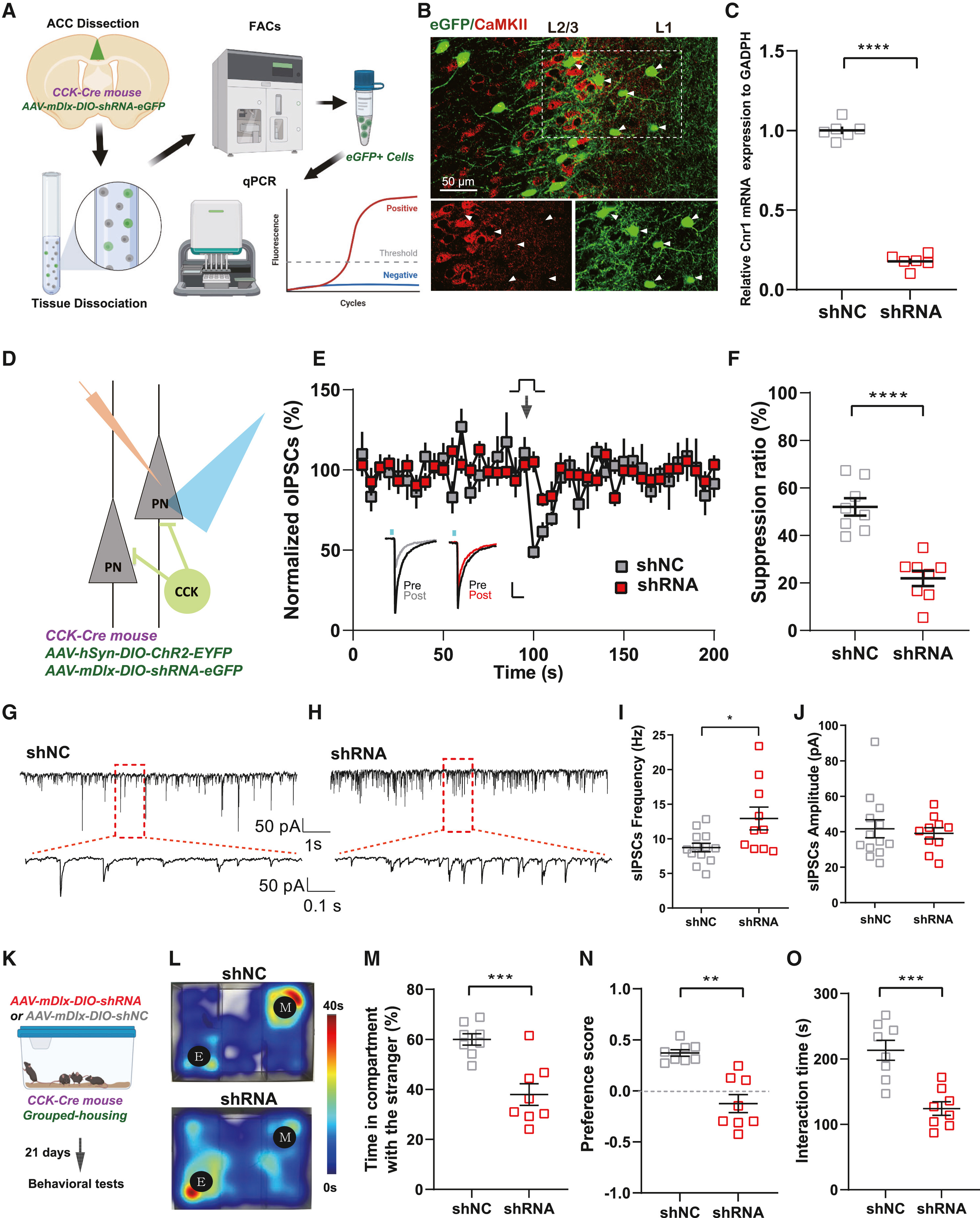
To test whether CB1R loss specifically in ACC CCK+ interneurons causes social deficits, the authors injected AAV-mDlx-DIO-shRNA(Cnr1)-eGFP bilaterally into the ACC of group-housed CCK-Cre mice. FACS sorting of eGFP+ interneurons followed by single-cell qPCR confirmed selective Cnr1 mRNA knockdown in ACC CCK+ interneurons.
Electrophysiological recordings showed that Cnr1 knockdown in ACC CCK+ interneurons impaired DSI and increased sIPSC frequency (without altering amplitude) in ACC pyramidal neurons of group-housed mice, which was sufficient to induce CSIS-like deficits in both three-chamber social preference and juvenile social interaction.
Source: et al. · Neuron · Original figure reproduced for academic literature review under fair scholarly commentary; copyright remains with the authors/publisher.
Original figure and English caption ↗
Optogenetic Activation of Chimeric opto-CB1R in ACC CCK+ Interneurons Reverses CSIS-Induced Social Deficits
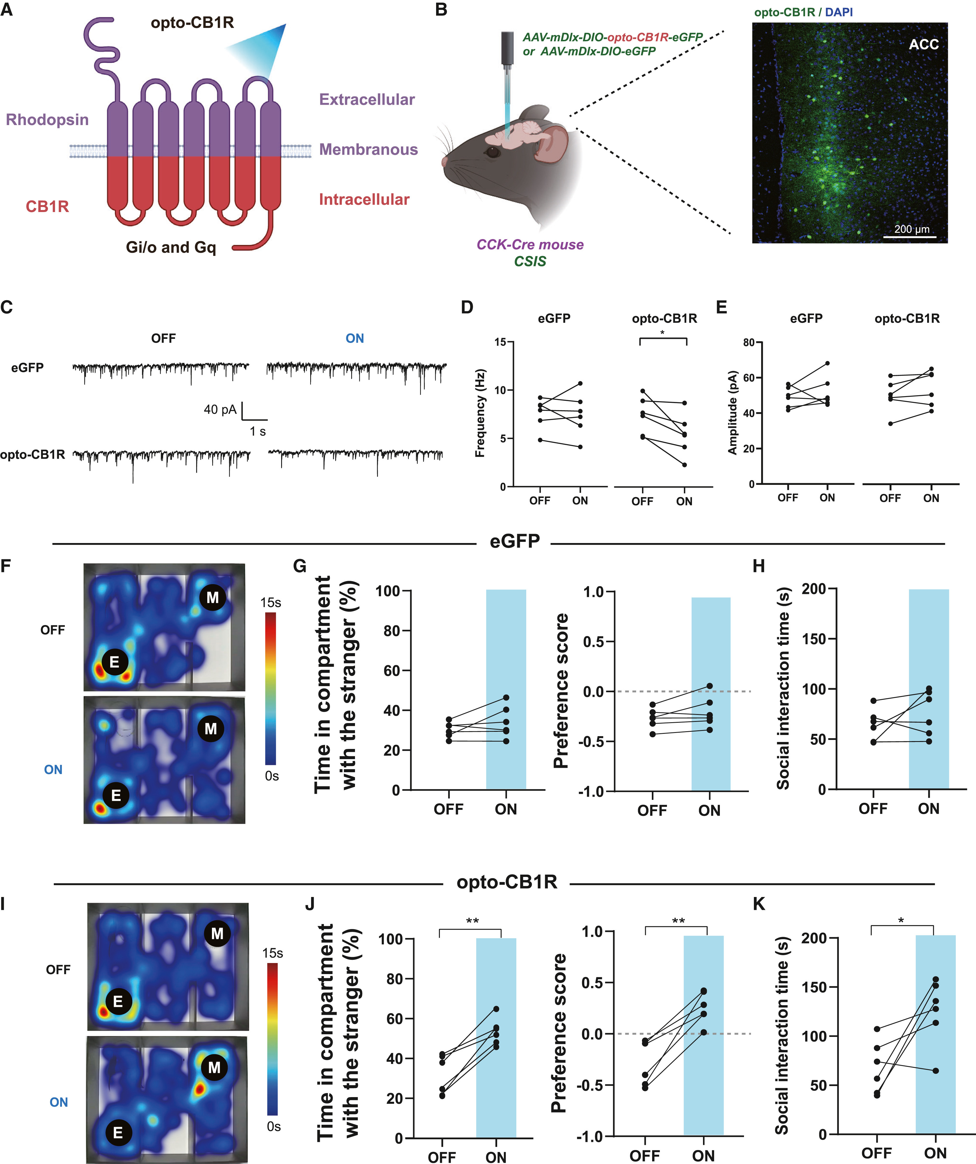
To restore CB1R intracellular signaling with cell-type and temporal precision, the authors engineered an interneuron-specific photosensitive chimeric receptor (AAV-mDlx-DIO-opto-CB1R-eGFP, replacing the intracellular loops and C-terminus of rhodopsin with human CB1R) and expressed it in ACC CCK+ interneurons of CSIS CCK-Cre mice. In acute slices, 473 nm blue-light stimulation (20 Hz) of opto-CB1R significantly suppressed the elevated sIPSC frequency in ACC pyramidal neurons.
In vivo 473 nm photostimulation of opto-CB1R in the ACC of CSIS mice significantly restored social chamber exploration and preference scores in the three-chamber test and rescued active interaction time in the juvenile social interaction assay without altering locomotor activity.
Source: et al. · Neuron · Original figure reproduced for academic literature review under fair scholarly commentary; copyright remains with the authors/publisher.
Original figure and English caption ↗
Systemic or Intra-ACC Microinfusion of CB1R Agonists and Positive Allosteric Modulators Restores Synaptic Homeostasis and Sociability
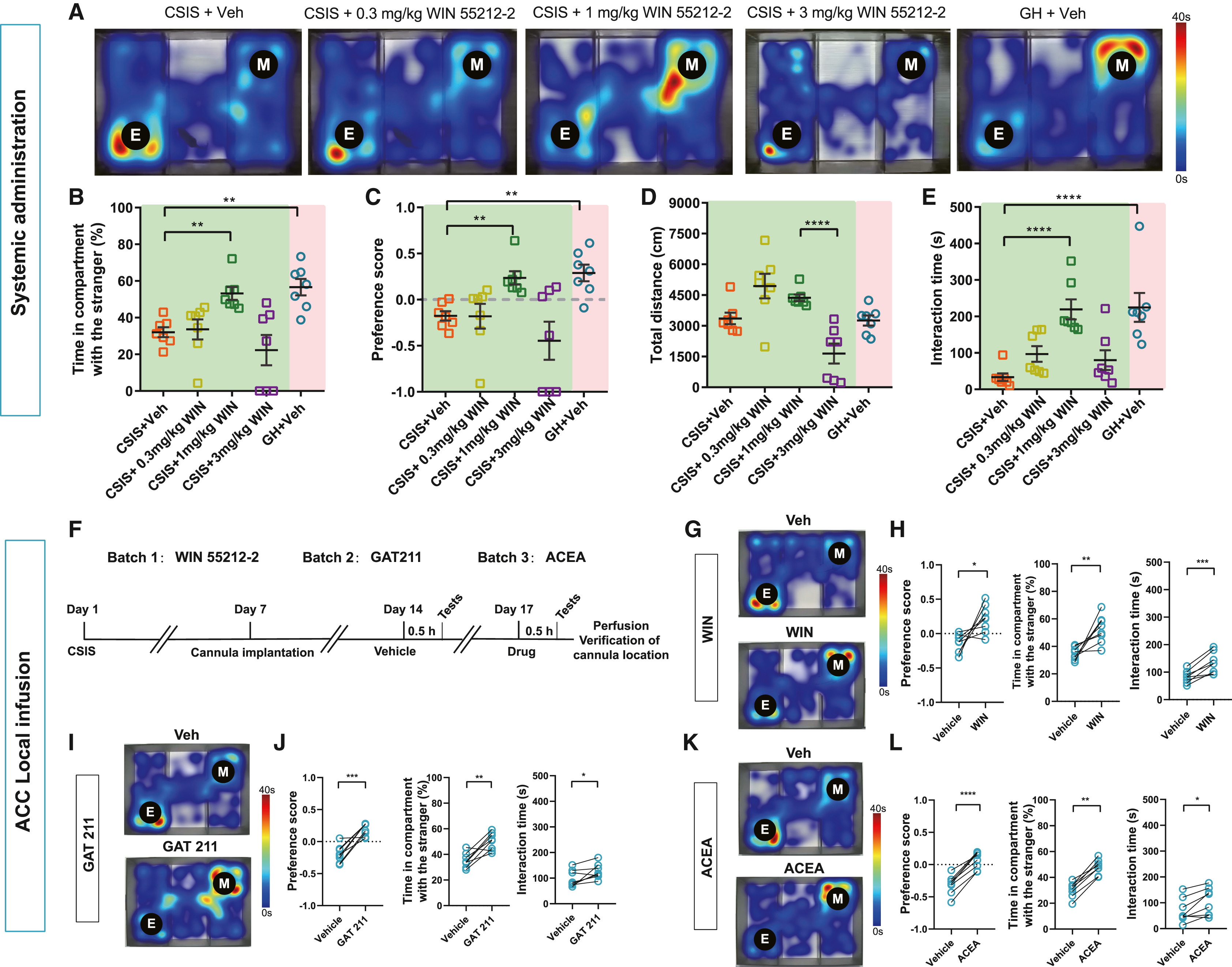
Evaluating pharmacological rescue, systemic administration of an intermediate dose of WIN 55,212-2 (1 mg/kg, i.p.) normalized ACC CCK+ axonal Ca2+ events and pyramidal neuron sIPSC frequency and rescued social preference and juvenile interaction in CSIS mice without suppressing motor cortex excitatory Ca2+ activity (recorded via rAAV-CaMKIIα-jRGECO1a fiber photometry at 561 nm/410 nm) or locomotion (unlike the subthreshold 0.3 mg/kg dose or the sedative 3 mg/kg dose).
Furthermore, bilateral intra-ACC cannula microinfusion of WIN 55,212-2 (1 μg/side), the CB1R positive allosteric modulator GAT211 (10 μg/side), or the selective CB1R agonist ACEA (0.73 μg/side) each reduced quantal aIPSC frequency in slices and robustly restored social preference and juvenile social interaction in CSIS mice.
Source: et al. · Neuron · Original figure reproduced for academic literature review under fair scholarly commentary; copyright remains with the authors/publisher.
Original figure and English caption ↗