Staying resilient after social defeat: how does an auditory thalamocortical circuit support stress coping?
A mouse study shows that transient hyperpolarization of medial geniculate neurons during social defeat triggers BDNF–TrkB plasticity, maintaining auditory cortex PV interneuron activity and resilience.
When confronted with severe chronic adversity, individuals diverge markedly into susceptible phenotypes exhibiting depressive-like behaviors and resilient phenotypes capable of rebounding through active neurobiological adaptations. Yet how the brain senses early stress episodes and initiates endogenous circuit-level resilience programs has remained elusive. On March 1, 2023, Xin-Hong Zhu, Yuan-Qing Li, and colleagues at Southern Medical University, Pazhou Lab, and South China University of Technology published a landmark article in Cell entitled 'A thalamic-primary auditory cortex circuit mediates resilience to stress' (with Huan-Yu Li, Min-Zhen Zhu, and Xin-Rui Yuan as co-first authors). Integrating chronic social defeat stress (CSDS), monosynaptic rabies tracing, in vivo electrophysiology and fiber photometry, transcriptomics, synaptic reconstruction, and temporally patterned optogenetics, the team uncovered a medial geniculate-to-primary auditory cortex parvalbumin interneuron circuit (MGA1→A1PV) that drives innate resilience to stress.
Key finding: Activation of parvalbumin interneurons in the primary auditory cortex (A1 PV-INs) by glutamatergic inputs from the ipsilateral medial geniculate body (MG, accounting for ~16.22% of monosynaptic inputs to A1 PV-INs) is required for resilience to chronic social defeat stress (CSDS). During early defeat episodes, resilient mice exhibit transient hyperpolarization in A1-projecting MG neurons (MGA1), which triggers presynaptic BDNF-TrkB-dependent synaptogenesis onto A1 PV-INs; optogenetically mimicking this short-term hyperpolarization—unlike simple tonic activation—awakens innate resilience mechanisms and produces sustained antidepressant-like effects across multiple animal models.
Contents · 5 sections
Sustained activity of parvalbumin interneurons in the primary auditory cortex is essential for resilience to chronic social defeat stress
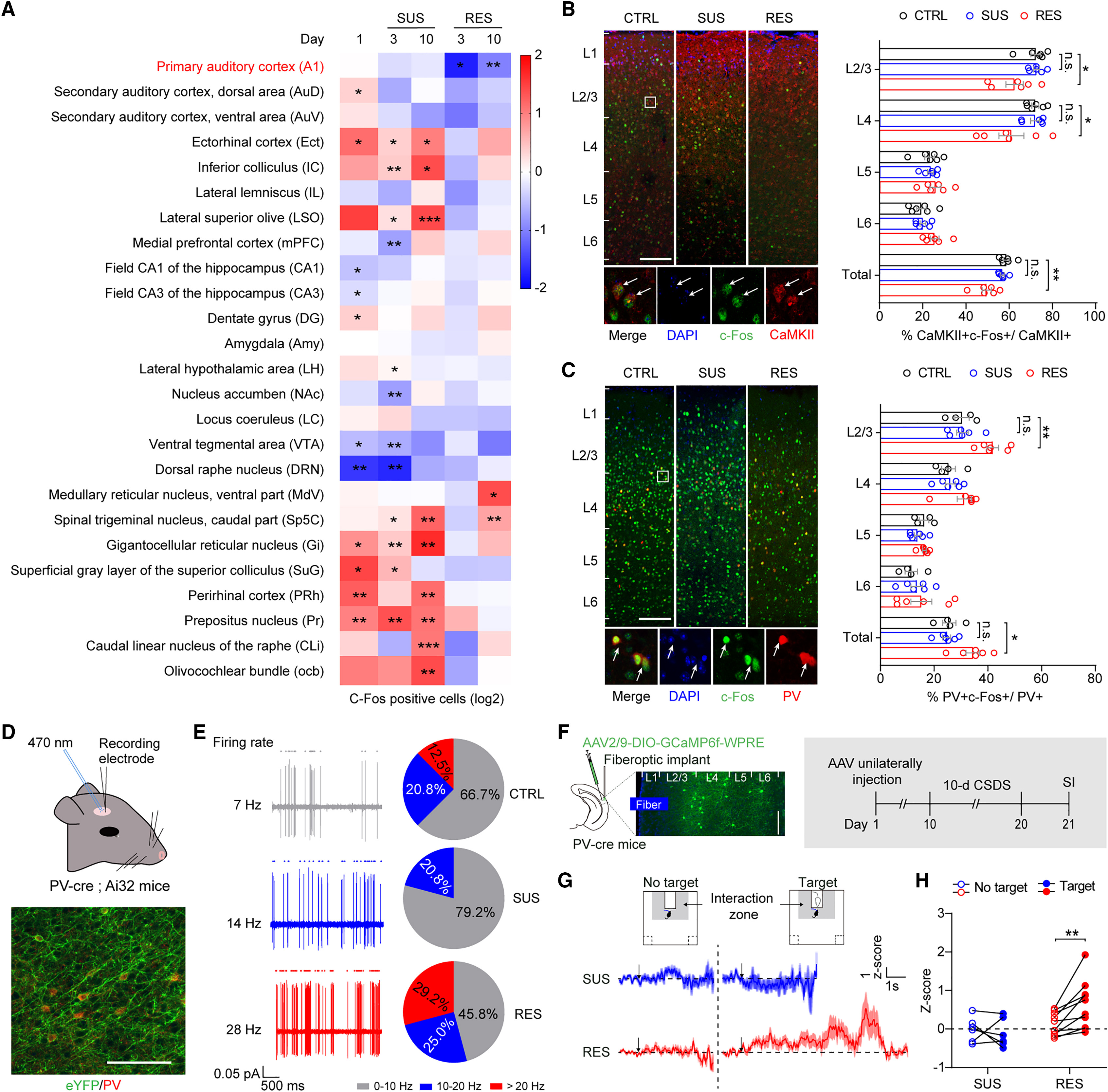
Starting from the observation that acoustic enrichment promotes resilience to 10-day chronic social defeat stress (CSDS) via the primary auditory cortex (A1) (Figures 1–2), the authors mapped c-Fos expression across 21 brain regions and performed in vivo recordings in susceptible (SUS) and resilient (RES) male mice. In RES mice, c-Fos co-labeling decreased in A1 L2/3 and L4 CaMKII+ excitatory neurons but significantly increased in A1 parvalbumin-expressing interneurons (PV-INs); opto-tagged single-unit recordings and fiber photometry confirmed heightened A1 PV-IN firing rates (averaging 28 Hz in RES vs. 7 Hz in CTRL and 14 Hz in SUS) and robust social-target-evoked Ca2+ transients.
Pharmacological and chemogenetic manipulations (Original Figures 2A–J) further demonstrated that selective ablation (taCasp3) or chemogenetic inhibition (hM4Di) of A1 PV-INs in PV-Cre mice rendered mice highly susceptible to subthreshold social defeat stress, whereas chemogenetic activation (hM3Dq) of A1 PV-INs alleviated social avoidance and behavioral despair, establishing A1 PV-INs as a critical cortical node for stress resilience.
Source: Huan-Yu Li et al. · Cell · © Elsevier Inc.
Original figure and English caption ↗
Monosynaptic rabies tracing identifies ipsilateral medial geniculate glutamatergic projections (MGA1) driving A1 PV-INs
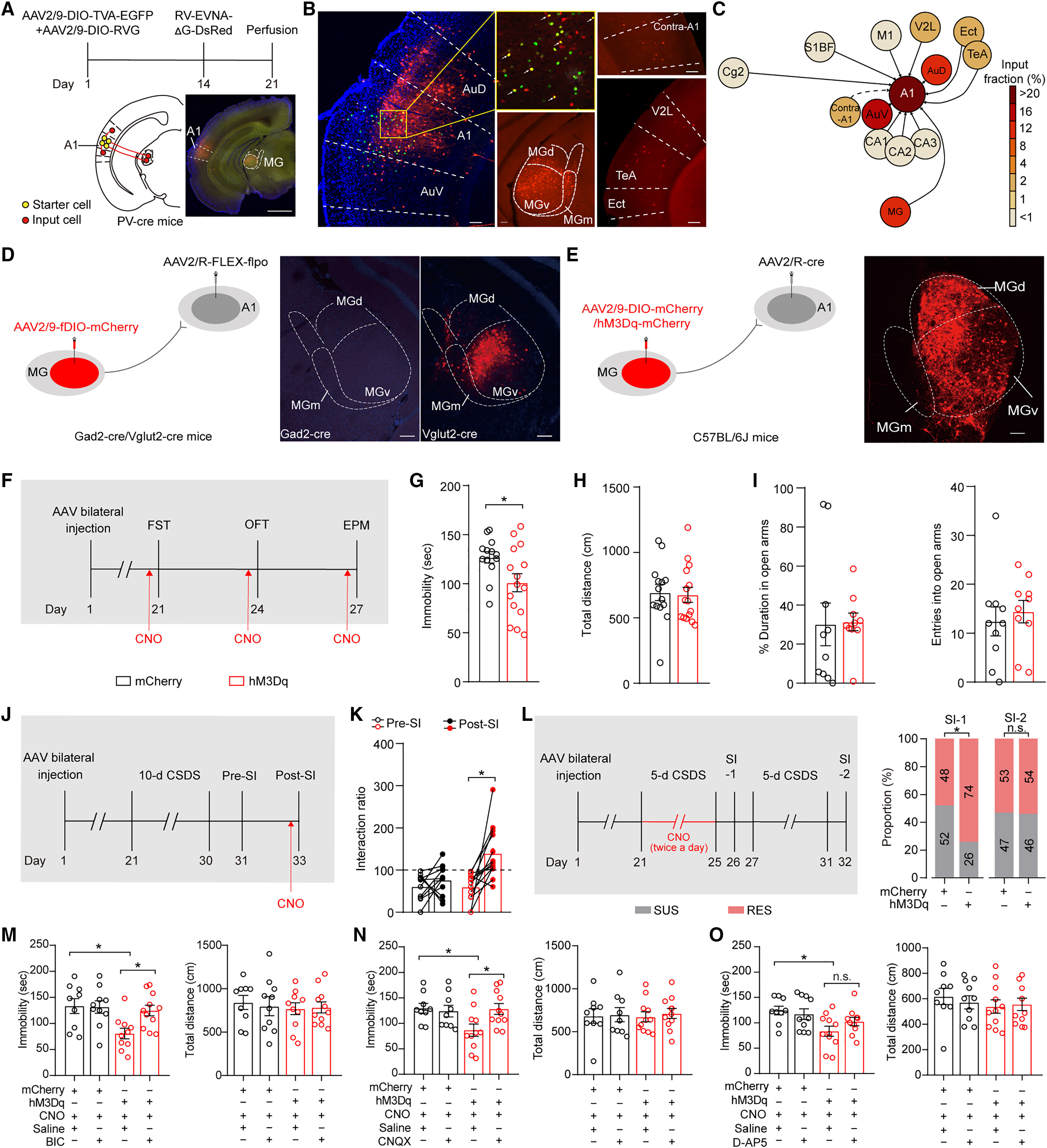
To identify the upstream inputs maintaining A1 PV-IN activity in resilient mice, the authors performed monosynaptic rabies virus tracing from A1 PV-INs in PV-Cre mice. Whole-brain quantification (Table S1) revealed that outside local auditory cortical areas (A1: 42.17 ± 3.31%; AuV: 16.69 ± 1.00%; AuD: 15.86 ± 1.47%), the ipsilateral medial geniculate body (MG) provided the largest subcortical monosynaptic input, accounting for 16.22 ± 0.65% of total input cells.
Intersection tracing and anterograde trans-synaptic manipulations (injecting AAV2/1-FLEX-flpo into the MG and Flp-dependent fDIO-hM3Dq, hM4Di, or taCasp3 into the A1 of PV-Cre mice; Original Figures 4A–O) confirmed that Vglut2+ MGA1 neurons directly drive postsynaptic A1MG-PV interneurons: chemogenetically activating MGA1 or postsynaptic A1MG neurons reduced FST immobility, reversed post-CSDS social avoidance, restored sucrose preference after 6-week CMS, and raised the resilient proportion to 73%–74%, whereas selective taCasp3 ablation of A1MG-PV neurons abolished these pro-resilient effects.
Source: Huan-Yu Li et al. · Cell · © Elsevier Inc.
Original figure and English caption ↗
Single-cell RNA-seq and 3D synaptic reconstruction reveal upregulated Gria2/Grip1 and increased excitatory synapses onto A1MG-PV neurons in resilient mice
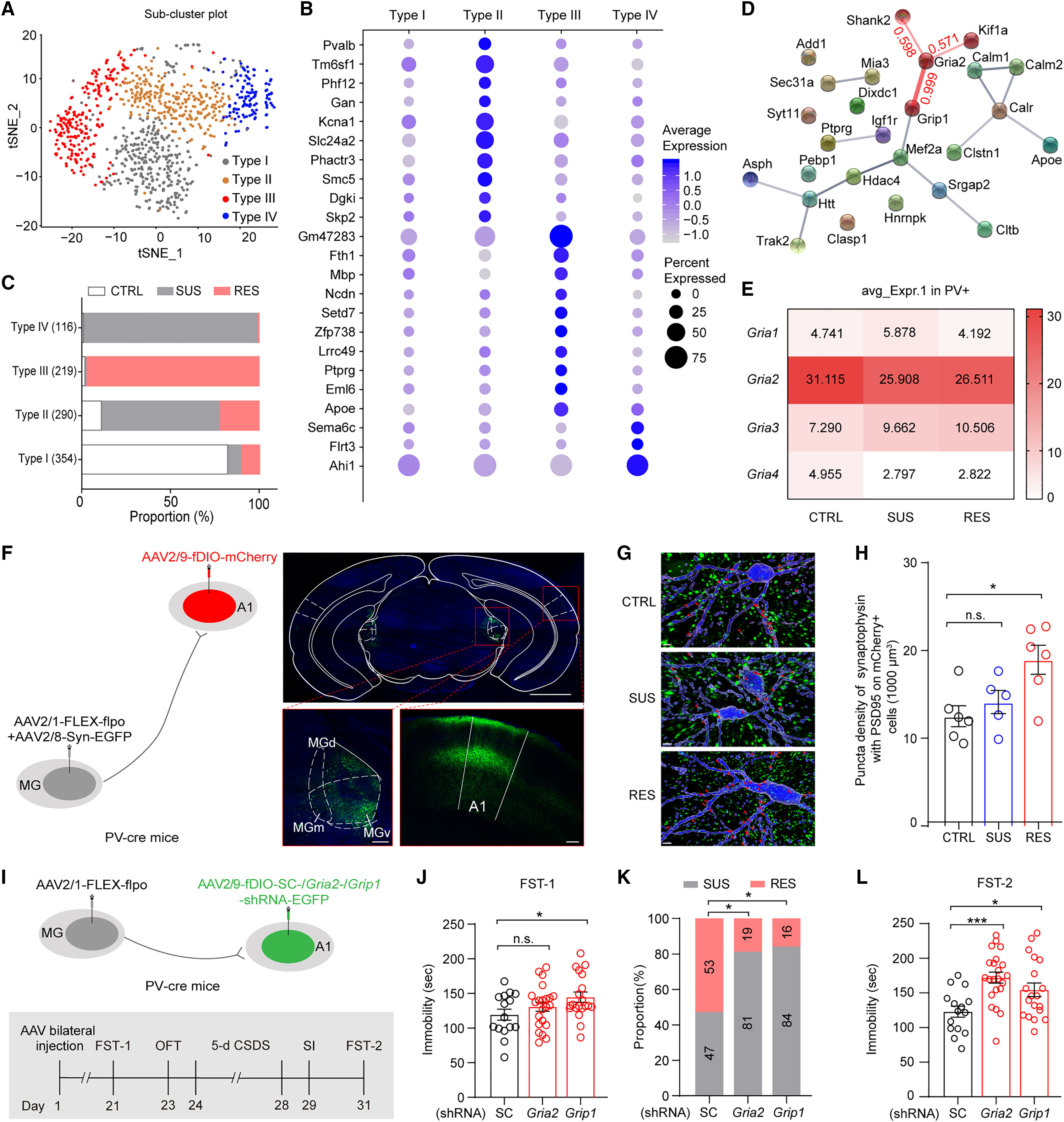
To uncover the molecular and structural basis of heightened A1 PV-IN excitability in resilient mice, the authors performed single-cell RNA-seq and GO enrichment analysis on A1 PV+ interneurons sorted from CTRL, SUS, and RES mice (Figures 5A–E, Table S2). tSNE clustering identified a resilience-enriched subcluster (Type III) in which the AMPA receptor subunit gene Gria2 and its scaffolding partner Grip1 served as upregulated hub genes.
High-resolution 3D synaptic reconstruction (AAV2/1-FLEX-flpo + AAV2/8-Syn-EGFP in MG and AAV2/9-fDIO-mCherry in A1, Figures 5F–H) demonstrated a significantly higher density of Syn-EGFP+/PSD95+ excitatory synaptic puncta on A1MG dendrites in RES mice. Targeted knockdown of Gria2 or Grip1 in A1MG neurons via AAV2/9-fDIO-Gria2/Grip1-shRNA-EGFP (Figures 5I–L) increased the SUS proportion after 5-day CSDS from 47% to 81% and 84%, respectively.
Source: Huan-Yu Li et al. · Cell · © Elsevier Inc.
Original figure and English caption ↗
Transient MGA1 hyperpolarization during social defeat attacks boosts subsequent approach-evoked circuit activity, and optogenetic mimicking awakens resilience
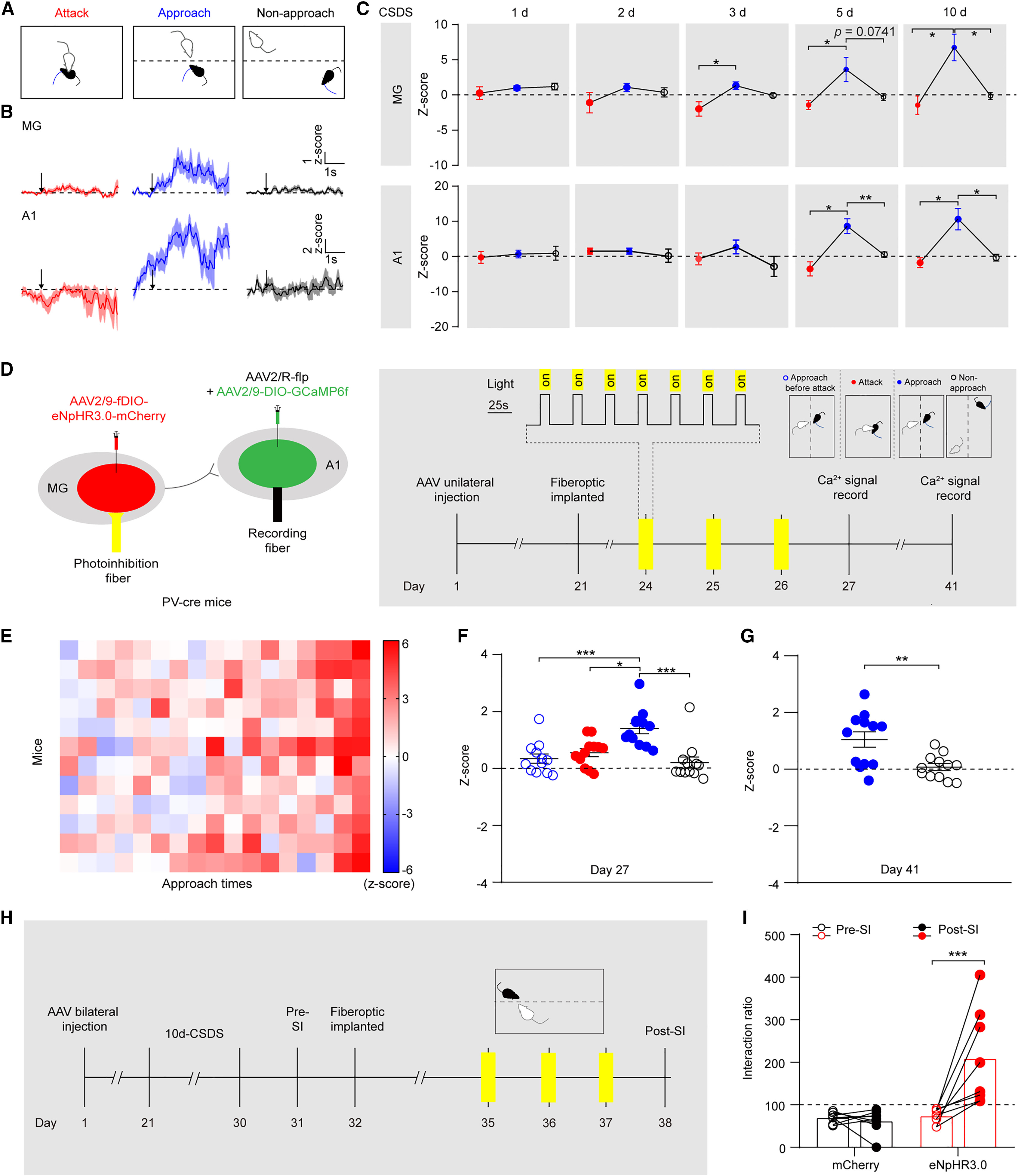
Whereas tonic chemogenetic activation of MGA1 neurons produced only transient symptom relief that dissipated after drug withdrawal, longitudinal fiber photometry and electrophysiological recordings across CSDS Days 1–10 (Figures 6A–C) revealed a distinct temporal signature in future resilient (RES) mice: MGA1 neurons exhibited transient hyperpolarization (reduced Ca2+ and firing activity) during 5-minute physical defeat attacks (Attack), followed by robust rebound Ca2+ activation in MGA1 and A1PV neurons during subsequent active social approach (Approach).
Guided by this temporal signature, the authors delivered patterned eNpHR3.0 optogenetic hyperpolarization to the MG→A1 pathway specifically during the attack window (Figures 6D–I). Mimicking transient hyperpolarization amplified subsequent approach-evoked Ca2+ transients and triggered enduring antidepressant-like resilience.
Source: Huan-Yu Li et al. · Cell · © Elsevier Inc.
Original figure and English caption ↗
Transient MGA1 hyperpolarization triggers retrograde A1 BDNF-to-presynaptic TrkB signaling to drive MG→A1PV synaptogenesis and lasting resilience
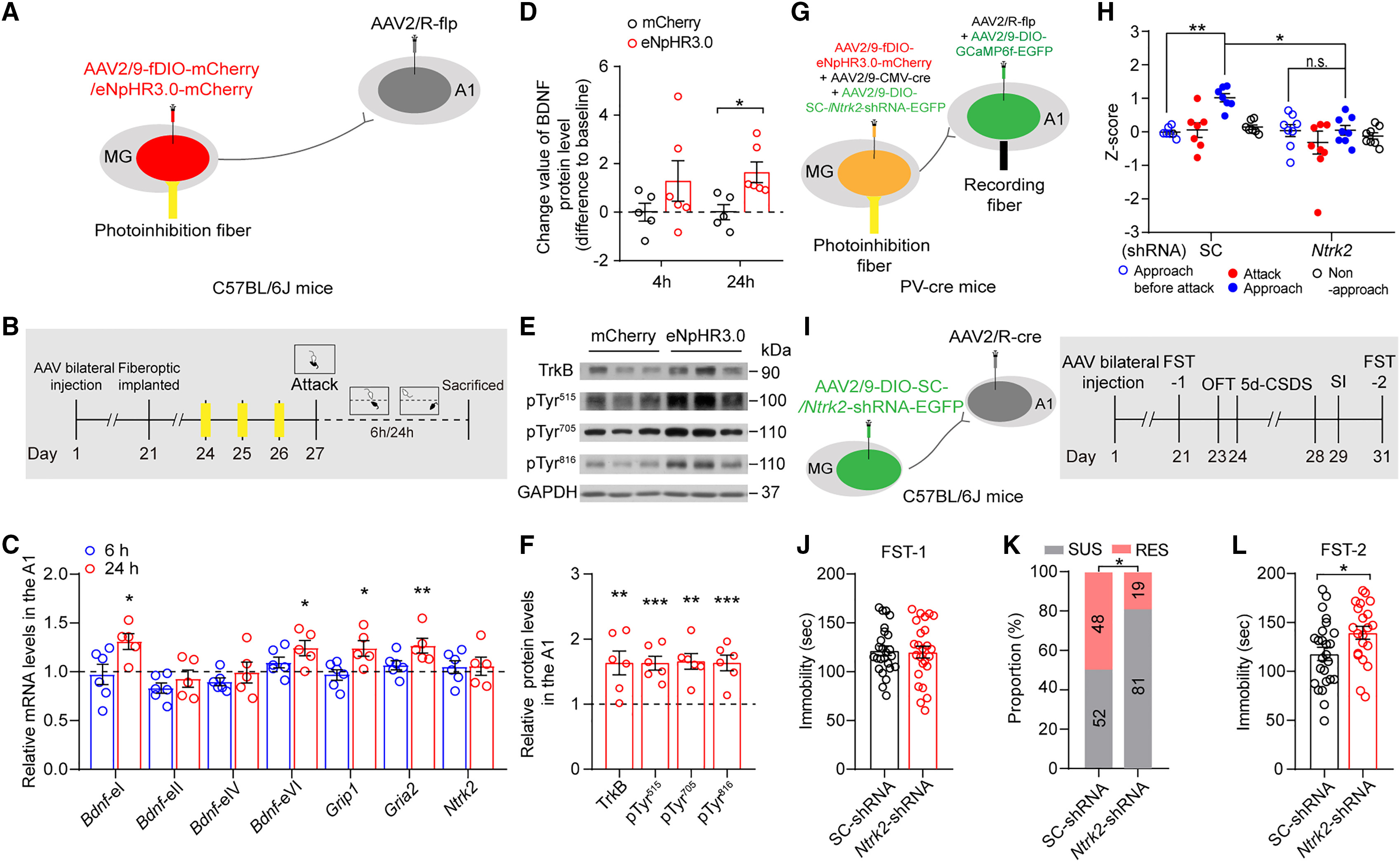
How does transient MGA1 hyperpolarization translate into durable MG→A1PV synaptic potentiation? Molecular and biochemical analyses (Figures 7A–F) demonstrated that short-term MGA1 hyperpolarization followed by active social approach upregulated A1 brain-derived neurotrophic factor expression (Bdnf-eI/eVI) and promoted retrograde activation of presynaptic TrkB receptors (elevated pTyr515, pTyr705, and pTyr816 phosphorylation) on MGA1 terminals, driving Gria2/Grip1 upregulation and synaptogenesis onto A1MG-PV neurons.
Selectively knocking down presynaptic TrkB receptors in MGA1 neurons via retrograde Cre and AAV-DIO-Ntrk2-shRNA (Figures 7G–L) abolished Syn-EGFP+/PSD95+ synaptogenesis, prevented Gria2/Grip1 upregulation, and abrogated the pro-resilient behavioral effects of patterned optogenetic hyperpolarization, establishing the presynaptic BDNF–TrkB–Gria2/Grip1 cascade as the molecular engine of circuit resilience.
Source: Huan-Yu Li et al. · Cell · © Elsevier Inc.
Original figure and English caption ↗