Smelling danger: a defensive route that bypasses the amygdala
A route linking olfactory input, cortex and brainstem cells contributes to odor-driven innate defense in male mice. Manipulating its activity also changes exploration following acute stress.

Some dangers can be detected before they are seen. A drifting odor makes a mouse leave an area or run toward shelter. How does information at the nose become a defensive action? Fear often brings the amygdala to mind, but a study published in Nature Neuroscience on February12,2024 traces an additional route from odor through a cortical region into specific brainstem cells and onward. Defensive responses remain possible after manipulation of selected amygdalar regions, adding a distinct branch to the neural picture of innate fear.
The study links olfactory bulb, dorsal peduncular cortex, anterior parabrachial Cck-marked cells and Tac1-marked cells in PSTh. Circuit activity affects odor-driven escape and avoidance, as well as anxiety-like exploration in defined stress conditions.
Contents · 6 sections
Where can odor recruit defense beyond the amygdala?
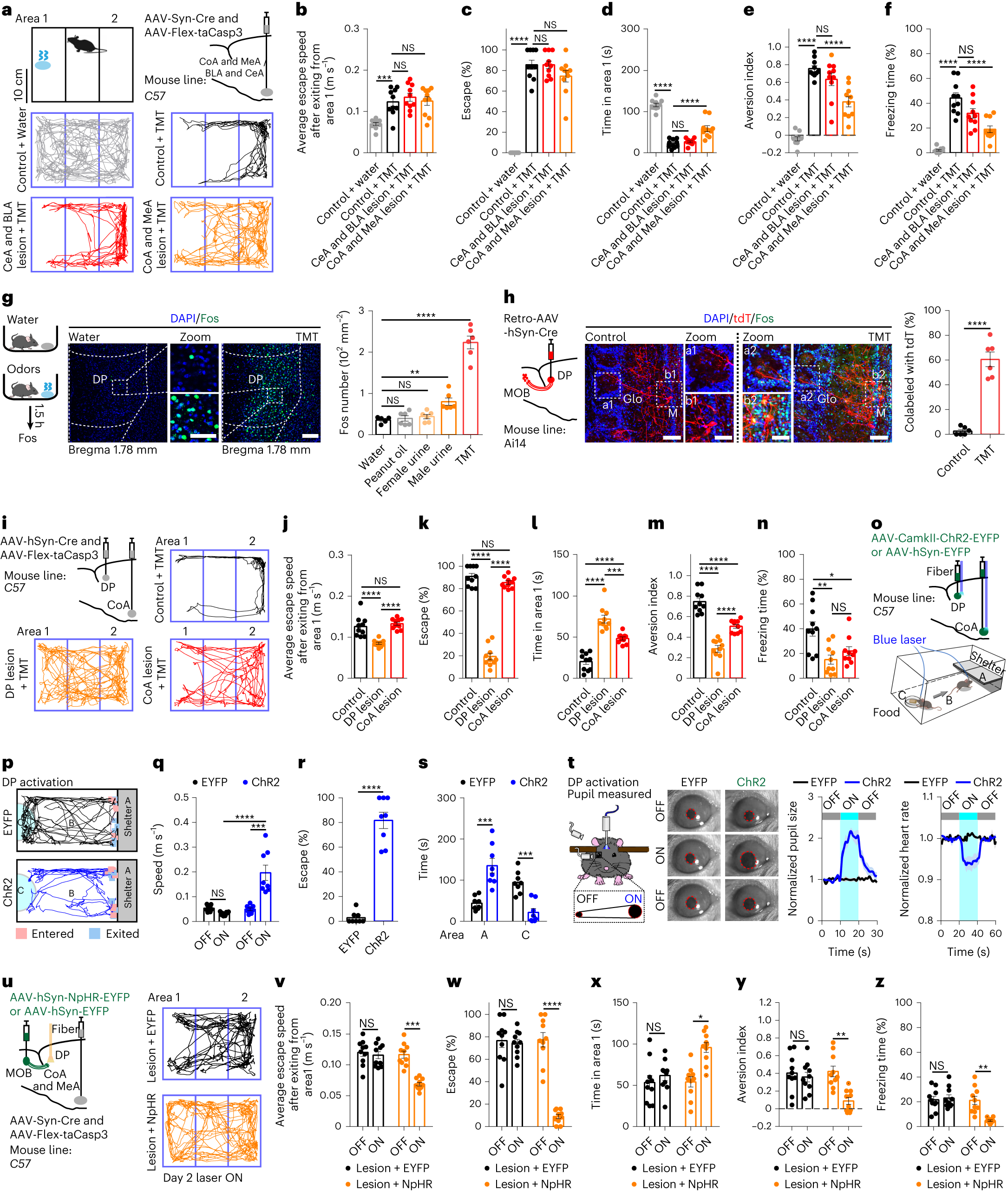
The researchers use the predator-associated odorant TMT to examine escape, area avoidance and freezing in male mice. Several responses remain after manipulation of basolateral and central amygdalar regions. Cortical and medial amygdalar manipulation attenuates some avoidance and freezing measures without a corresponding significant reduction in escape speed or probability. Defensive readouts do not all move together.
Manipulating the dorsal peduncular cortex (DP), by contrast, strongly attenuates several TMT responses. DP activation drives flight toward shelter and changes pupil size and heart rate. Suppressing olfactory-bulb input to DP also attenuates defense in a selected amygdalar-lesion background. DP emerges as an important entry for this odor-associated response, with distinct contributions of amygdalar regions still evident.
Source: Hao Wang et al. · Nature Neuroscience ·
Original figure and English caption ↗
From olfactory bulb to molecularly identified downstream cells
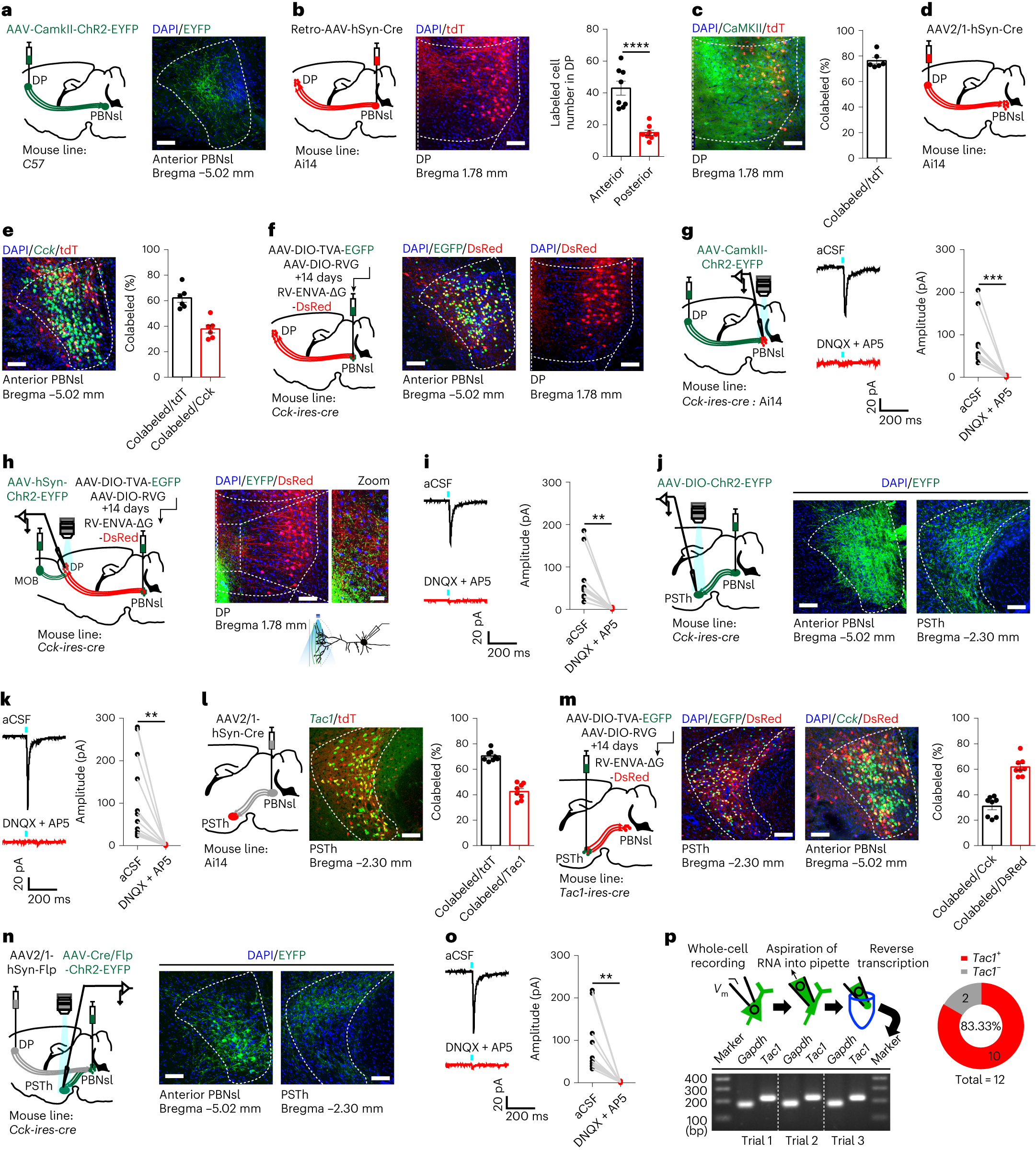
Tracing onward from DP identifies the anterior superior lateral parabrachial region, anterior PBNsl. DP provides excitatory input to cells including a Cck-marked population, which projects onward to the parasubthalamic nucleus (PSTh) and contacts Tac1-marked cells. The olfactory, cortical and downstream nodes become a more specific sequence.
Cellular labeling and evoked currents complement one another: tracing identifies connected populations, while light-evoked excitatory currents support functional links. Marker-overlap proportions are not universally100%. Molecular labels define the populations under study without implying that an entire region contains only that cell type or sends every output to one destination.
Source: Hao Wang et al. · Nature Neuroscience ·
Original figure and English caption ↗
Odor activates some cells rather than every cell uniformly
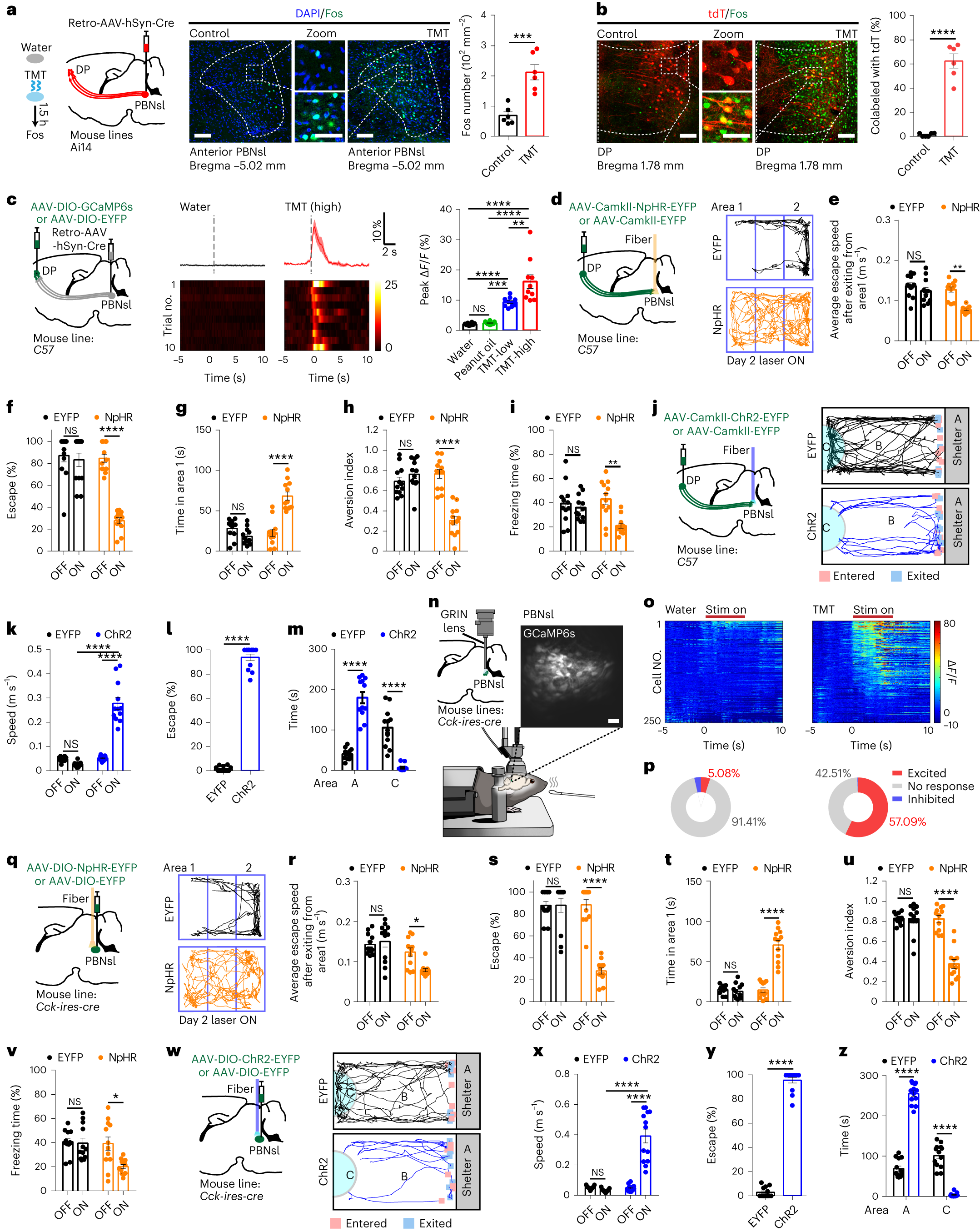
TMT increases activity in DP cells projecting to anterior PBNsl, with larger calcium signals at the higher tested concentration. Peanut oil does not differ significantly from water in the same comparison. Cck-marked cells in anterior PBNsl also respond heterogeneously: some increase activity, while others are inhibited or show no clear response.
Suppressing DP terminals in anterior PBNsl attenuates escape, avoidance and freezing. Activating this input or the relevant Cck-marked cells drives movement toward shelter. Odor-evoked activity and manipulation-related behavior together support this circuit segment in defense. Comparisons involving visual and auditory threats additionally suggest sensory-context selectivity in the observed responses.
Source: Hao Wang et al. · Nature Neuroscience ·
Original figure and English caption ↗
The downstream signal also influences escape and avoidance
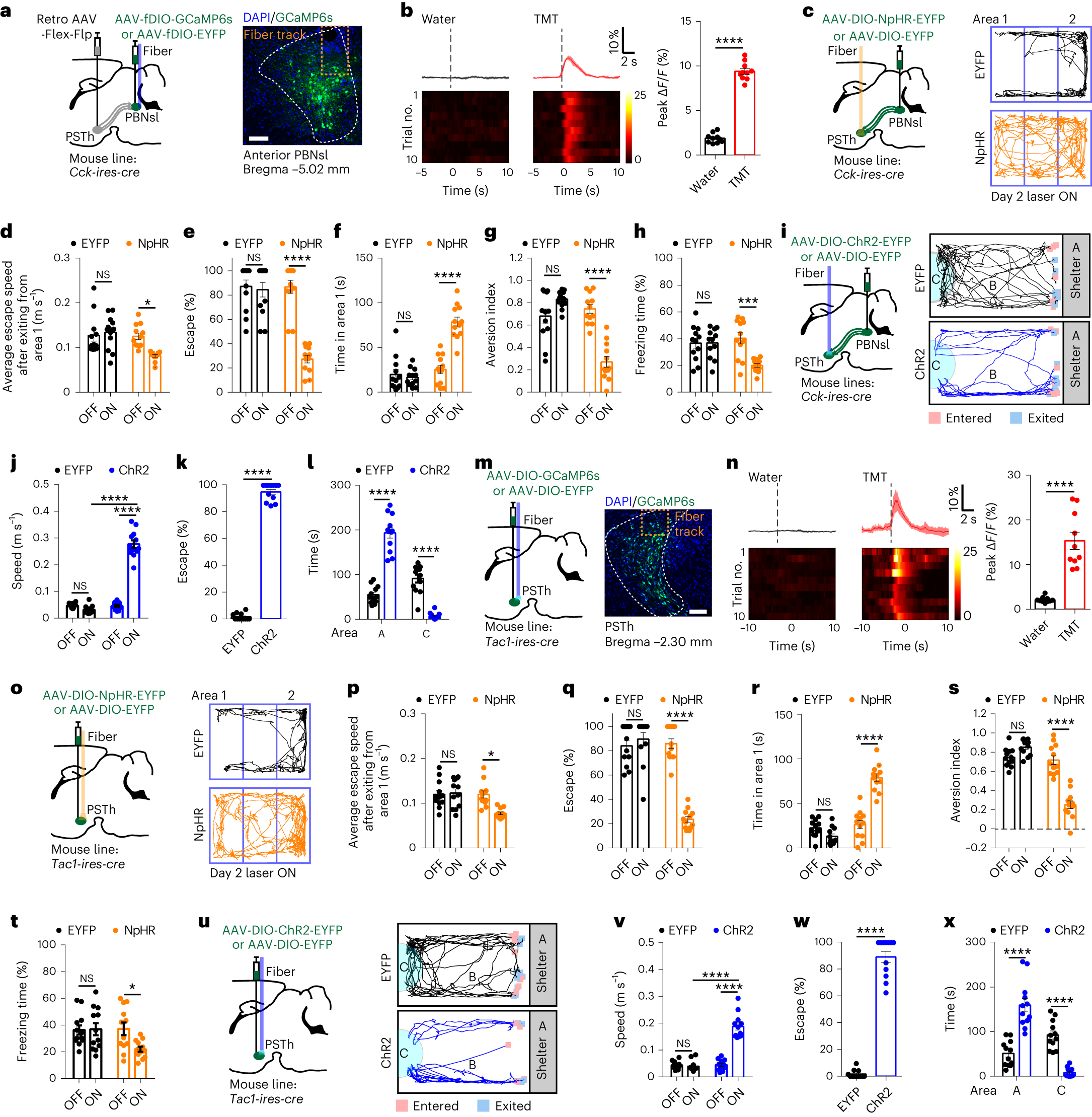
Cells linking anterior PBNsl to PSTh, and Tac1-marked cells in PSTh, show increased calcium signals during TMT exposure. Suppressing anterior PBNsl terminals in PSTh attenuates escape probability, area avoidance and freezing, while activating these terminals drives movement toward shelter.
Directly manipulating Tac1-marked PSTh cells produces similarly directed defensive changes. Several lines of evidence thus connect input with downstream cells and behavior. They identify a defined route under specific cellular and stimulus conditions, sharpening the broader association between parabrachial activity and negative information into particular inputs, outputs and responses.
Source: Hao Wang et al. · Nature Neuroscience ·
Original figure and English caption ↗
DP-linked cells can drive defense after selected amygdalar lesions
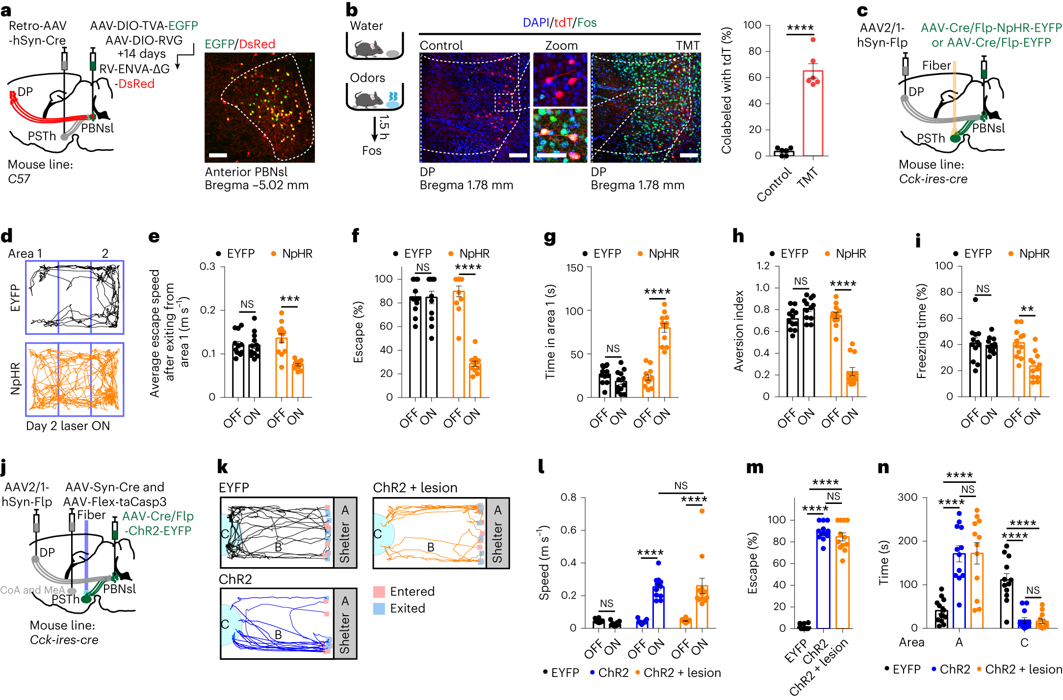
The team further identifies anterior PBNsl Cck-marked cells receiving DP input, connecting this selection with the established PSTh output. Suppressing the selected cells attenuates TMT-related escape, avoidance and freezing; activating them produces shelter-directed defense. Segment-level evidence converges again in manipulation of a more selectively defined population.
Activation also drives escape in mice with cortical and medial amygdalar lesions. Several escape and occupancy readouts do not differ significantly from the activation group without those lesions. This supports a bypass of the selected amygdalar regions under the tested conditions, alongside the earlier finding that distinct amygdalar manipulations affect different defensive measures.
Source: Hao Wang et al. · Nature Neuroscience ·
Original figure and English caption ↗
Beyond odor avoidance, the circuit changes exploration after stress
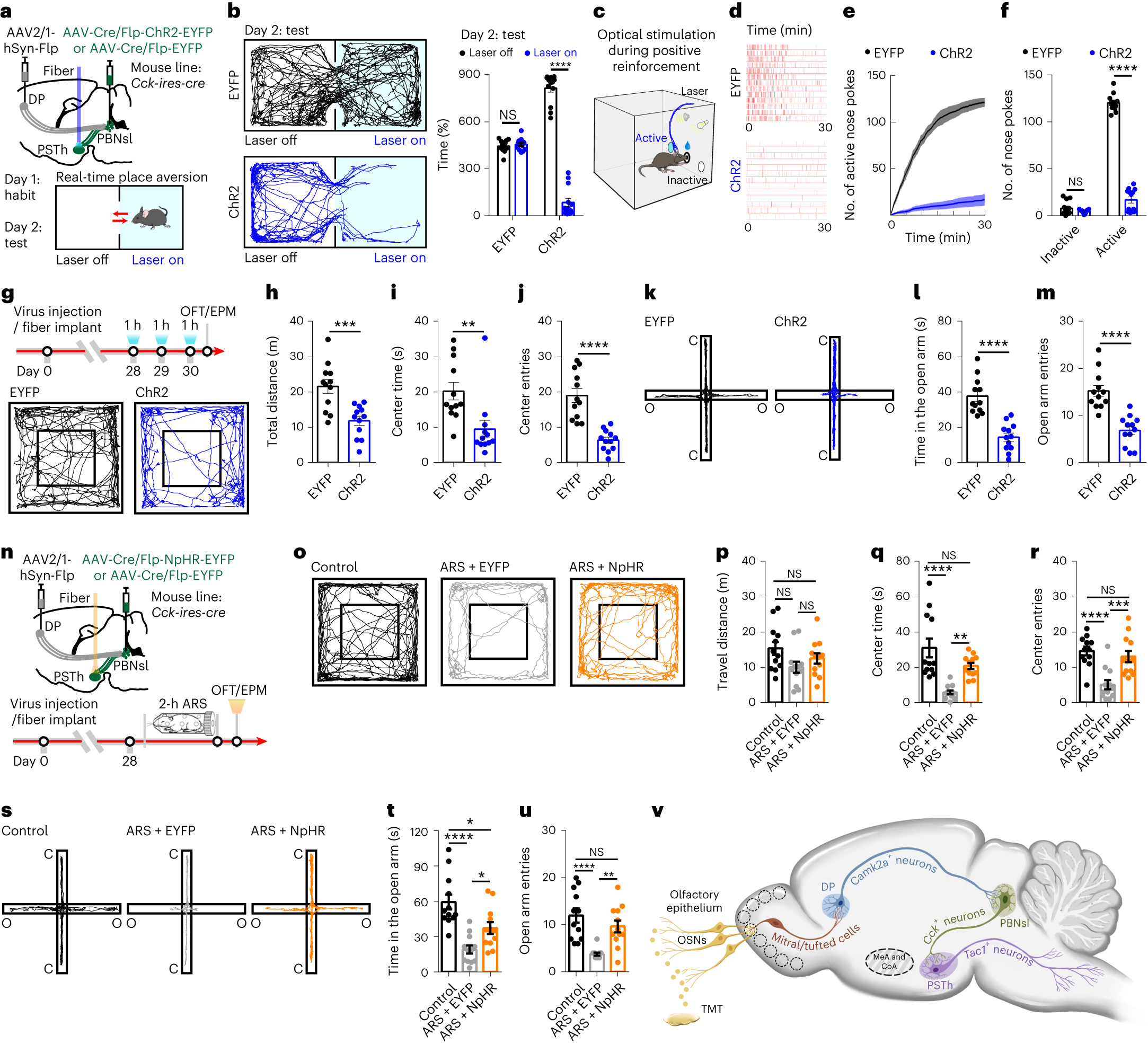
Activating the selected population also makes mice avoid a stimulation-paired area and reduces active nose poking in a rewarded task. After one hour of activation on each of three days, central open-field and open-arm exploration decrease, alongside reduced total locomotion. Several behavioral measures change together in this activation condition.
Suppressing the selected circuit following acute restraint stress improves center and open-arm exploration without a significant between-group difference in total locomotion. Open-arm time nevertheless remains different from the unstressed control. From rapid odor-related escape to exploration after stress, this male-mouse study brings innate defense and negative-state responses together in a molecularly identified route while preserving their distinct behavioral readouts.
Source: Hao Wang et al. · Nature Neuroscience ·
Original figure and English caption ↗