Remembering a dangerous room or a warning sound: how do two parallel amygdala outputs divide fear memory?
A mouse study dissects parallel inhibitory projections from central lateral amygdala PKCδ and SST neurons to the ventral bed nucleus, showing how they separately regulate contextual and auditory fear.
Differentiating and appropriately maintaining contextual versus discrete auditory-cued fear memories is essential for adaptive survival, yet how molecularly distinct subpopulations within the extended amygdala segregate these fear modalities has remained unclear. On September 9, 2023, Xiao-Ming Li, Jiadong Chen, and colleagues at Zhejiang University School of Medicine published a research article in Biological Psychiatry entitled 'Distinct Circuits From the Central Lateral Amygdala to the Ventral Part of the Bed Nucleus of Stria Terminalis Regulate Different Fear Memory' (with Yi Zhu, Shi-Ze Xie, and Ai-Bing Peng as co-first authors). Combining rabies virus monosynaptic retrograde tracing, anterograde transsynaptic HSV labeling, slice electrophysiology, in vivo optogenetics, and local pharmacology, the authors delineated how parallel PKCδCeL→vBNST and SSTCeL→vBNST inhibitory pathways differentially govern contextual and tone fear memories through opioid receptor-mediated modulation.
Key finding: Both PKCδ+ and SST+ GABAergic neurons in the central lateral amygdala (CeL) form monosynaptic inhibitory synapses onto GABAergic and glutamatergic neurons in the ventral bed nucleus of the stria terminalis (vBNST). Prolonged optogenetic activation or inhibition of the PKCδCeL→vBNST pathway selectively reduces contextual fear memory, whereas manipulating the SSTCeL→vBNST pathway predominantly reduces auditory tone fear memory; furthermore, local blockade of δ and κ opioid receptors in the vBNST restores fear memory expression following prolonged pathway activation.
Contents · 5 sections
Rabies tracing and slice electrophysiology map monosynaptic inhibitory inputs from CeL PKCδ+ and SST+ neurons to the vBNST
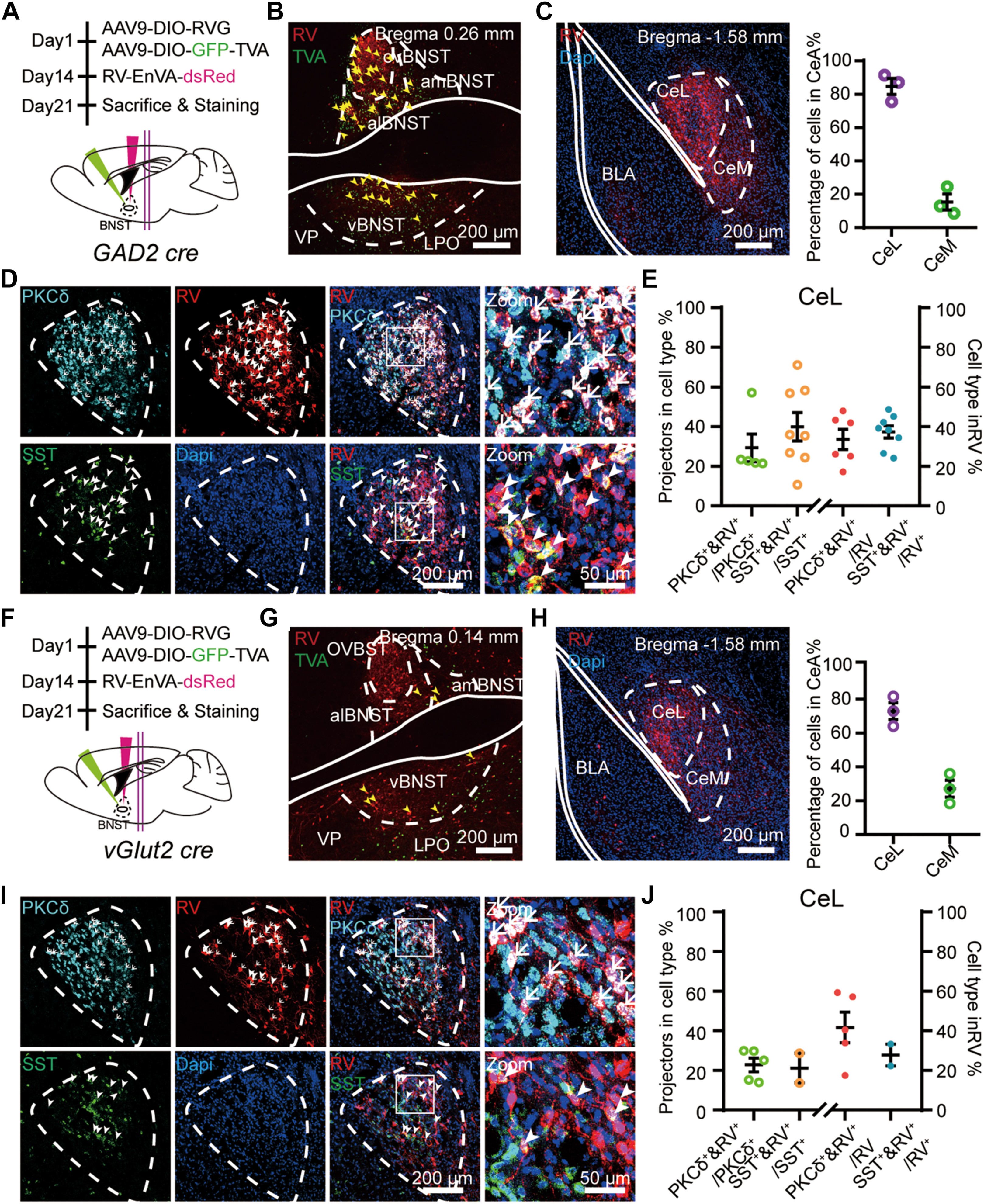
To map cell-type-specific projections from the CeL to the vBNST, the authors injected retrograde AAV2/2Retro Plus-Syn-Cre into the vBNST and Cre-dependent helper viruses plus RV-EnvA-ΔG-dsRed into the CeL (Figures 1A–D). Immunostaining revealed that 49.82% of vBNST-projecting CeL neurons were PKCδ+ and 35.67% were SST+. Anterograde tracing with AAV9-DIO-mCherry in PKCδ-Cre and SST-Cre mice confirmed dense terminal arborizations of both cell types within the vBNST (Figures 1E–J).
Dual-retrograde tracing (CTB-488 in vBNST and CTB-555 in CeM) showed that CeL neurons projecting to the vBNST (43.30%) and CeM (41.78%) were largely segregated, with only 14.92% collateralizing to both targets (Figures 1K–M). Whole-cell patch-clamp recordings in acute vBNST slices from PKCδ-Cre and SST-Cre mice expressing AAV9-DIO-ChR2-mCherry in the CeL demonstrated that blue-light stimulation of either PKCδCeL or SSTCeL terminals evoked robust, short-latency (<5 ms) optically evoked inhibitory postsynaptic currents (oIPSCs, >1000 pA) in 100% of recorded vBNST neurons, which were completely abolished by the GABAA receptor antagonist picrotoxin (Figures 1N–Q).
Source: Yi Zhu et al. · Biological Psychiatry · © Elsevier Inc.
Original figure and English caption ↗
Optogenetic manipulation of PKCδCeL→vBNST versus SSTCeL→vBNST pathways selectively modulates contextual versus tone fear memory
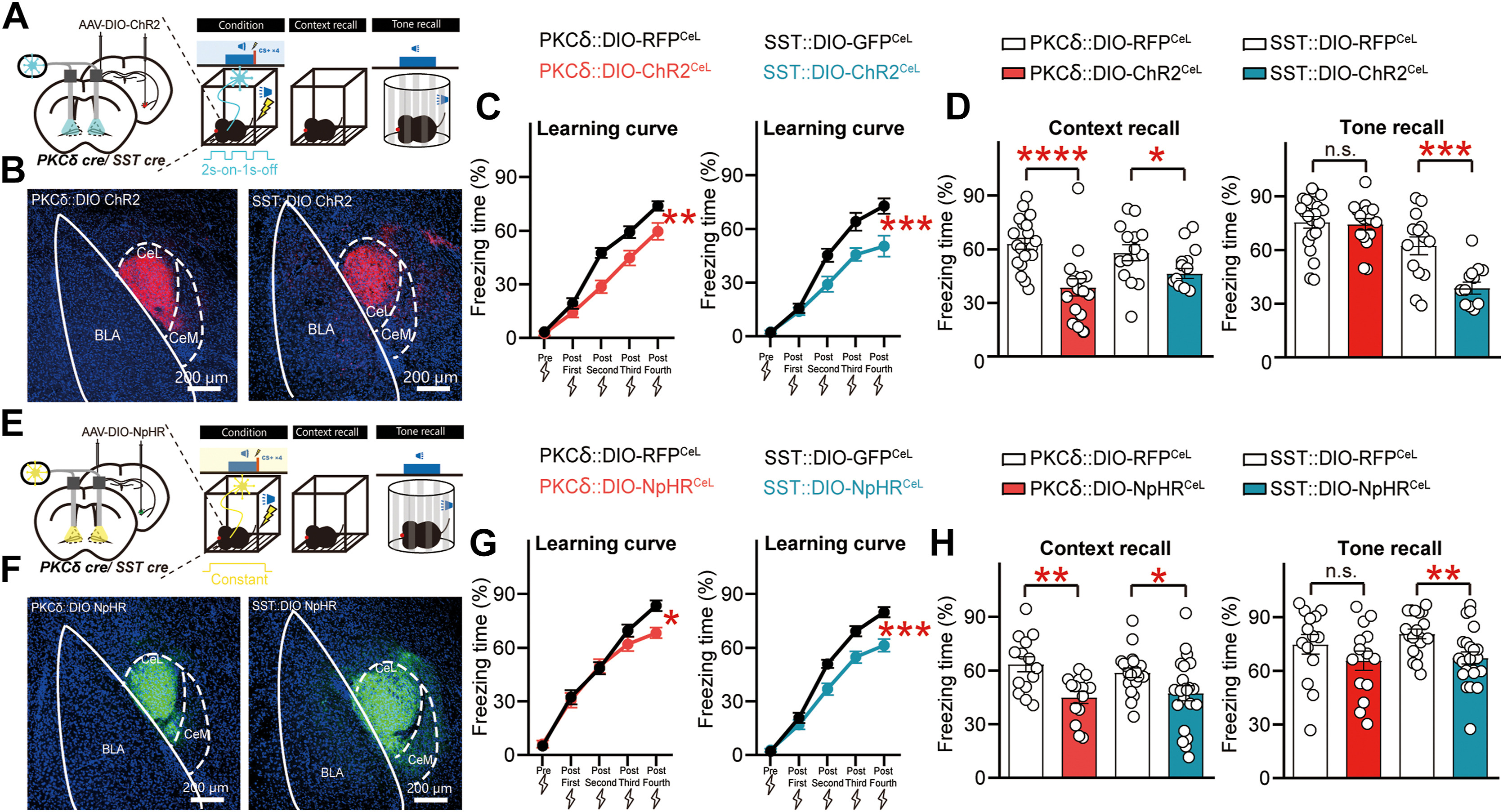
To determine the behavioral roles of these parallel projections during fear memory formation, the authors expressed Cre-dependent ChR2 or NpHR in the bilateral CeL of PKCδ-Cre and SST-Cre mice and photostimulated axon terminals in the vBNST throughout auditory-cued fear conditioning (Figures 2A–H). Delivering either pulsed blue-light activation (ChR2, 2s-on-1s-off) or continuous yellow-light inhibition (NpHR) of PKCδCeL→vBNST terminals during conditioning selectively reduced freezing during subsequent contextual fear recall without altering tone fear recall, whereas whole-session manipulation of SSTCeL→vBNST terminals during cued conditioning reduced both contextual and tone fear recall.
Dissecting specific temporal windows of photostimulation (Original Figures 3A–I) further clarified this functional segregation: during pure contextual fear conditioning (or inter-tone post-shock intervals), activating or inhibiting PKCδCeL→vBNST terminals selectively impaired contextual fear recall whereas manipulating SSTCeL→vBNST terminals had no effect on contextual fear (Figures 3A–F); conversely, restricting SSTCeL→vBNST ChR2 activation strictly to the four 30-s tone-CS presentations selectively enhanced auditory tone fear recall without altering contextual fear (Figures 3G–I).
Source: Yi Zhu et al. · Biological Psychiatry · © Elsevier Inc.
Original figure and English caption ↗
Anterograde transsynaptic targeting of postsynaptic vBNST neurons enables bidirectional regulation of contextual and tone fear
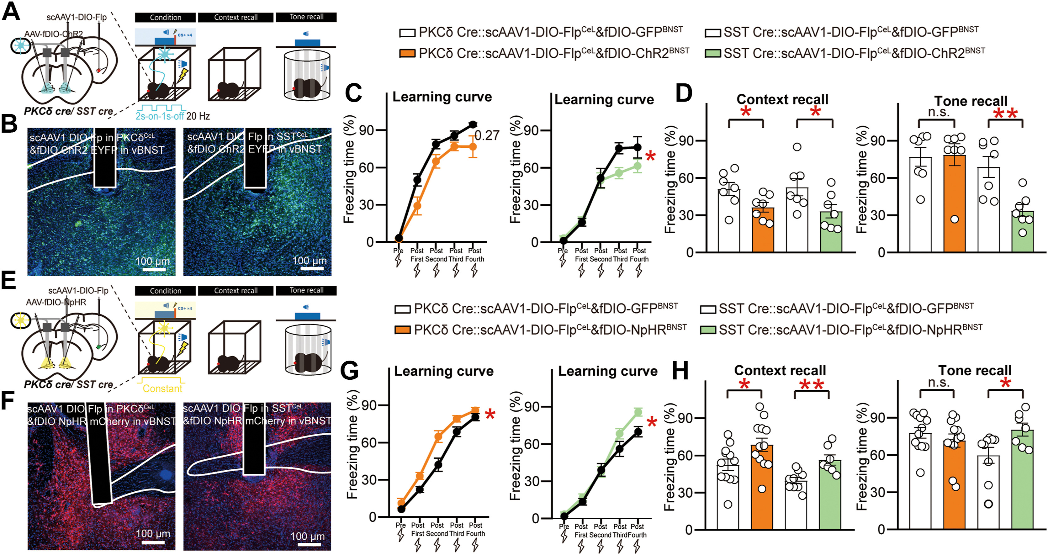
To directly examine the causal role of postsynaptic vBNST neurons, the authors injected anterograde trans-synaptic scAAV1-DIO-Flp into the CeL of PKCδ-Cre or SST-Cre mice and Flp-dependent AAV-fDIO-ChR2 or AAV-fDIO-NpHR into the vBNST (verified by monosynaptic rabies tracing) to selectively manipulate vBNST neurons postsynaptic to either PKCδCeL or SSTCeL inputs (Figures 4A–H).
Direct optogenetic activation (fDIO-ChR2) of vBNST neurons postsynaptic to PKCδCeL inputs decreased contextual fear recall whereas their inhibition (fDIO-NpHR) enhanced contextual fear without affecting tone fear (Figures 4A–D). Meanwhile, activating or inhibiting vBNST neurons postsynaptic to SSTCeL inputs bidirectionally suppressed or enhanced both contextual and tone fear memories (Figures 4E–H), demonstrating that postsynaptic vBNST neuronal activity inversely gates fear memory expression.
Source: Yi Zhu et al. · Biological Psychiatry · © Elsevier Inc.
Original figure and English caption ↗
δ and κ opioid receptor expression in CeL→vBNST circuits accounts for prolonged activation effects and enables pharmacological rescue
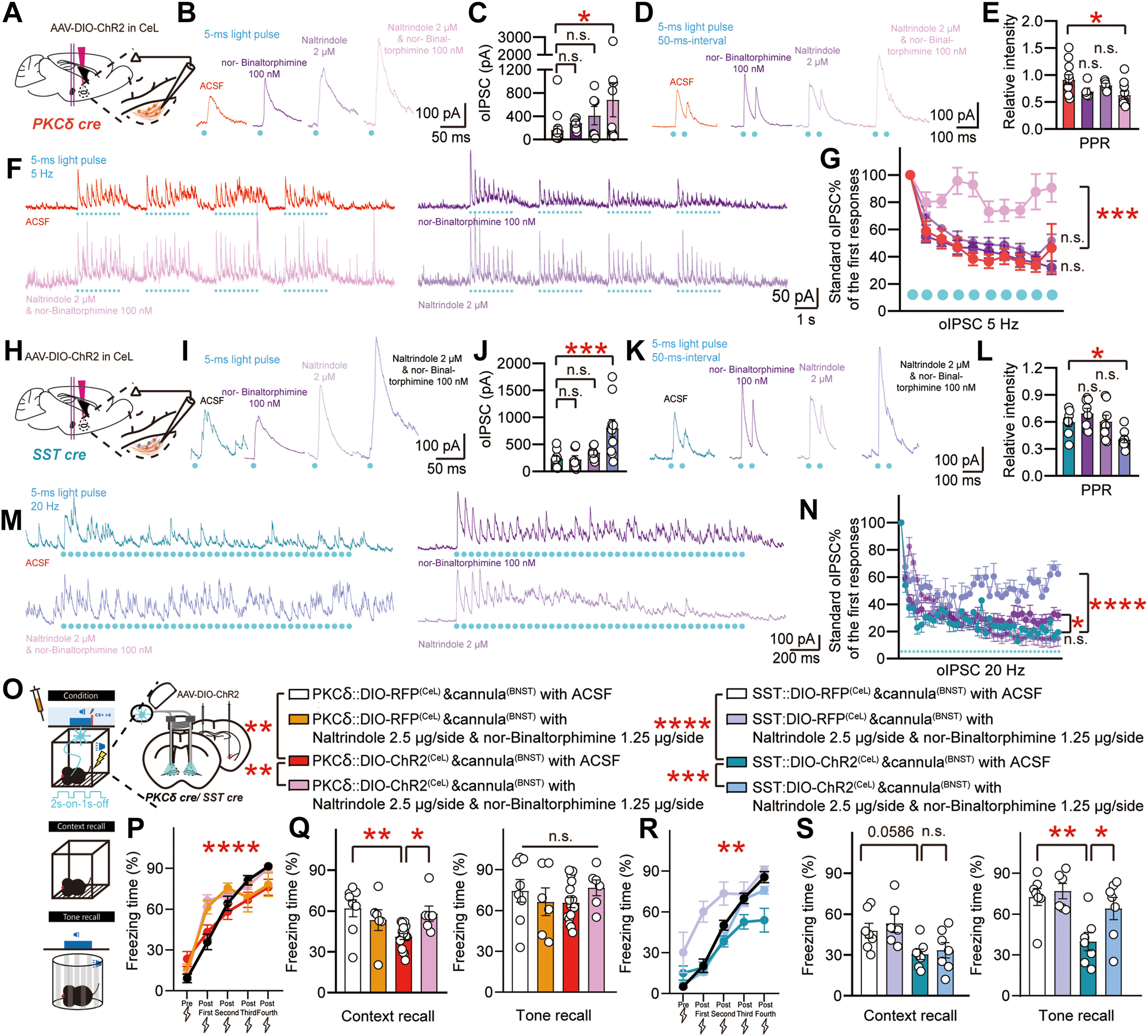
High-resolution confocal immunostaining (Original Figures 5A–N) revealed that δ-opioid receptors (δOR) are preferentially enriched in PKCδCeL somata (~54.8%) and their Synaptophysin+ putative boutons in the vBNST, whereas κ-opioid receptors (κOR) are preferentially enriched in SSTCeL somata (~45.2%) and their vBNST terminals. Slice patch-clamp recordings (Figures 6A–N) demonstrated that the δOR antagonist Naltrindole (2 μM) and κOR antagonist nor-Binaltorphimine (100 nM) increased single-pulse oIPSC amplitude and decreased 50-ms paired-pulse ratio (PPR); crucially, Naltrindole selectively prevented 5 Hz train oIPSC depression at PKCδCeL→vBNST synapses (Figures 6F–G), whereas nor-Binaltorphimine selectively attenuated 20 Hz train oIPSC depression at SSTCeL→vBNST synapses (Figures 6M–N).
To validate this mechanism in vivo, the authors infused an opioid receptor antagonist cocktail (Naltrindole 2.5 μg/side + nor-Binaltorphimine 1.25 μg/side) into the bilateral vBNST prior to conditioning with terminal ChR2 photostimulation (Figures 6O–S). Local opioid receptor blockade completely rescued contextual fear memory in PKCδ::DIO-ChR2CeL mice (Figures 6P–Q) and restored tone fear recall in SST::DIO-ChR2CeL mice (Figures 6R–S).
Source: Yi Zhu et al. · Biological Psychiatry · © Elsevier Inc.
Original figure and English caption ↗
Beyond Local CeL Reciprocal Inhibition: Parallel Projections Independently Encode Cued and Contextual Fear
Classical models of fear processing posit that somatostatin-positive (SOM+) and protein kinase C-delta-positive (PKC-delta+) neurons within the central lateral amygdala (CeL) gate fear expression primarily via local reciprocal inhibition ('Fear-On' versus 'Fear-Off' seesaw dynamics) relayed through the central medial amygdala (CeM). By contrast, this study demonstrates that both SOMCeL->vBNST and PKCdeltaCeL->vBNST pathways form functional monosynaptic inhibitory connections onto vBNST GABAergic neurons, exerting opposing control over fear behaviors directly at their downstream target.
Crucially, because δ and κ opioid receptors are differentially enriched across the two CeL→vBNST pathways, endogenous opioid peptide co-release recruited during intense terminal activation engages presynaptic δOR/κOR autoinhibition to depress subsequent GABA release. This parallel long-range projection and GABA–opioid co-modulation framework explains how contextual and discrete auditory-cued threats are differentially processed along the CeL-to-vBNST axis.